2: intro into ret
1/24
There's no tags or description
Looks like no tags are added yet.
Name | Mastery | Learn | Test | Matching | Spaced | Call with Kai |
|---|
No analytics yet
Send a link to your students to track their progress
25 Terms
intro into retinoscopy
what is retinoscopy
it is means of objectively determining the size of refractive error
can determine if the eye has astig,atism and can correct it
millodots definition of retinoscopy
the determination of the refractive state of the eye by means of a retinscope
why is retinscopy objective
patient is not required to respond as no questions are being asked
why do we ask pateints to look into the distance
to relax their eyes focusing myscles (ciliary muscles), making it easier to read the true prescription. Eyes stop accomodating giving a more accurate measurement
what does retinscope look like
light emitted by streak ret shows rectangle beam, spot ret is circle
sleeve/ collar must always be at lowest position, turning the collar changes the orientation of the streak
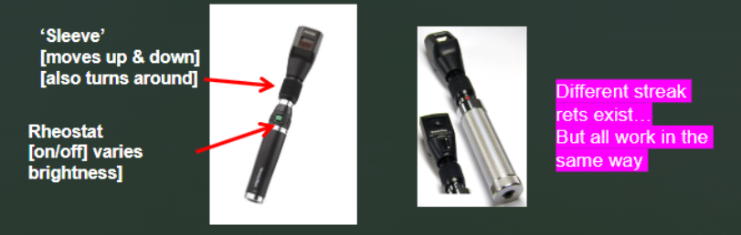
what does changing orientation of streak using sleeve mean
tells us that the eye has diff focusing powers in diff directions - astigmatism
streak of light moves differently depending on the axis of astig
by rotating sleeve and watching how light relfex changes doctor can figure out:
axis of astig
amount of astig
advantages of retinoscopy
objective means establishing refractive error
speeds up refractive error determination
quick to perfomr
accurate
in some patients specs can be prescribed directlty ffrom retinoscopy
disadvantages
can be difficult to learn
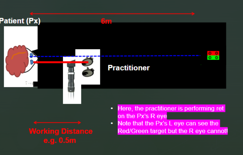
basic set up for retinoscopy
patient wears trial frame to hold lenses
patient views distant target 6m, so refractive eror can be determined when accom is relaxed
direct them to spotlight target needed toholf eyes steady and ensure looking a fixed distance away
fairly level with the eyes
dim lights
observe relfex of one eye using BOTH eyes
working distance
should take under a minute
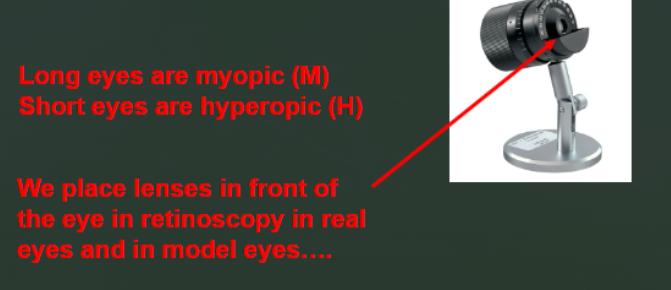
how ca we generate different amounts of myopia or hyperopia
by rotating/ pulling back the back of the eye
the scale is usually calibrated :
e.g. -4 to+4 unit in D
minus signs
simulated myopia
if scale reads -3.00 Dt he model eye is myopic by -3.00 D
plus signs
simulated hyperopia e.g if the cale reads +2.00 D the model eye is hyperopic by +2.00 D
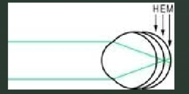
how do you alter axial length
pull retina away from the front of the eye or by pushing it towards the front of the eye
long eyes are myopic
short eyes are hyperopic
leses are placed in front of the eye
dioptre
a unit proposed by monoyer to evalate the refractive power of the lens or of an optical system
duochrome
a subjective refraction test in which the sibject compares the sharpness of black targets of similar sizes on a red background on one side and a green on the other
what are we doing when we alter axiallength
we are generating axial ametropia
when light located at E light from the distant object is fomred on the retina
when retina is moved further away from the front of the eye ( longer axial length) eye becomes myopic M
when retina moved closer then eye becomes hyperopic H
how does retinoscopy work
involves you comparing the movement of retinal reflex with the movement of light outside the patients eye, when the retinoscopy light is moved
whats retinal reflex
the light that bouncs back from the eye of patient youre examining relfex might move with or againts you
with and against movements
if you move light to th right:
if relfex moves left its against: myopic; means far point is in front of infinity - somehwere closer to examiner
if behind the far point during retinoscopy (usually are unless really close) the light leaving eye diverges and moves in opp direction
if reflex moves right its with; hyperopic, youre in front of the far point
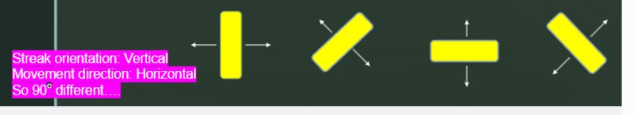
always move the light at right angles to the prientation of streak e.g if streak is vertical, move the light horizontally
in myopia the far point is closer to the eye so you end up being behind it causing against movement
speed of relfex
shows how fast light moves across the pupil
fast reflex: eye is closer to emmetropia
slow reflex: eye is far from focused, high myopia or hyperopia
tells how weak the refractive error is
brightness of reflex
bright reflex: eyes focusing is stronger or closer to neutral , easy to see
dim reflex: may indicate high refractive error , small pupil or media opacity like cataracts
how does dim reflex indicate cataracts
catarcats is clouding of the lens
cloudy lens scatters and absorbs some of the light, so less light reaches the retina and reflected back
what are we trying to achieve in retinoscopy
we are trying to identify the lens power where the relfex movement is neither with or against but instead flashes ( reversak) .
shows an endpoint of retinoscopy