Comprehensive Guide to Thyroid and Parathyroid Ultrasound and Pathology
1/77
There's no tags or description
Looks like no tags are added yet.
Name | Mastery | Learn | Test | Matching | Spaced | Call with Kai |
|---|
No analytics yet
Send a link to your students to track their progress
78 Terms
Where is the thyroid gland located?
In the anteroinferior neck at the level of the thyroid cartilage.
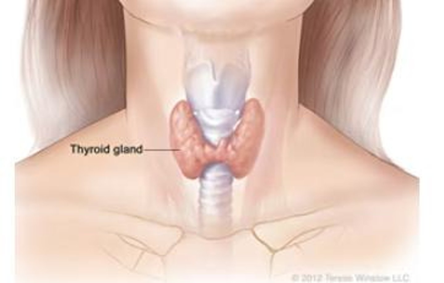
What connects the right and left lobes of the thyroid gland?
The isthmus.
What anatomical variant may be seen in the thyroid gland?
Pyramidal lobe.
What is the normal length of an adult thyroid gland?
4-6 cm.
What is the normal width of an adult thyroid gland?
1.5-2 cm.
What is the normal anteroposterior diameter of an adult thyroid gland?
2-3 cm.
What is the normal isthmus diameter of the thyroid gland?
4-6 mm.
What is the normal mean thyroid volume?
10-12 ± 3 mL.
What is the common method to calculate thyroid volume?
Length x Width x Thickness x 0.52 for each lobe.
What muscles are located anterior to the thyroid gland?
Strap muscles including sternothyroid, omohyoid, and sternohyoid.
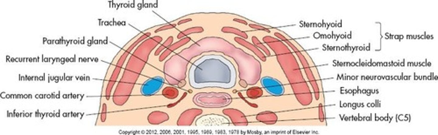
What structures are found posterior/laterally to the thyroid gland?
Common carotid artery, internal jugular vein, and vagus nerve.
What is a common cause of hypothyroidism?
Undersecretion of thyroid hormones, possibly due to low iodine intake.
What are some symptoms of hypothyroidism?
Weight gain, hair loss, lethargy, cold intolerance, and constipation.

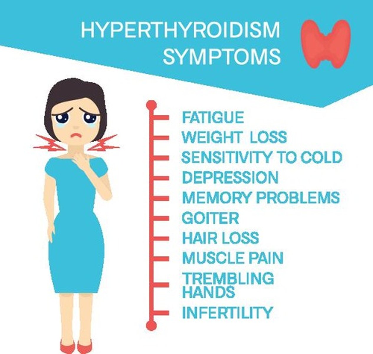
What condition results from oversecretion of thyroid hormones?
Hyperthyroidism.
What are some symptoms of hyperthyroidism?
Weight loss, increased appetite, tremors, and exophthalmos.
What tests are used to determine thyroid function?
Iodine uptake scan and thyroid scan.
What indicates hyperthyroidism on a thyroid scan?
Increased radioactivity, known as a hot spot.
What indicates hypothyroidism on a thyroid scan?
Decreased radioactivity, known as a cold spot.
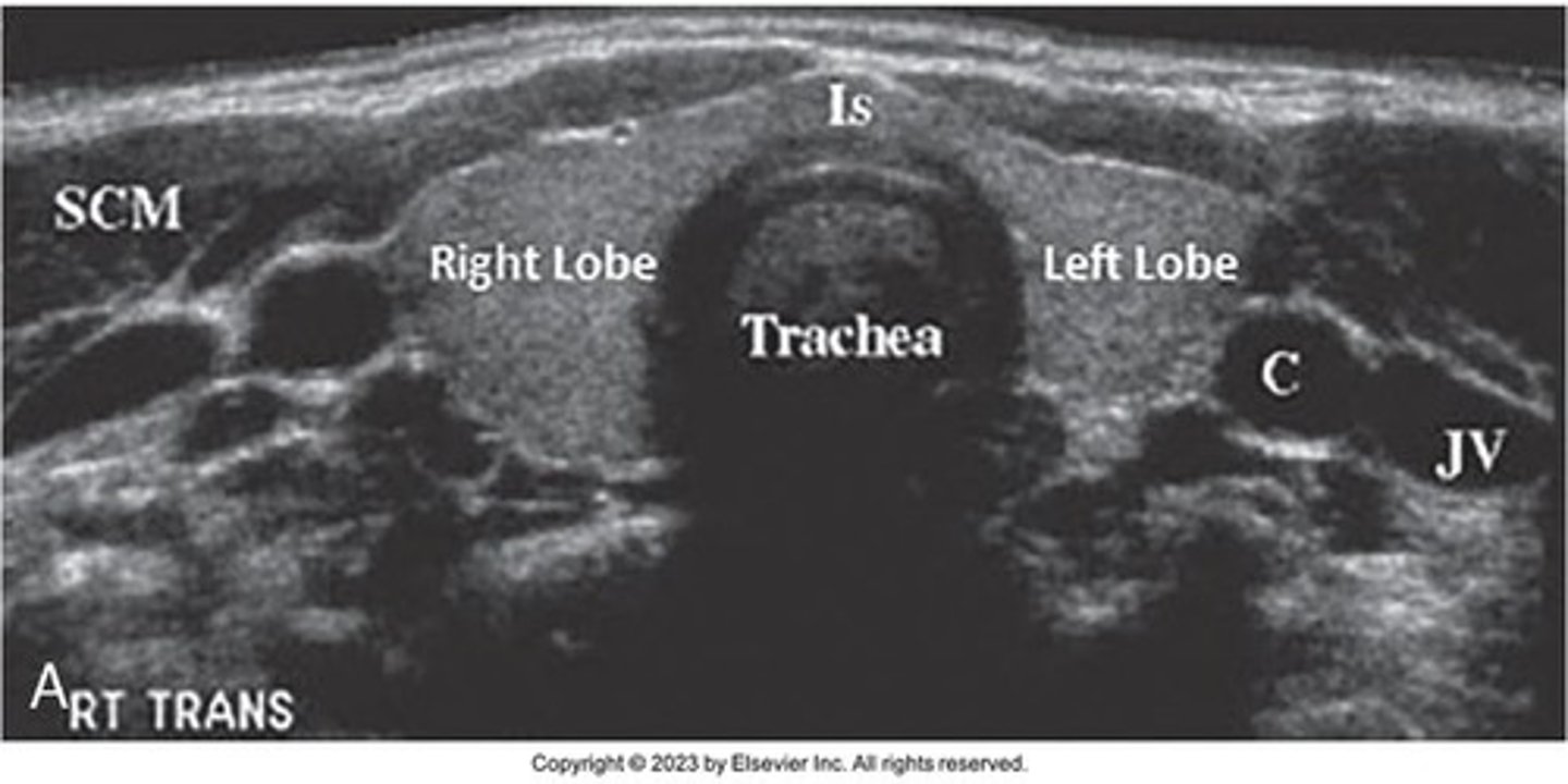
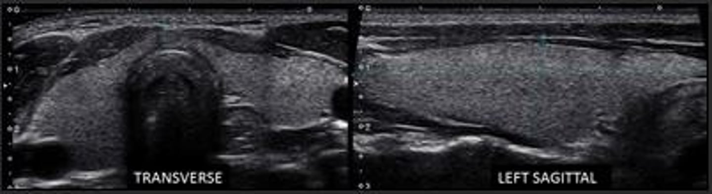
What is the sonographic appearance of the thyroid gland?
Fine, homogenous echotexture slightly more echogenic than surrounding musculature.
What is the purpose of the Thyroid Imaging Reporting & Data System (T-RADS)?
To determine thyroid cancer risk based on ultrasound characteristics.
What is goiter?
Enlargement of the thyroid gland due to compensatory hypertrophy and hyperplasia.
What is a multinodular goiter?
A hyperthyroid condition resulting from hyperactivity of the thyroid gland.
What is endemic goiter?
Goiter occurring in geographic areas with low iodine levels in soil, food, and water.
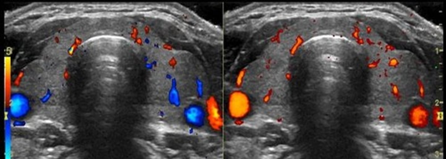
What is the primary blood supply to the thyroid gland?
Two superior thyroid arteries from external carotids and two inferior thyroid arteries from the thyrocervical trunk.
What is the Doppler peak systolic velocity range for major thyroid arteries?
20 to 40 cm/sec.
What is the Doppler peak systolic velocity range for intraparenchymal arteries?
15 to 30 cm/sec.
What is the appearance of the esophagus on ultrasound?
Identified by a target appearance in transverse plane and peristaltic movements when swallowing.
What is the most common cause of thyroid disorders worldwide?
Iodized salt as a dietary supplement.
What are the sonographic findings of a goiter?
Overall gland enlargement or discrete nodules, which may be poorly circumscribed or well-defined with a thin peripheral hypoechoic halo.
What characterizes a multinodular goiter?
An inhomogeneous enlarged tissue mass with increased vascularity.
What is a thyroid cyst?
A cystic degeneration of follicular adenoma that can be purely cystic, contain colloid fluid, or be hemorrhagic.
What are the sonographic findings of a thyroid cyst?
Anechoic cyst within the thyroid gland or a colloid cyst with an echogenic focus.
What defines a benign thyroid adenoma?
A benign neoplasm characterized by complete fibrous encapsulation.
What are the sonographic characteristics of thyroid adenomas?
Adenomas are often solitary, homogeneous, and can vary in size and echogenicity, with a peripheral halo.
What is Graves' disease?
An autoimmune disorder that attacks the thyroid, leading to elevated T3 and T4 levels, and is the most frequent cause of hyperthyroidism.
What are the clinical findings of Graves' disease?
Hypermetabolism, diffuse toxic goiter, and exophthalmos (bulging eyes).
What are the sonographic findings in Graves' disease?
Enlarged, inhomogeneous gland with increased vascularity, especially in overactive cases.
What is thyroiditis?
Inflammation of the thyroid due to infections, autoimmune diseases, or other causes.
What are the types of thyroiditis?
Acute suppurative, subacute granulomatous (de Quervain disease), and chronic lymphocytic (Hashimoto disease).
What are the symptoms of Hashimoto's thyroiditis?
Painless, diffusely enlarged gland, often in young or middle-aged females, with elevated TSH and decreased T3 and T4.
What are the sonographic findings of Hashimoto's thyroiditis?
Enlarged, heterogeneous gland with ill-defined hypoechoic areas and variable flow patterns.
What characterizes malignant thyroid lesions?
Solid thyroid nodules with cervical adenopathy, often presenting as painless, palpable, hard nodules.
What is papillary carcinoma?
The most common thyroid malignancy, characterized by round, laminated calcifications and lymphatic spread.
What are the sonographic features of papillary carcinoma?
Hypoechoic nodules, taller than wide, with microcalcifications and increased vascularity.
What defines follicular carcinoma?
The second most common type of thyroid cancer, aggressive, and spreads through the bloodstream.
What are the sonographic findings of follicular carcinoma?
Thick irregular halo, tortuous internal blood vessels, and cervical lymphadenopathy.
What is medullary carcinoma?
A type of thyroid cancer that accounts for 5% of cases, often familial and associated with multiple endocrine neoplasia.
What are the sonographic findings of medullary carcinoma?
Solid, hypoechoic mass with calcifications.
What is anaplastic carcinoma?
A rare, undifferentiated, and lethal thyroid tumor that occurs mostly in men over 60.
What are the characteristics of lymphoma in the thyroid?
Non-Hodgkin's lymphoma accounts for 4% of thyroid malignancies, often seen in older women with a rapidly growing neck mass.
What is the typical presentation of anaplastic carcinoma?
A hard, fixed mass with rapid growth, dyspnea, dysphagia, and a high mortality rate.
What is the primary function of the parathyroid glands?
To produce parathyroid hormone (PTH) and monitor serum calcium levels.
Where are the parathyroid glands located?
On the posterior medial surface of the thyroid gland.
How many parathyroid glands are typically present?
Typically four, but some individuals may have three or five.
What is the size of each parathyroid gland?
Approximately 5 x 3 x 1 mm, flat, and disc-shaped.
What happens to PTH levels when serum calcium decreases?
PTH levels increase.
What laboratory findings are associated with primary hyperparathyroidism?
Elevated serum calcium (hypercalcemia), elevated urine calcium (hypercalciuria), and low phosphorus (hypophosphatemia).
What is the most common cause of primary hyperparathyroidism?
Parathyroid adenoma.
What is secondary hyperparathyroidism caused by?
Chronic hypocalcemia due to renal failure, vitamin D deficiency, or intestinal malabsorption syndromes.
What are the sonographic characteristics of a parathyroid adenoma?
Solid, oval, homogenous, hypoechoic masses usually <3 cm in size.
What is a key feature of parathyroid carcinoma on ultrasound?
Large mass with irregular or lobulated borders and increased vascularity.
What is a thyroglossal duct cyst?
A congenital anomaly that appears as a palpable mass in the midline of the neck, typically between the hyoid bone and the isthmus.
What are branchial cleft cysts?
Cystic formations usually located lateral to the thyroid in the submandibular region, resulting from embryonic development.
What is the sonographic appearance of an abscess?
Complex cystic mass with low-level echogenicity and irregular walls.
What is cervical adenopathy?
Abnormal enlargement of lymph nodes, appearing hypoechoic with a thick outer cortex.
What is the typical appearance of normal lymph nodes on ultrasound?
Symmetrical, oval shape, echogenic hilum, and thin outer cortex.
What is the significance of unexplained hypercalcemia?
It is a common referral for parathyroid sonography.
What is the role of PTH in the body?
To increase calcium absorption in the blood from bone, kidney, and intestine.
What imaging technique is used for evaluating parathyroid glands?
High-resolution ultrasound with a 7.5- to 15-MHz linear transducer.
What is the typical age group for primary hyperparathyroidism?
More common in women over the age of 40.
What is the treatment for parathyroid adenoma?
Surgical removal of the adenoma.
What are the sonographic findings of chronic abscesses?
Isoechoic with surrounding tissue and indistinct margins.
What is the appearance of parathyroid hyperplasia on ultrasound?
Enlarged parathyroid glands, which may be >1 cm.
What is a common symptom of a parathyroid adenoma?
Most patients are asymptomatic at the time of diagnosis.
What is the relationship between vitamin D deficiency and parathyroid function?
Vitamin D deficiency can lead to secondary hyperparathyroidism.
What is the significance of the longus colli muscle in parathyroid ultrasound?
It appears as a discrete area posterior to the thyroid and helps in identifying parathyroid glands.
What is the typical size of parathyroid adenomas?
Usually <3 cm, with larger adenomas >5 cm.
What is the appearance of parathyroid carcinoma on ultrasound?
Irregular mass with lobulated borders and increased vascularity.