The brain (Lab 2)
1/48
There's no tags or description
Looks like no tags are added yet.
Name | Mastery | Learn | Test | Matching | Spaced | Call with Kai |
|---|
No analytics yet
Send a link to your students to track their progress
49 Terms
Describe the organization of the brain
The brain is made up of a central gray matter region surrounding a fluid-filled cavity. Surrounding the gray matter are large white matter tracts (commissural, projection, and association) for carrying ascending and descending information. Then, superficial to the white matter is another region of gray matter (the cerebral cortex), which is where sensory inputs converge to allow us to make sense of our environment.
What is the neural tube?
It is an embryonic precursor to the central nervous system. Aka it eventually develops to form the brain and spinal cord.
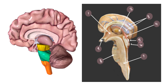
What are the five main parts of the brain
1. Ventricles
- lateral, third, fourth
2. Cerebrum
- 2 cerebral hemispheres
- 5 different lobes
3. Diencephalon
- pineal gland/body
- thalamus
- hypothalamus
4. Brainstem
- midbrain
- pons
- medulla oblongata
5. Cerebellum (“the little brain”)
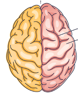
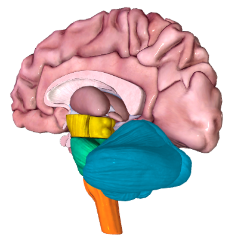
What is this picture showing?
The cerebrum. Orange is the left hemisphere and red is the right hemisphere.
Purple region: name and description
Diencephalon
Composed of the pineal gland/body, thalamus, and hypothalamus
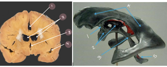
Describe the ventricles of the brain
The ventricles are a series of chambers traveling down the brain’s embryological center to the spinal cord. They contain cerebrospinal fluid (CSF), and help to distribute nourishment and provide protection to the brain.
#3: Name and function
Choroid Plexus
A specialized blood vessel that produces CSF for the ventricles of the brain
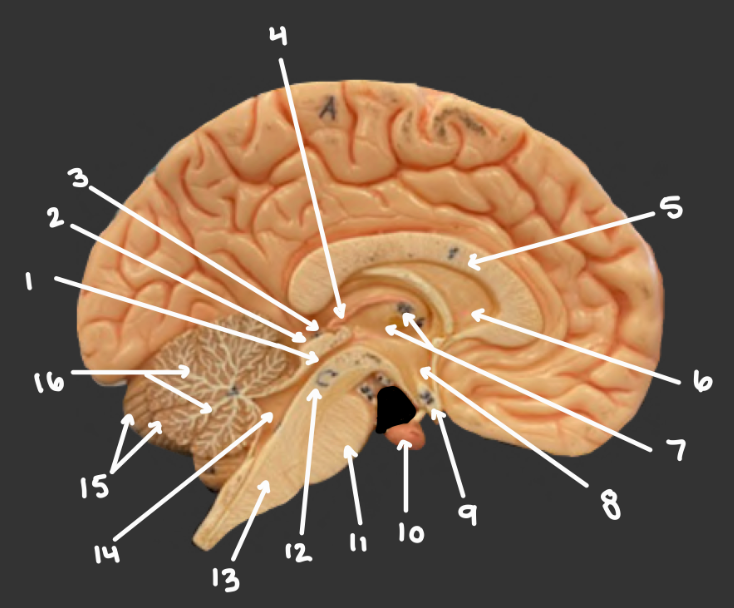
#2: Name and function
Lateral ventricles
Lie within the cerebral hemisphere. Anteriorly, they come in close contact and are separated by a thin membrane called the septum pellucidum
#6: Name and function
Septum pellucidum
A thin membrane that separates the two lateral ventricles
#1: Name and function
Interventricular foramen
Each lateral ventricle leads into an interventricular foramen, which opens to the third ventricle.
#3 and #2
Third Ventricle
The 3rd ventricle is found within the diencephalon. Each interventricular foramen opens to the third ventricle, which then narrows inferiorly to form the cerebral aqueduct.
#1 and #3: Name and function
Cerebral Aqueduct
Formed by the narrowing of the 3rd ventricle, which sits superior to it. The cerebral aqueduct travels through the midbrain and connects with the 4th ventricle.

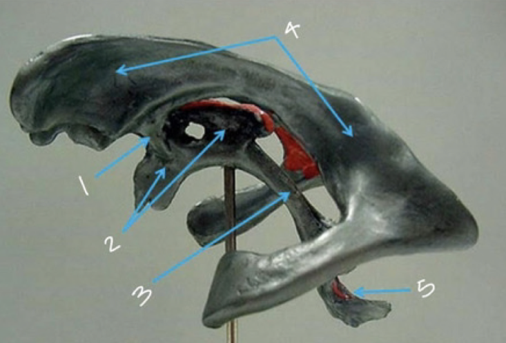
#14 and #5: Name and function
4th ventricle
Found between the pons and cerebellum, it is a large, diamond-shaped expansion. It is connected to the cerebral aqueduct superiorly and the central canal of the medulla and spinal cord inferiorly.
What is the connection sequence of the ventricles in the brain?
The lateral ventricles connect to the interventricular foramen which connect to the third ventricle which connect to the cerebral aqueduct which connect to the fourth ventricle which connects to the central canal of the medulla and spinal cord.
#3: Name and function
Arachnoid granulations
They are outpockets of the arachnoid mater that allow the dural venous sinuses to absorb CSF
What is the role of the dural venous sinuses
The dural venous sinuses surround the brain and absorb excess CSF via arachnoid granulations
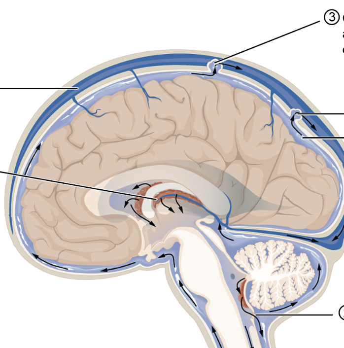
Describe the circulation of CSF through the brain
CSF is produced by the choroid plexus and enters the lateral ventricles. It then moves through the interventricular foramen to the 3rd ventricle. From there, it travels through the cerebral aqueduct, which is surrounded by the midbrain. From there, it travels into the 4th ventricle, which is surrounded by the pons (anteriorly) and cerebellum (posteriorly). Small foramina allow the CSF to enter the subarachnoid space surrounding the brain and spinal cord (this is where it is most important). Excess CSF then moves from the subarachnoid space through the arachnoid granulation into the dural venous sinuses surrounding the brain.
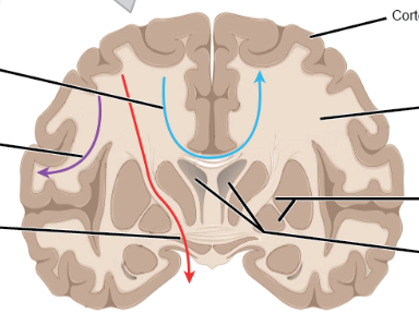
What are the three main tracts within the white matter of the brain, and what do they do? ID them in the pic
Blue: Commissural tract - connects the right and left sides of the brain
Red: Projection tract - connects higher and lower regions of the central nervous system. They can be ascending or descending. For example, the interneurons within the corticospinal and spinothalamic pathways are projection tracts
Purple: Association tract - connects one part of the cortex to adjacent regions of the same hemisphere. Generally found in the cerebrum

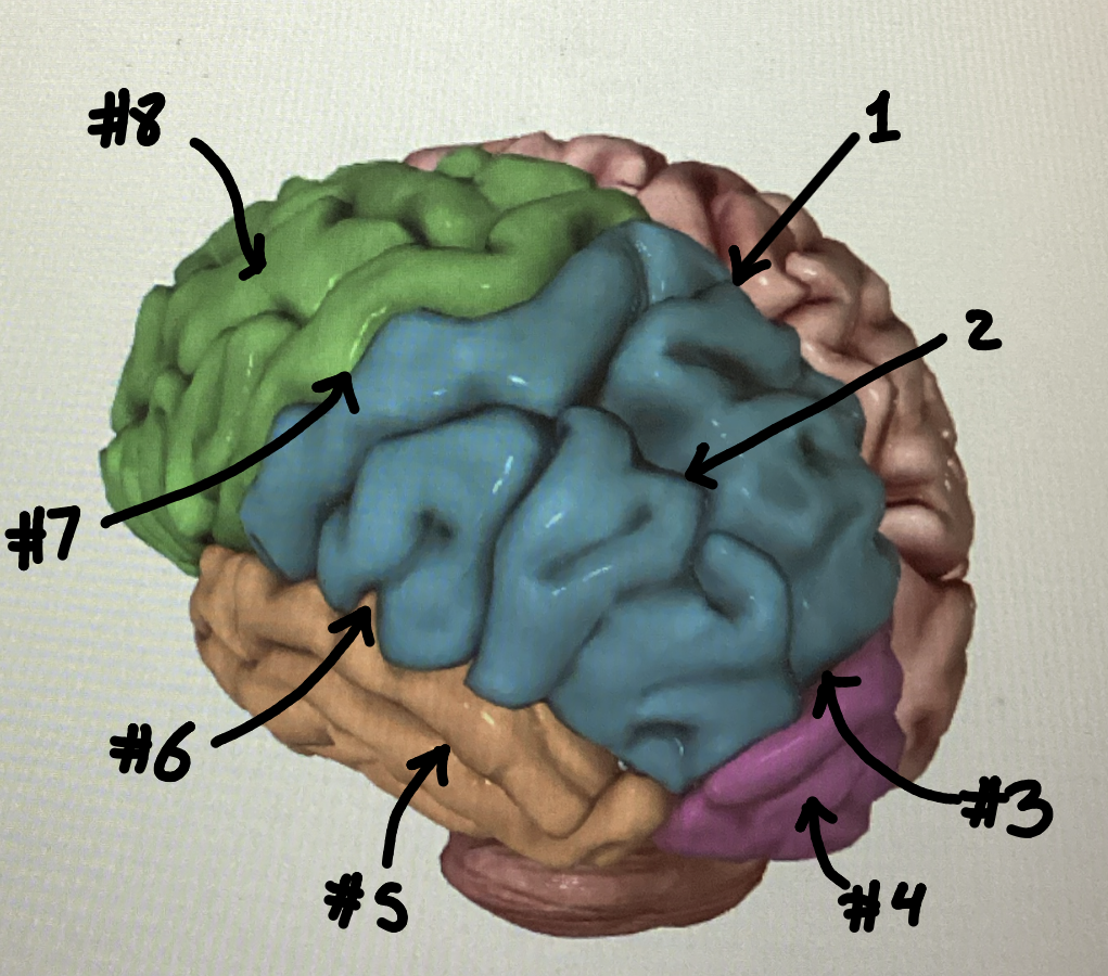
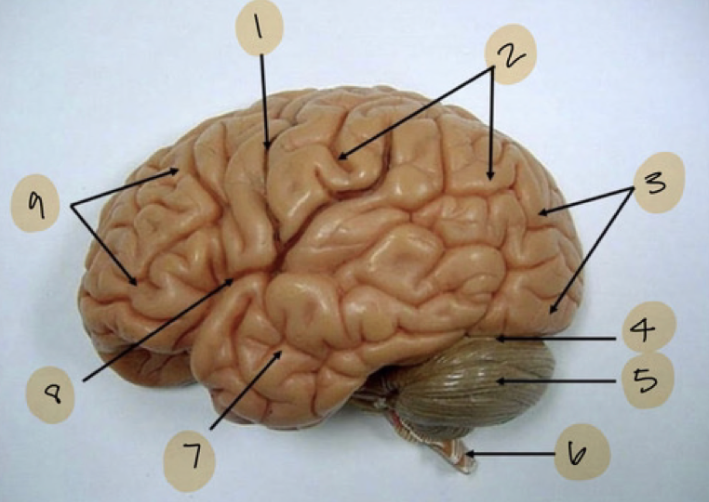
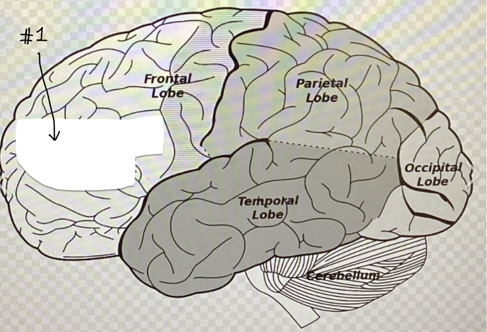
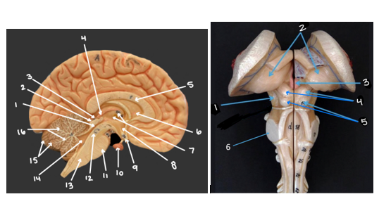
What are the five lobes of the brain? ID them in the picture.
#2 is the parietal lobe. #3 is the occipital lobe. #7 is the temporal lobe. #9 is the frontal lobe.
The fifth lobe is the insular lobe. It is sucked away within the lateral sulcus (#8) and hard to visualize
What are the ridges, grooves, and major grooves on the surface of the cerebrum called?
The ridges are called gyri (gyrus, singular). Grooves are called sulci (sulcus, singular). Major grooves are called fissures.
What are the four essential gyri/sulci/fissures to know and what do they do? ID them in the pictures
#1: Longitudinal fissure - separates the right and left hemispheres
#7: Central sulcus - divides the frontal and parietal lobes
#6: Lateral sulcus - divides the temporal from the frontal and parietal lobes
#3: Parieto-occipital sulcus - divides the parietal and occipital lobes

Identify the red region (name and function)
Precentral gyrus/ primary motor cortex
There is a precentral gyrus on the frontal lobe of each hemisphere, and it is the site of the primary motor cortex. The primary motor cortex initiates and controls voluntary movements by sending neural commands to muscles on the opposite side of the body
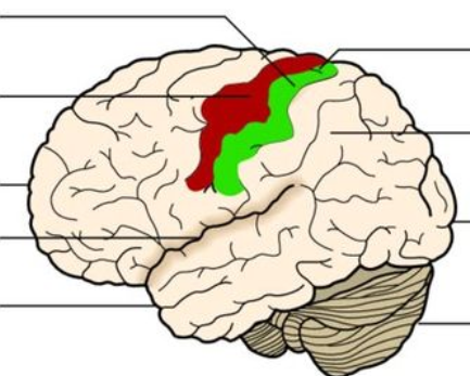
Identify the green region (name and function)
Postcentral gyrus/ primary somatosensory cortex
There is a postcentral gyrus on the parietal lobe of each hemisphere, and it is the site of the primary somatosensory cortex. The primary somatosensory cortex processes sensory information from the body, such as touch, temperature, and pain
What are the four main cortices for the special senses? What part of the cerebral cortex do they project to? ID the region in the picture
Primary visual cortex: projects to the occipital lobe (#3)
Primary auditory cortex: projects to the temporal lobe (#7)
Primary olfactory cortex: projects to the temporal lobe (#7)
Primary gustatory (taste) cortex: parietal and insular lobes (parietal is #2, insular is hidden)
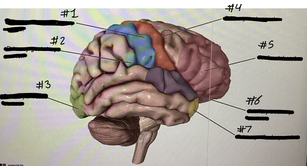
Identify each cortex and its location
1: Primary somatosensory cortex (located in the postcentral gyrus)
2: Primary gustatory cortex (located in the parietal and insular lobes)
3: Primary visual cortex (located in the occipital lobe)
4: Primary motor cortex (located in the precentral gyrus)
5: Prefrontal cortex (located in the frontal lobe)
6: Primary auditory cortex (located in the temporal lobe)
7: Primary olfactory cortex (located in the temporal lobe)
#1: Name and function
The prefrontal cortex
A multimodal association area, meaning it integrates inputs from multiple senses and allows us to make meaning of the information we receive. The prefrontal cortex is important in social interactions and personality.
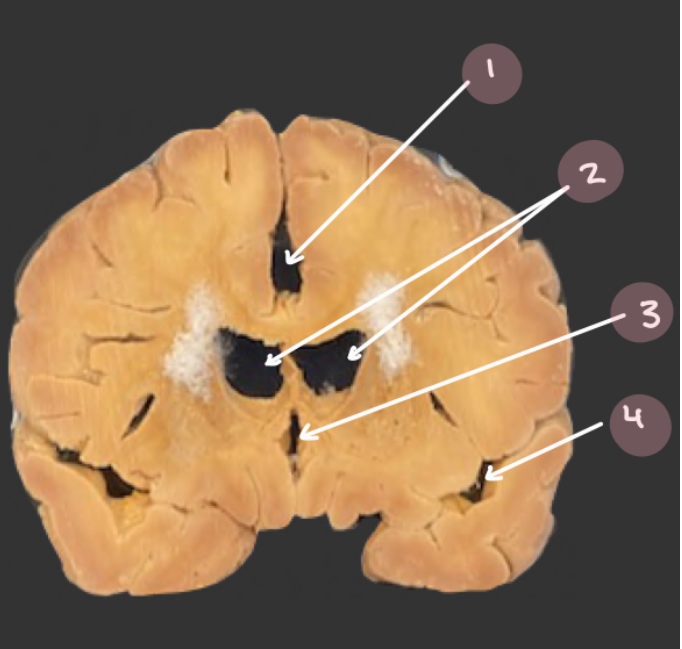
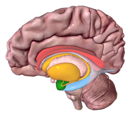
What do the basal nuclei do
The basal nuclei are grey matter clusters into two main bodies and separated by the internal capsule. The basal nuclei are responsible for the regulation of movement initiation and coordinated control of antagonistic muscle pairs
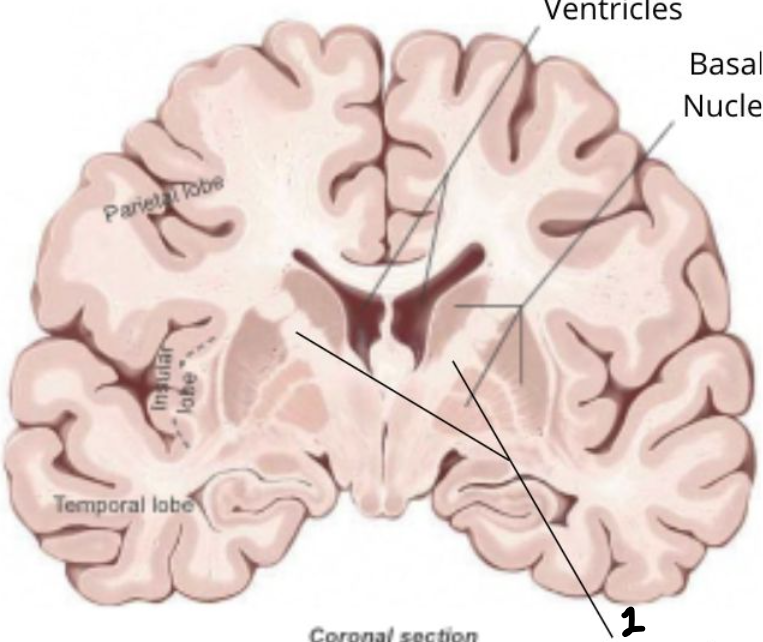
#1: Name and function
Internal capsule
A large bundle of projection tracts that contains axons heading to and from the cerebral cortex. It is located between/ separates the two basal nuclei
What does the amygdala do?
The amygdala is involved in memory, decision-making, and emotional responses.
What does the hippocampus do?
The hippocampus has a role in spatial memory and navigation
Dark pink: Name and function
Corpus callosum
The largest commissural tract, where neurons cross the hemispheres
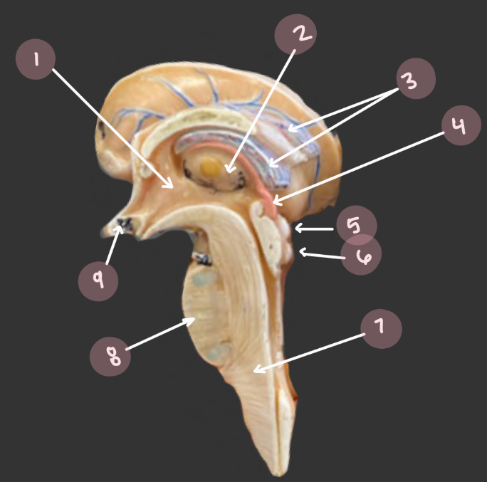
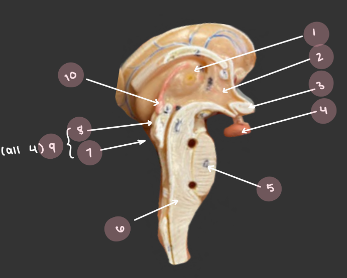
#4 (left) and #3 (right): Name and function
Pineal gland/body
Part of the diencephalon. It produces melatonin, which prepares the brain for sleep

#7 (left) and #2 (right): Name and function
Thalamus
Part of the diencephalon. Made up of two egg-shaped halves with the third ventricle sandwiched between them. Often called the relay station of the brain because it is the synapse site for nearly all sensory pathways (signals are routed from the spinal cord or lower brain to the appropriate region of the cerebral cortex)
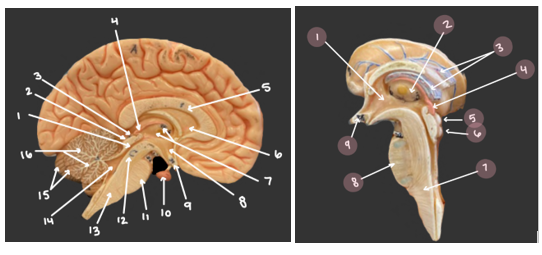
#8 (left) and #1 (right): Name and function
Hypothalamus
Part of the diencephalon. Has several nuclei containing autonomic, pituitary, body temperature, sleep cycle, and emotion control centers. The hypothalamus also responds to stimuli in the brain by releasing hormones that control the pituitary gland
#10 (left) and #4 (right)
Pituitary gland
Controlled by and suspended from the hypothalamus. This gland produces hormones that control the thyroid, adrenal cortex, liver, and reproductive functions.
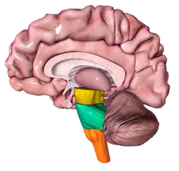
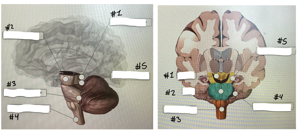
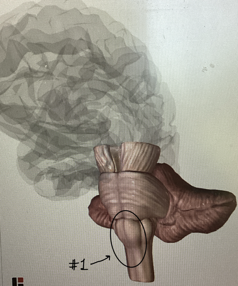
Yellow region: name and function
The midbrain
A narrow region between the pons and thalamus. It contains some nuclei and a lot of white matter. Made up of the cerebral peduncles and the corpora quadrigemina
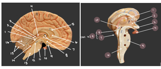
#2 (left) and #5 (right): Name and function
Cerebral peduncles
(peduncle = foot) These large bundles are part of the midbrain, and they contain projection tracts. They permit communication between the cerebral hemisphere and lower parts of the central nervous system
#7, 8, and 9: Names and functions
#7 is the inferior colliculus. #8 is the superior colliculus. #9 is the corpora quadrigemina
The corpora quadrigemina is composed of the superior colliculi and the inferior colliculi
Superior colliculi: nuclei on the posterior side of the midbrain that initiate visual reflexes, such as the coordination of eye and head movements
Inferior colliculi: nuclei on the posterior midbrain that receive auditory information and coordinate reflexive actions in response to sounds.

#3 (left) and #4 (right): Names and functions
Cerebellar peduncle
Part of the pons, the cerebellar peduncles contain tracts passing in or out of the cerebellum
#1: Name and function
Medullary pyramids
Large ridges on the anterior surface of the medulla oblongata. They contain the corticospinal tracts, which contain axons directing voluntary movements of the body
What are the three main centers of the medulla oblongata?
These centers send and receive information for the autonomic nervous system…
1. Cardiovascular centers: regulate heart rate and blood vessel diameter
2. Respiratory centers: control depth and rate of breathing
3. Various autonomic centers: reflex centers for the gastrointestinal tract
Orange structure and #7: Name and function
Medulla oblongata
Main features are the medullary pyramids. Also contains cardiovascular centers, respiratory centers, and various autonomic centers
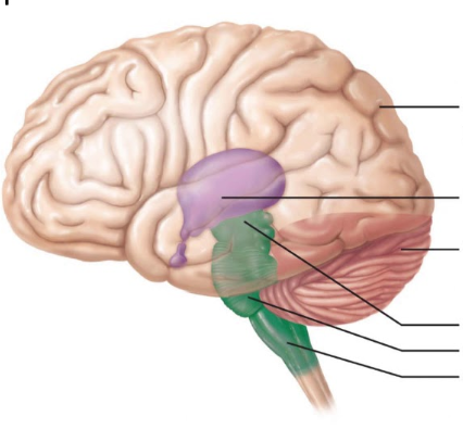
Blue structure: Name and function
Cerebellum
Cerebellum means “little brain.” It is made up of two cerebellar hemispheres. There is an outer cortex and inner white matter, which exhibits a branching pattern called arbor vitae. It also has a midline lobe between the hemispheres called the vermis (which means worm).
The cerebellum receives information from above and below. From the cerebral hemispheres, it is informed about the intent to initiate movement. From below, it receives information from sensory receptors in joints and muscles, providing information about the body’s position. Together, it calculates coordinated movements of the body.
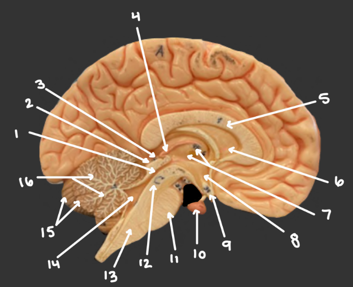
#16: Name and definition
Arbor vitae
A distinct tree-like branching pattern of the white matter in each cerebellar hemisphere

#5: Name and definition
Vermis
A midline lobe at the center of the cerebellum
Describe the limbic system
A multi-region system including parts of the cerebral cortex, thalamus, and hypothalamus. These structures coordinate to recognize social cues and elicit emotions and memory
Describe the reticular formation
A multi-regional system responsible for limiting the amount of sensory information that reaches your conscious mind.

Green structure: Name and function
The pons is part of the brainstem; it is located above the medulla oblongata, below the midbrain, and in front of the cerebellum. It contains mostly tracts of white matter and acts as a bridge, connecting the cerebrum, cerebellum, and spinal cord.
Epithalamus vs Thalamus vs Hypothalamus
Epithalamus: contains the pineal gland and choroid plexus
Thalamus: two lobes that are connected by the interthalamic adhesion. Site of neural synapse.
Hypothalamus: Responds to stimuli by releasing hormones