Exploring Medical Language Chapter 5
1/256
There's no tags or description
Looks like no tags are added yet.
Name | Mastery | Learn | Test | Matching | Spaced | Call with Kai |
|---|
No analytics yet
Send a link to your students to track their progress
257 Terms
nose
lined with mucous membrane and fine hairs; it acts as a filter to moisten and warm the entering air
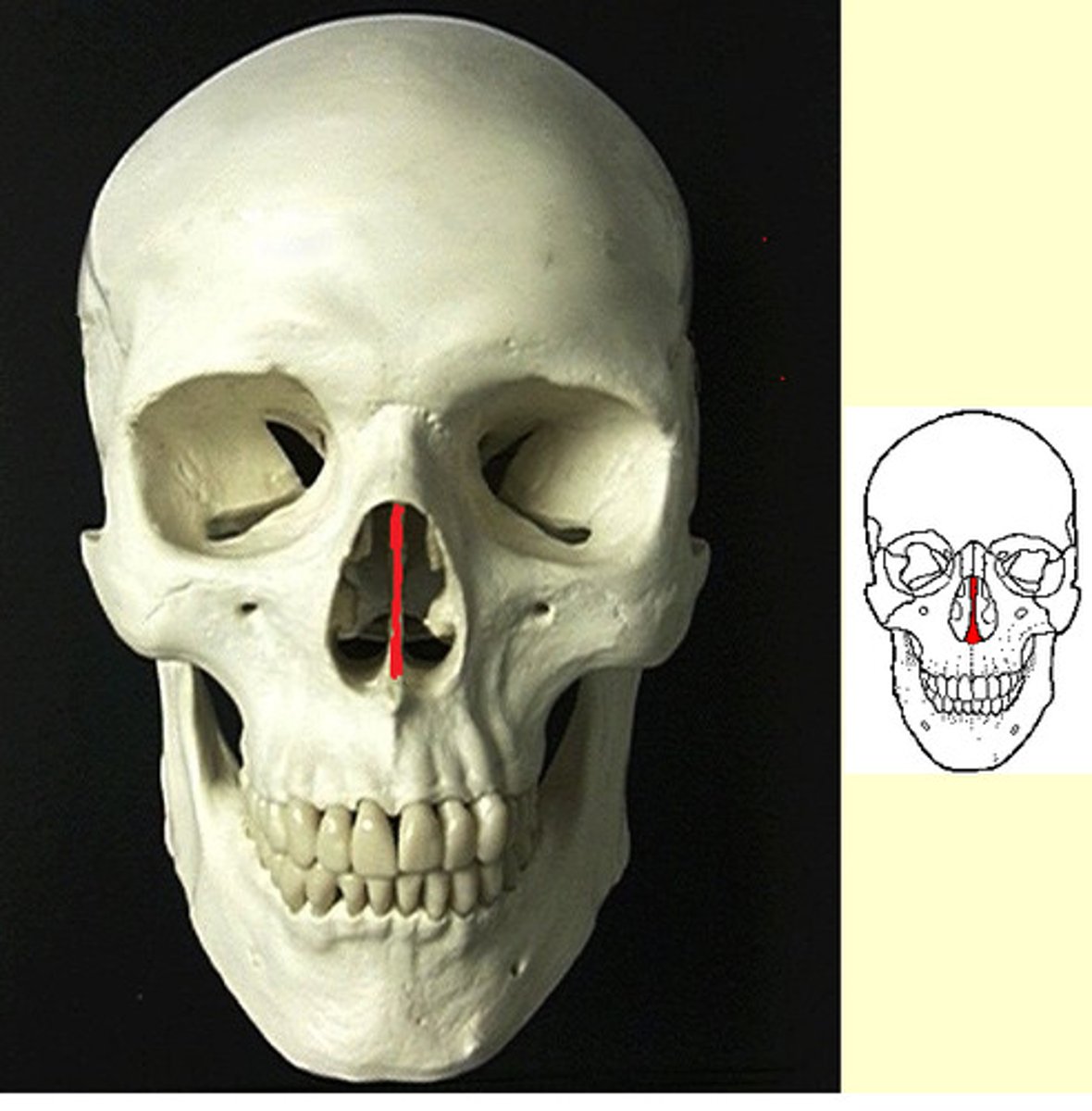
nasal septum
partition separating the right and left nasal cavities
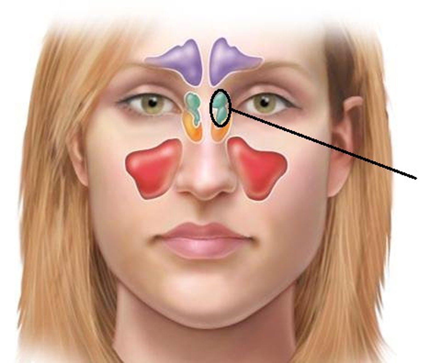
paranasal sinuses
air cavities within the cranial bones that open into the nasal cavities
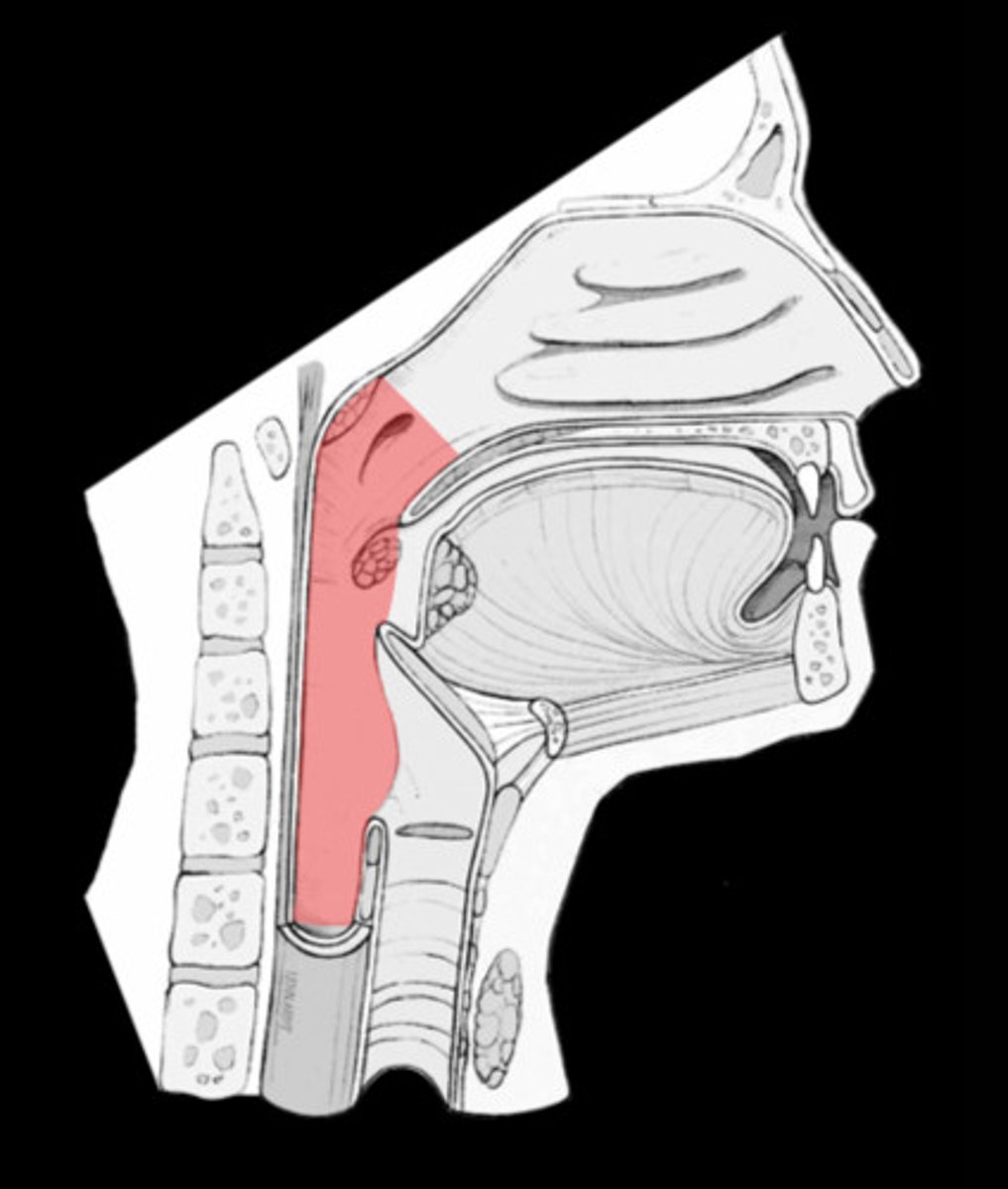
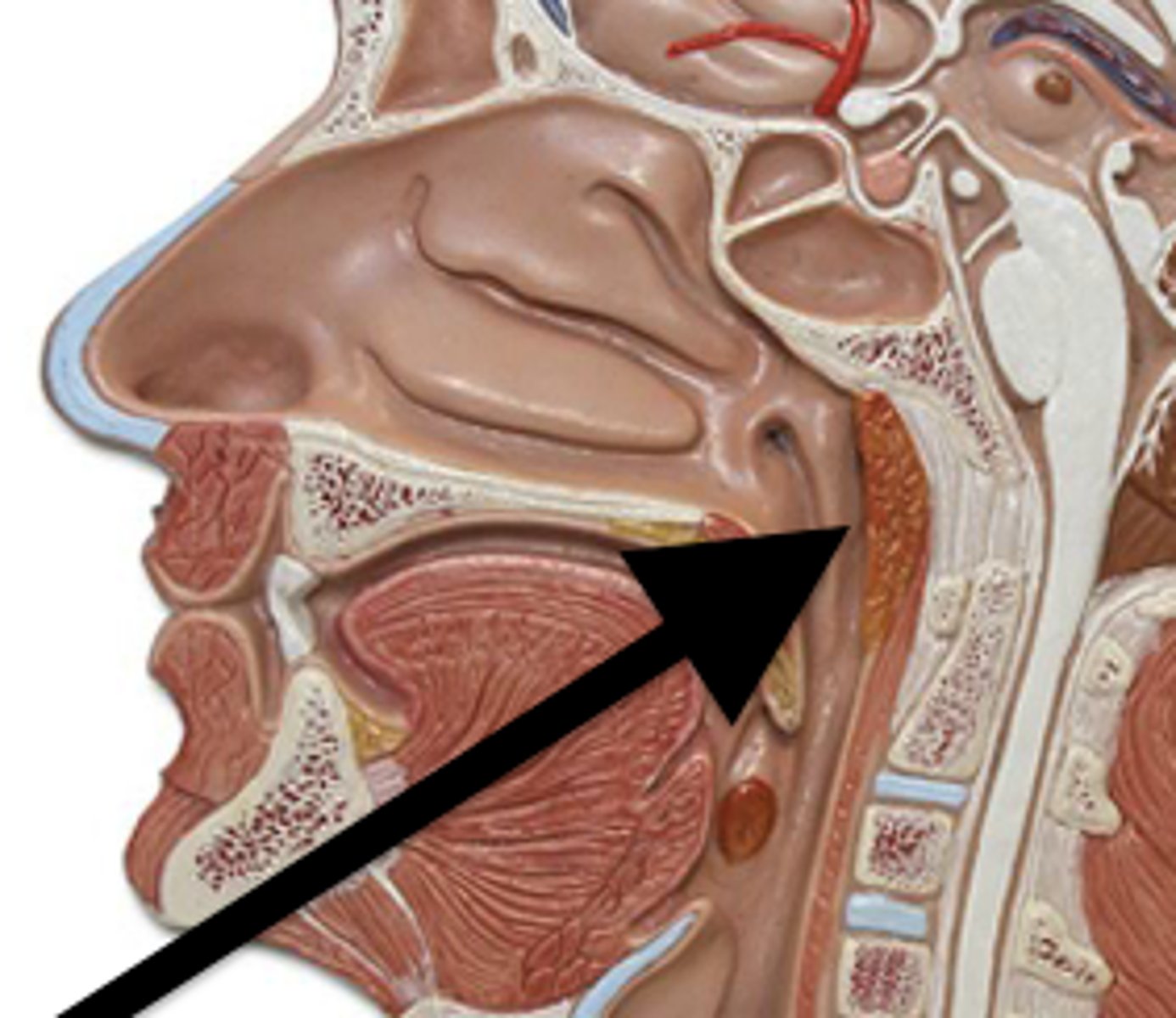
pharynx
serves as a food and air passageway. Air enters from the nasal cavities and/or mouth and passes through the pharynx to the larynx. Food enters the pharynx from the mouth and passes into the esophagus. (also called the throat)
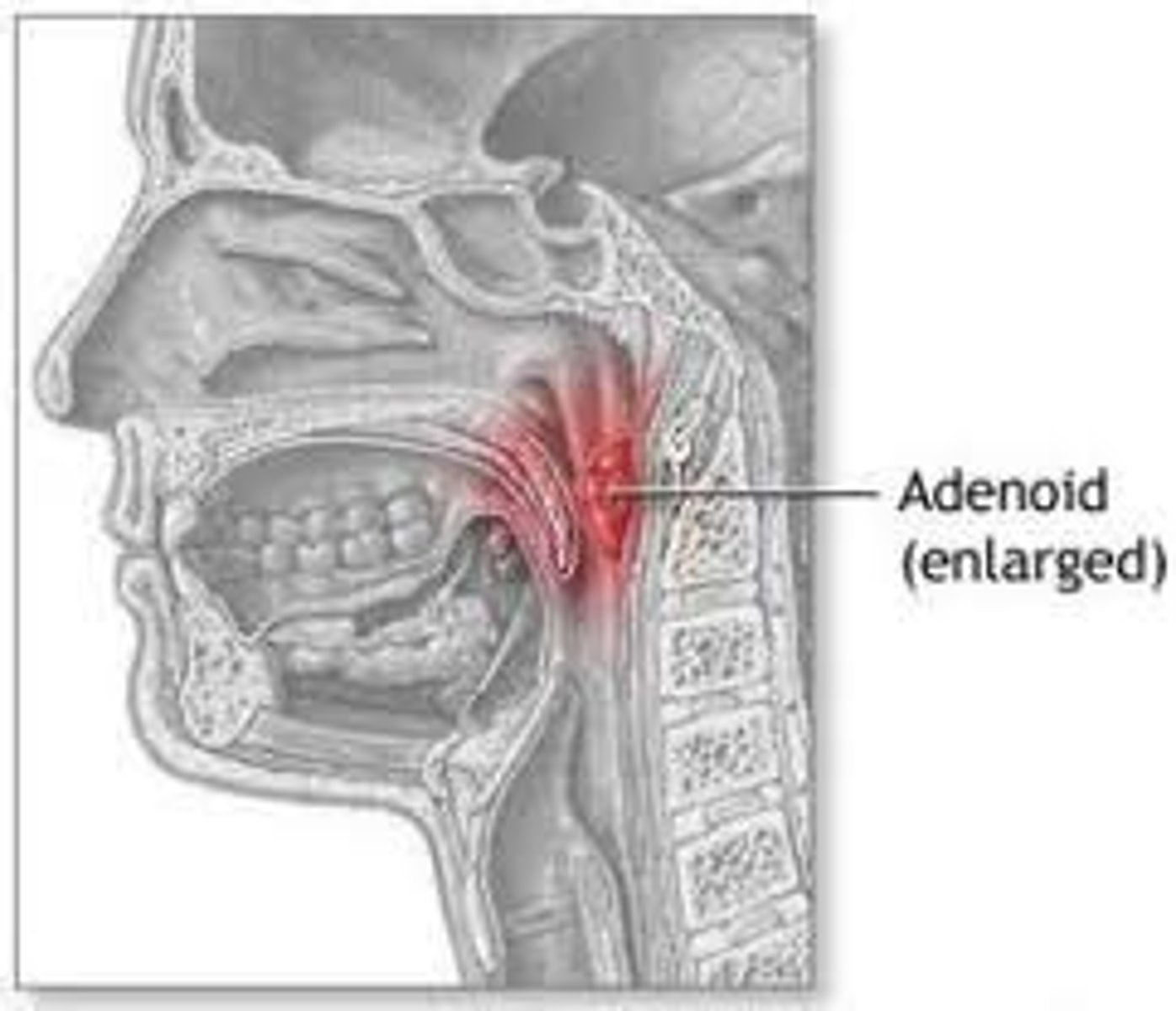
adenoids
lymphoid tissue located on the posterior wall of the nasal cavity (also called pharyngeal tonsils)
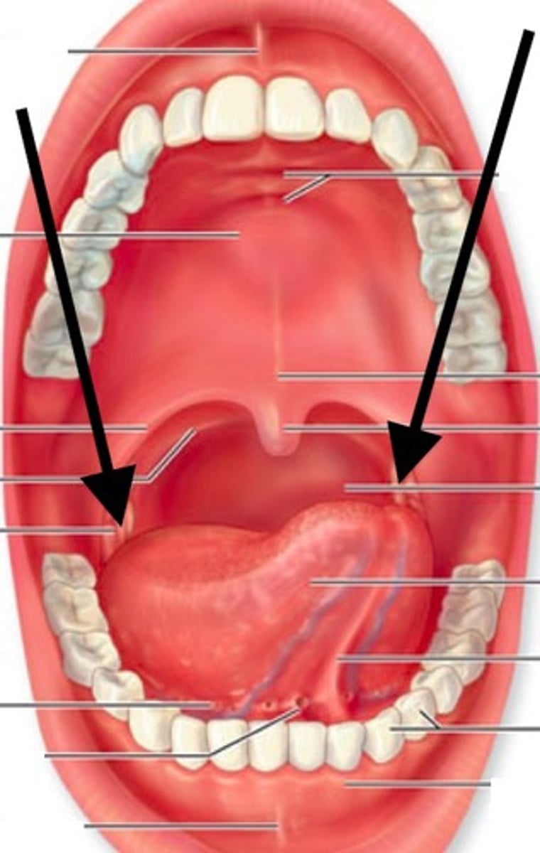
tonsils
lymphoid tissue located on the lateral wall at the junction of the oral cavity and oropharynx
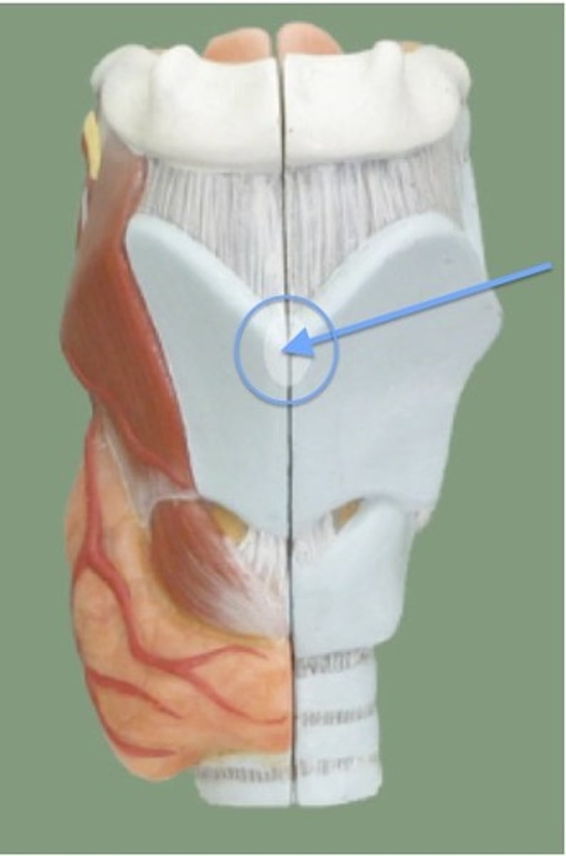
larynx
location of the vocal cords. Air enters from the pharynx. (also called the voice box)
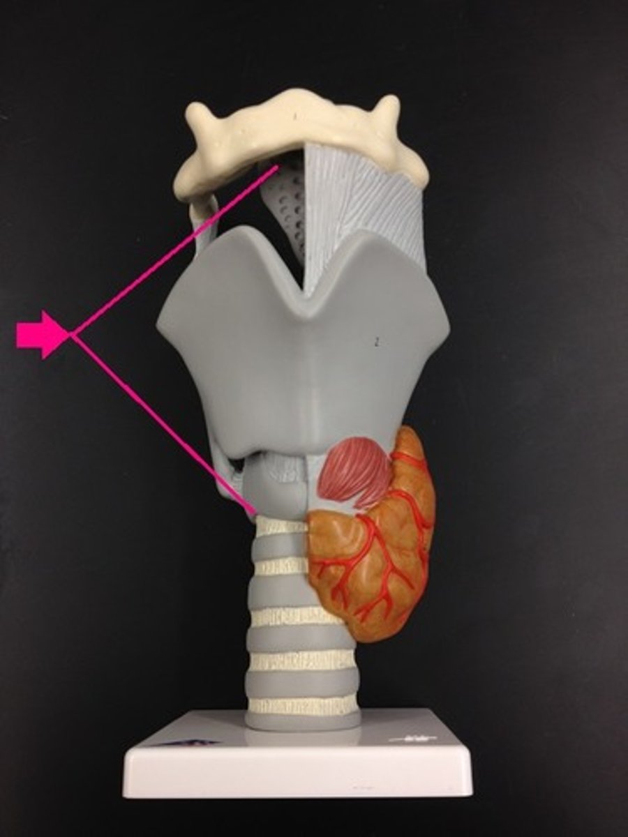
epiglottis
flap of cartilage that automatically covers the opening of the larynx and keeps food from entering the larynx during swallowing
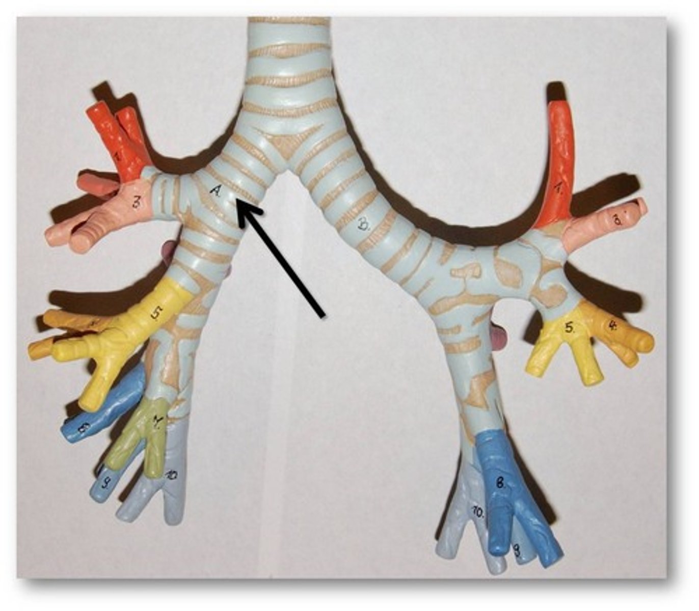
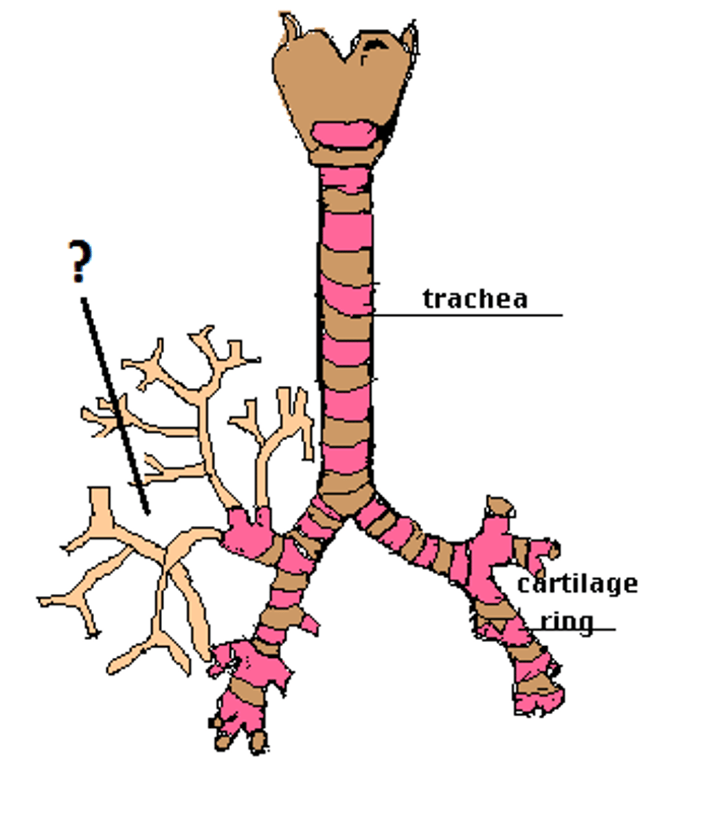
trachea
passageway for air to the bronchi from the larynx; (also called the windpipe)
bronchus (pl. bronchi)
one of two branches from the trachea that conducts air into the lungs, where it divides and subdivides. The branchings resemble a tree; therefore, they are referred to as a bronchial tree.
bronchioles
smallest subdivision of the bronchial tree
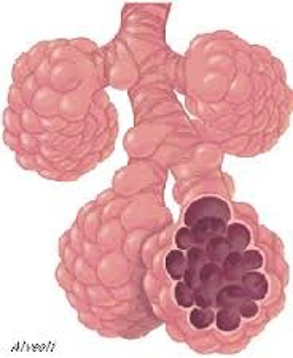
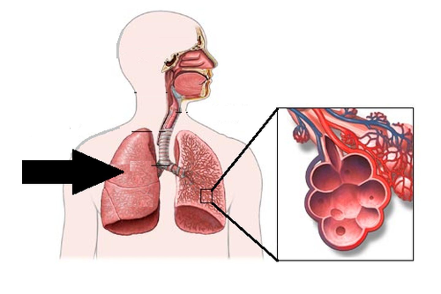
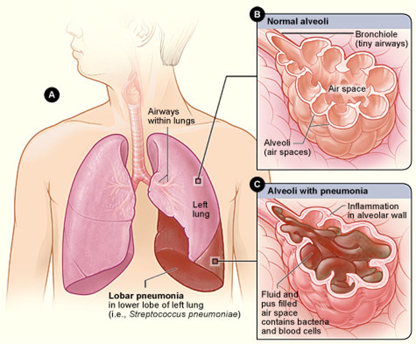
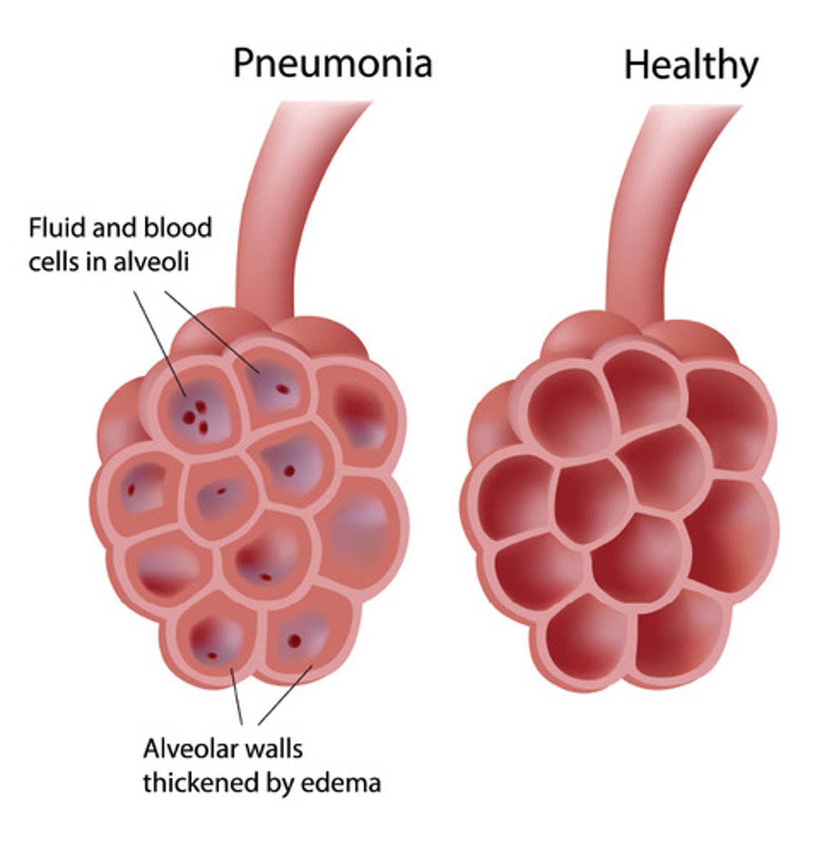
alveoli (s. alveolus)
air sacs at the end of the bronchioles. Oxygen and carbon dioxide are exchanged through the alveolar walls and the capillaries (also a term for the sockets in the jaw bones into which the teeth fit).
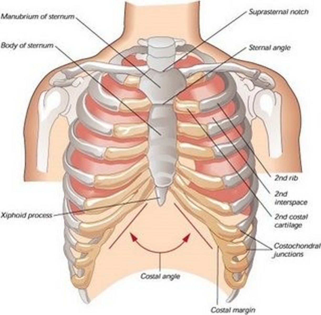
thorax
chest, the part of the body between the neck and the diaphragm encased by the ribs. Thoracic cavity is the hollow space between the neck and diaphragm.
lungs
two spongelike organs in the thoracic cavity. The right lung consists of three lobes, and the left lung has two lobes.
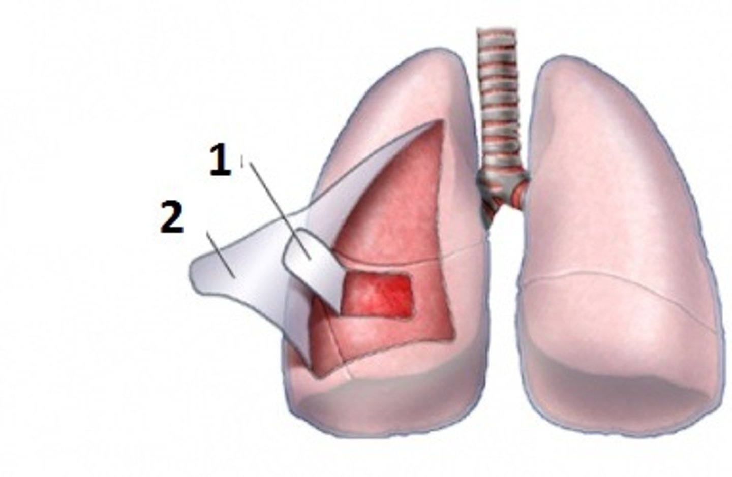
pleura
double-folded serous membrane covering each lung (visceral pleura) and lining the thoracic cavity (parietal pleura) with a small space between, called the pleural cavity, which contains serous fluid
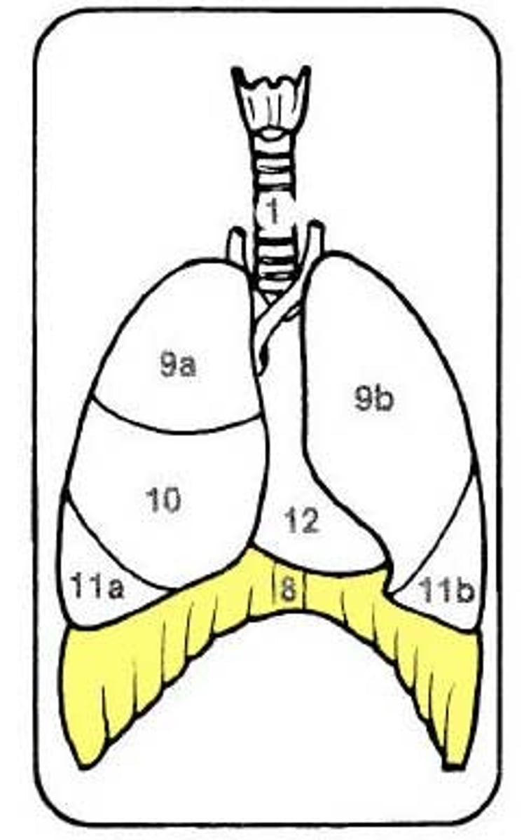
diaphragm
muscular partition that separates the thoracic cavity from the abdominal cavity. It aids in the breathing process by contracting and pulling air in, then relaxing and pushing air out.
mediastinum
space between the lungs. It contains the heart, esophagus, trachea, great blood vessels, and other structures.

adenoiditis
inflammation of the adenoids
alveolitis
inflammation of the alveoli (pulmonary or dental)
atelectasis
incomplete expansion (of the lung or portion of the lung)
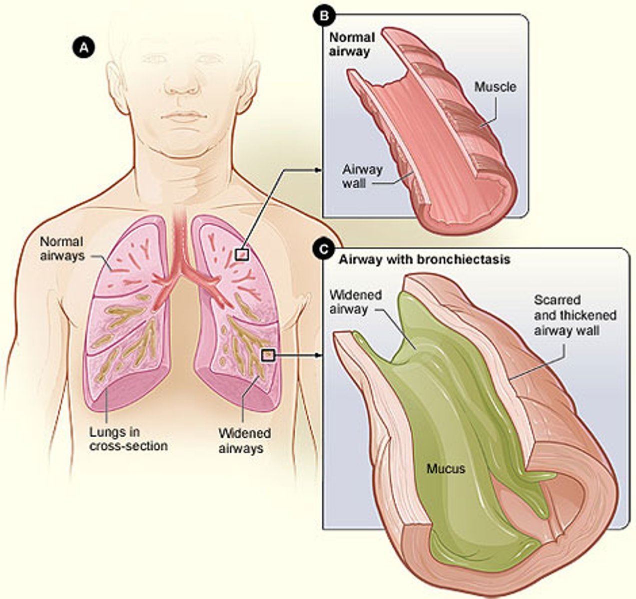
bronchiectasis
dilation of the bronchi
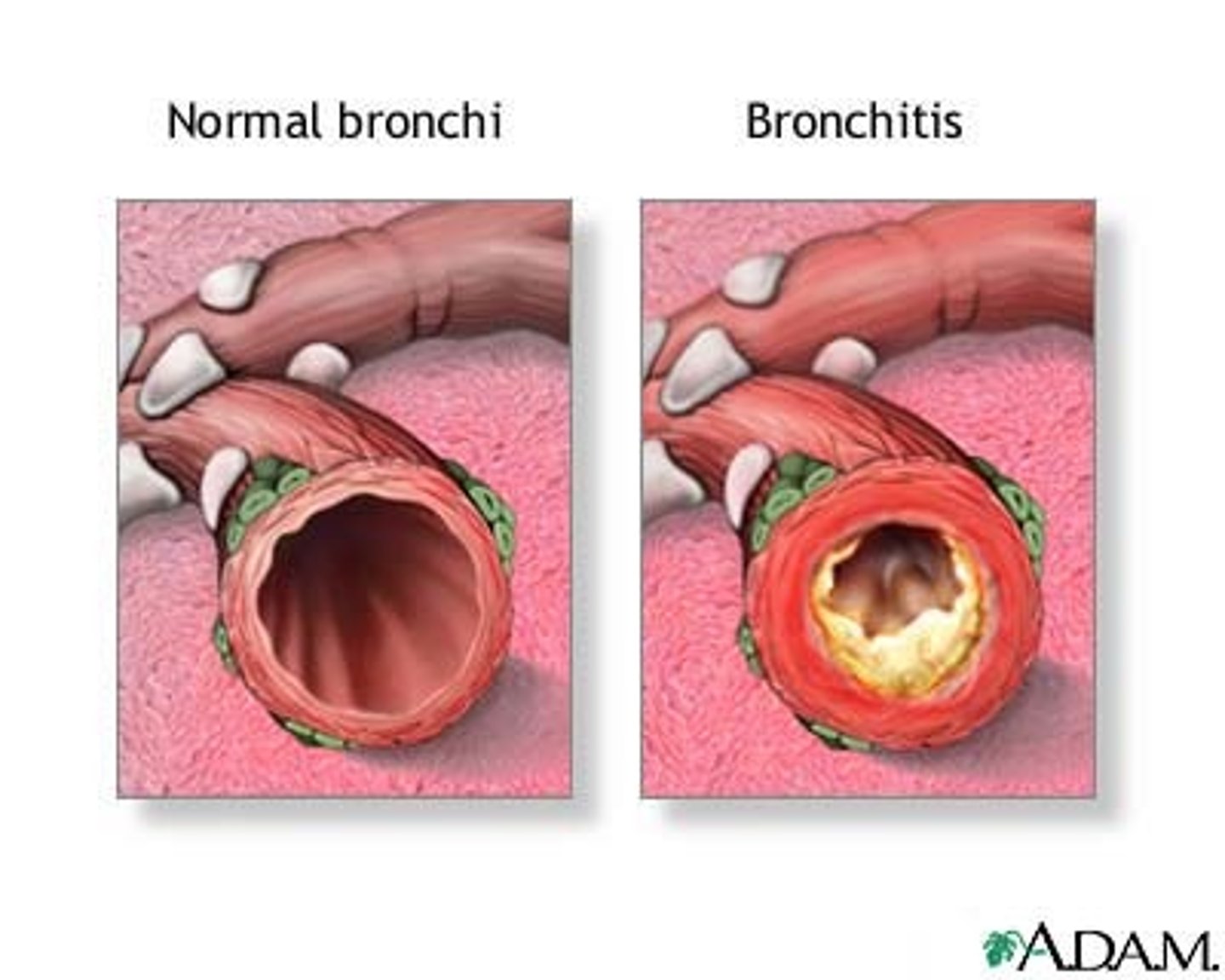
bronchitis
inflammation of the bronchi
bronchogenic carcinoma
cancerous tumor originating in a bronchus (also referred to as lung cancer)
bronchopneumonia
diseased state of the bronchi and lungs (an inflammation of the lungs that begins in the terminal bronchioles)
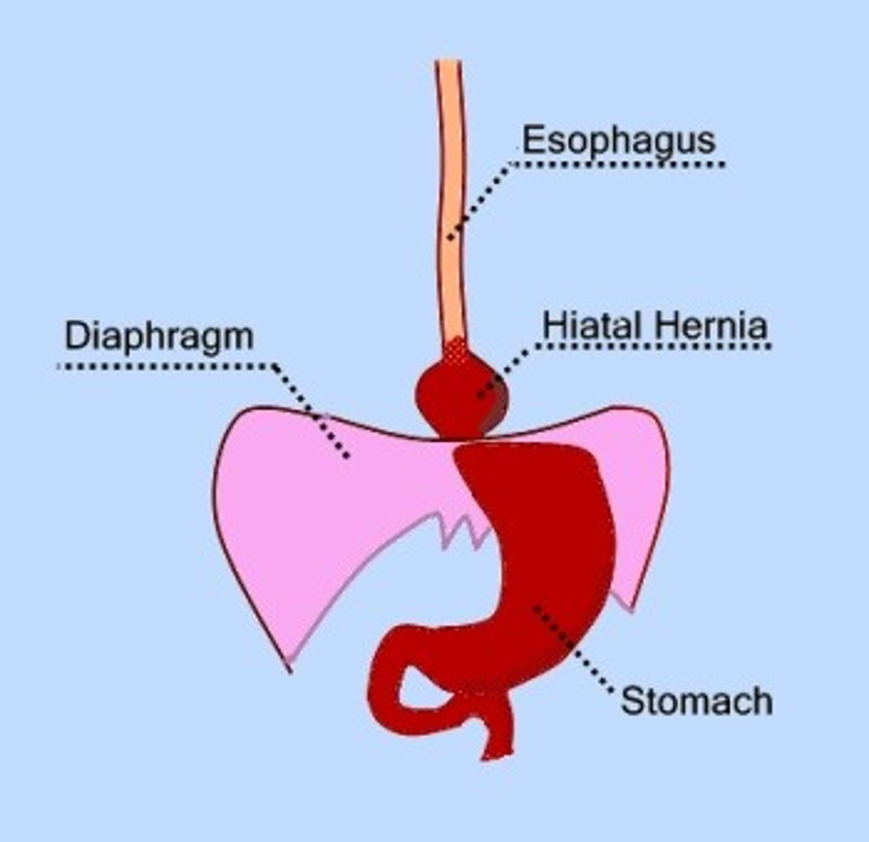
diaphragmatocele
hernia of the diaphragm
epiglottitis
inflammation of the epiglottis
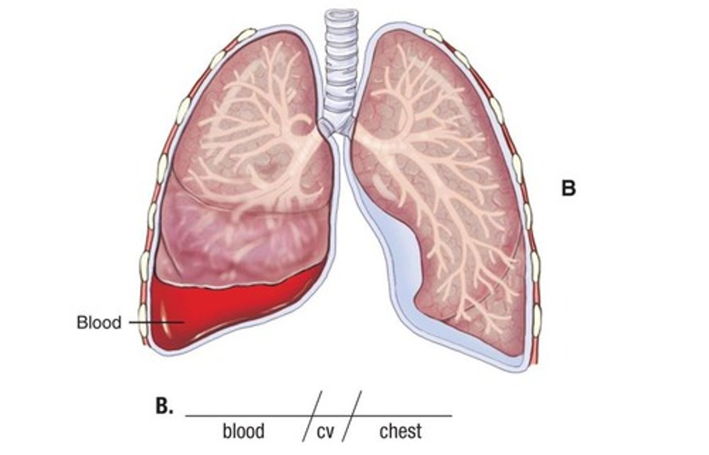
hemothorax
blood in the chest cavity (pleural space)
laryngitis
inflammation of the larynx
laryngotracheobronchitis (LTB)
inflammation of the larynx, trachea, and bronchi (the acute form is called croup)
lobar pneumonia
pertaining to the lobe(s); diseased state of the lung (infection of one or more lobes of the lung)
nasopharyngitis
inflammation of the nose and pharynx
pharyngitis
inflammation of the pharynx
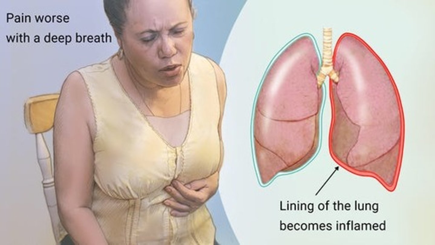
pleuritis
inflammation of the pleura (also called pleurisy)
pneumatocele
hernia of the lung (lung tissue protrudes through an opening in the chest)
pneumoconiosis
abnormal condition of dust in the lungs (pneumoconiosis is the general name given for chronic inflammatory disease of the lung caused by excessive inhalation of mineral dust. When the disease is caused by a specific dust, it is named for the dust. For example, the disease caused by silica dust is called silicosis).
pneumonia
diseased state of the lung (the infection and inflammation are caused by bacteria such as Pneumococcus, Staphylococcus, Streptococcus, and Haemophilus; viruses; and fungi)
pneumonitis
inflammation of the lung
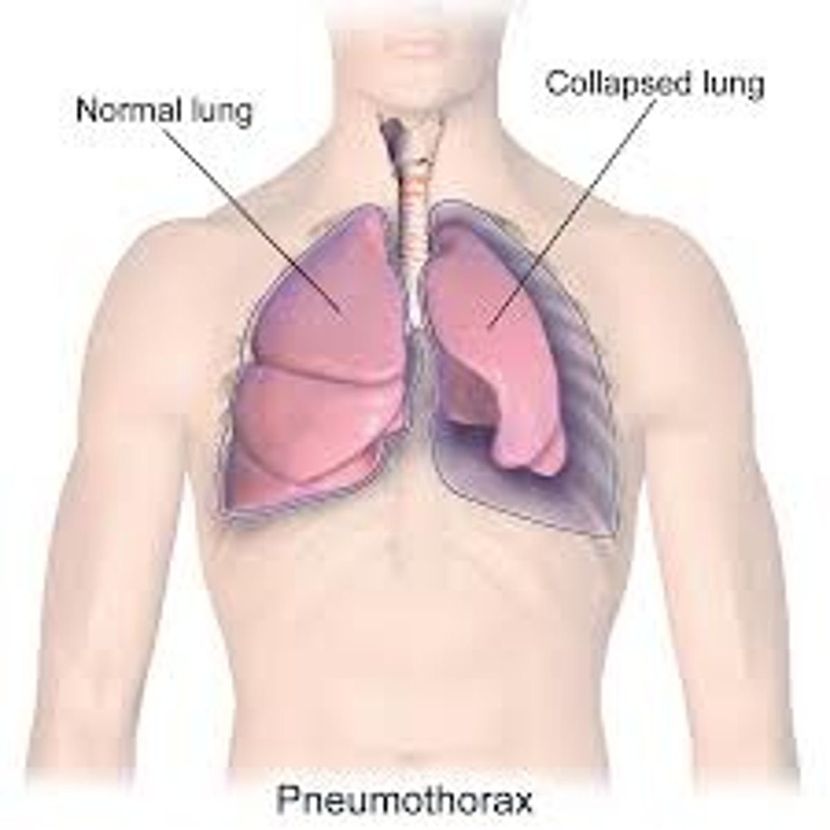
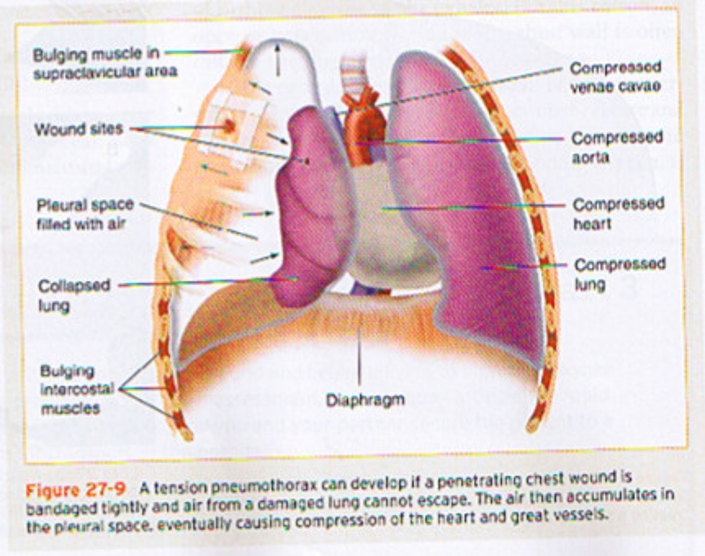
pneumothorax
air in the chest cavity (specifically, the pleural space, which causes collapse of the lung and is often a result of an open chest wound)
pulmonary neoplasm
pertaining to (in) the lung, new growth (tumor)
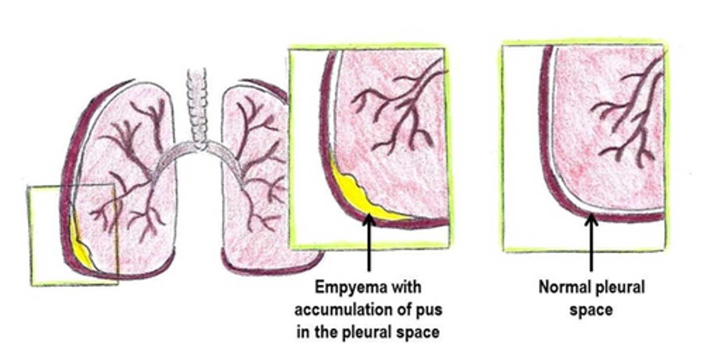
pyothorax
pus in the chest cavity (pleural space) (also called empyema)
rhinitis
inflammation of the nose (mucous membranes)
rhinomycosis
abnormal condition of fungus in the nose
rhinorrhagia
rapid flow of blood from the nose (also called epistaxis)
sinusitis
inflammation of the sinuses
thoracalgia
pain in the chest
tonsillitis
inflammation of the tonsils
tracheitis
inflammation of the trachea
tracheostenosis
narrowing of the trachea
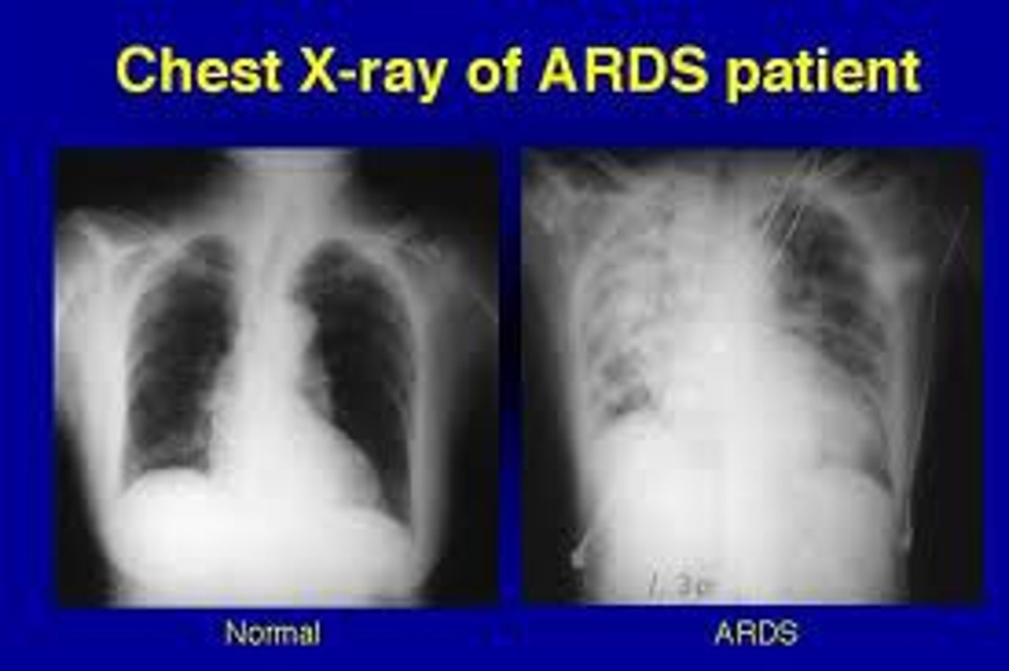
acute respiratory distress syndrome (ARDS)
respiratory failure as a result of disease or injury. ARDS is respiratory failure in an adult.Symptoms include dyspnea, tachypnea, and cyanosis. (also called adult respiratory distress syndrome)
asthma
respiratory disease characterized by coughing, wheezing, and shortness of breath, caused by constriction and inflammation of airways that is reversible between attacks
chronic obstructive pulmonary disease (COPD)
progressive lung disease obstructing air flow, which makes breathing difficult. Chronic bronchitis and pulmonary emphysema are the two main components of COPD. Most COPD is a result of cigarette smoking.
coccidioidomycosis
fungal disease affecting the lungs and sometimes other organs of the body (also called valley fever)
croup
condition resulting from acute obstruction of the larynx, characterized by a barking cough, hoarseness, and stridor. It may be caused by viral or bacterial infection, allergy, or foreign body. Occurs mainly in children. (also called laryngotracheobronchitis)
cystic fibrosis (CF)
hereditary disorder of the exocrine glands characterized by excess mucus production in the respiratory tract, pancreatic deficiency, and other symptoms
deviated septum
one part of the nasal cavity is smaller because of malformation or injury of the nasal septum
epistaxis
nosebleed (also called rhinorrhagia)
idiopathic pulmonary fibrosis (IPF)
chronic progressive lung disorder characterized by increasing scarring, which ultimately reduces the capacity of the lungs; etiology unknown. Most often affects adults over the age of 50. Smoking, pollutants, and heredity may play a role in its genesis. Symptoms include exertional dyspnea and a dry cough. Lung transplant may be indicated in severe cases; there is no cure.
influenza (flu)
highly contagious and often severe viral infection of the respiratory tract
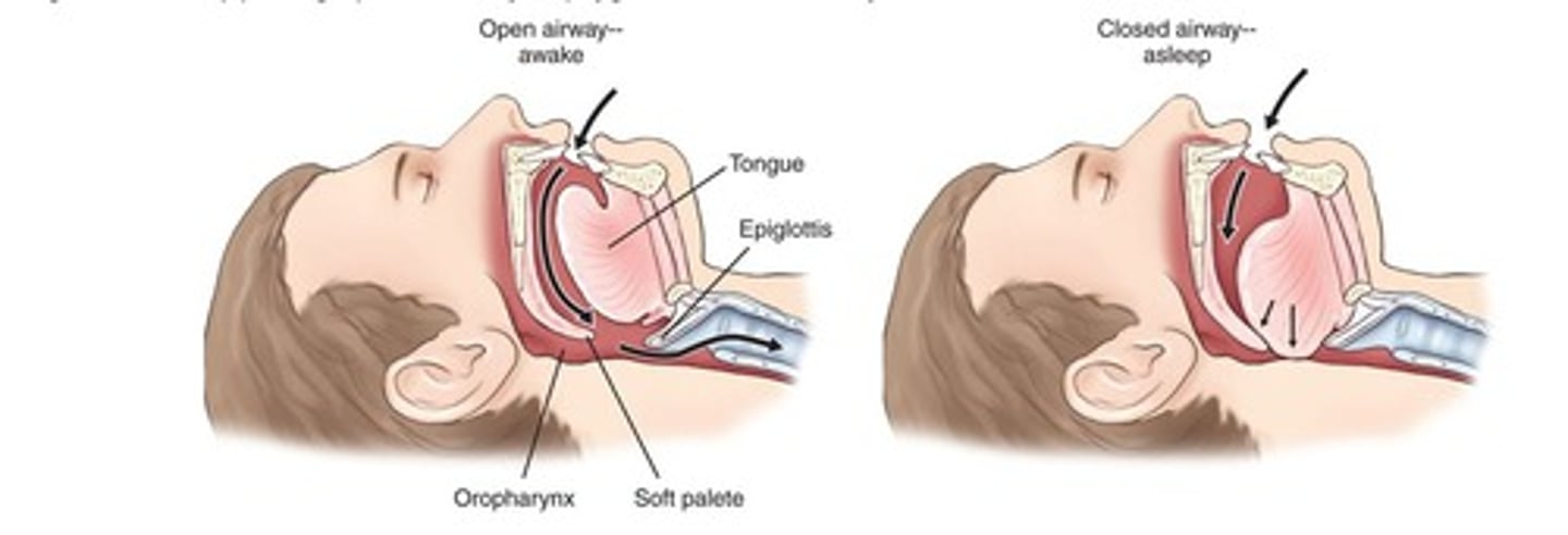
obstructive sleep apnea (OSA)
repetitive pharyngeal collapse during sleep, which leads to transient periods of apnea (absence of breathing); can produce daytime drowsiness and elevated blood pressure
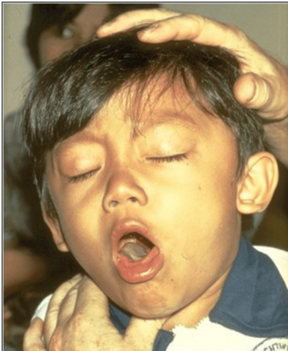
pertussis
highly contagious bacterial infection of the respiratory tract characterized by an acute crowing inspiration, or whoop (also called whooping cough)
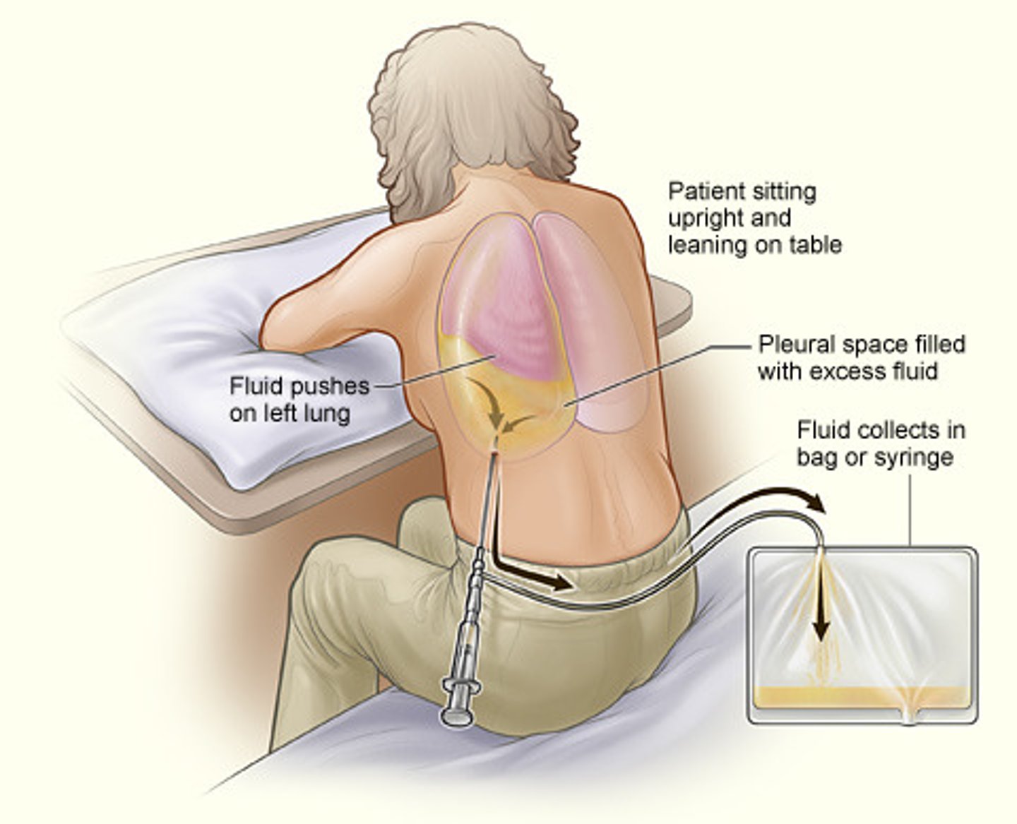
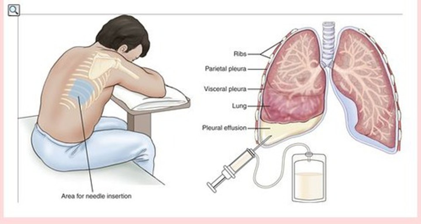
pleural effusion
fluid in the pleural space caused by a disease process or trauma
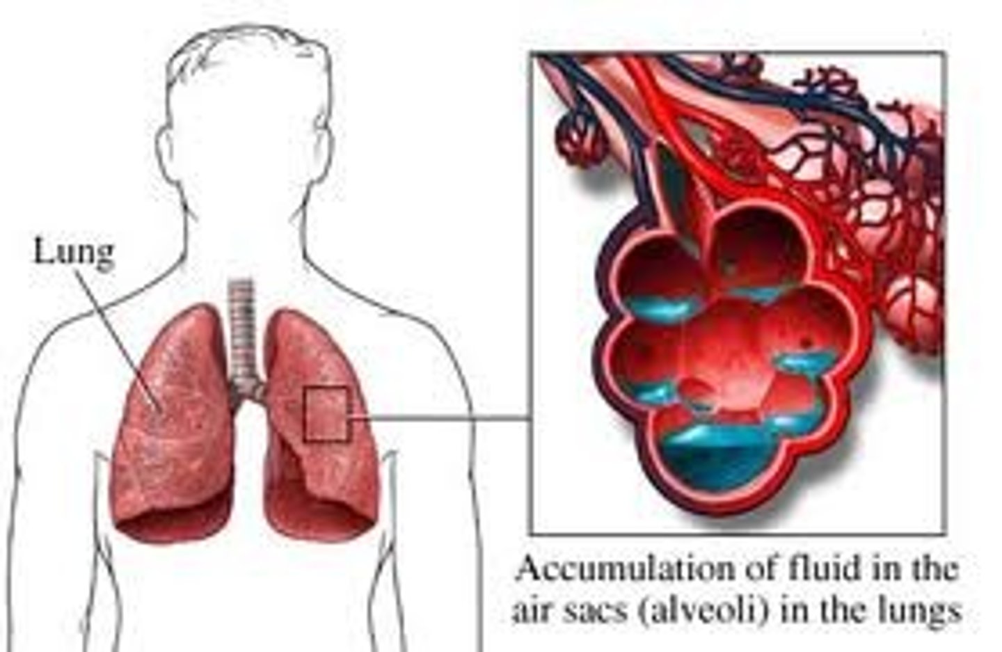
pulmonary edema
fluid accumulation in the alveoli and bronchioles, most often a manifestation of heart failure
pulmonary embolism (PE)
matter foreign to the circulation, carried to the pulmonary artery and its branches, where it blocks circulation to the lungs and can be fatal if of sufficient size or number. Blood clots broken loose from the deep veins of the lower extremities are the most common source.
pulmonary emphysema
loss of elasticity of the alveoli resulting in distention causing stretching of the lung. As a result, the body does not receive enough oxygen. (component of COPD)
tuberculosis (TB)
infectious bacterial disease, most commonly spread by inhalation of small particles and usually affecting the lungs; may spread to other organs
upper respiratory infection (URI)
infection of the nasal cavity, pharynx, or larynx usually caused by a virus (commonly called a cold)
adenoidectomy
excision of the adenoids
adenotome
instrument used to cut the adenoids
bronchoplasty
surgical repair of a bronchus
laryngectomy
excision of the larynx
laryngoplasty
surgical repair of the larynx
laryngostomy
creation of an artificial opening into the larynx
laryngotracheotomy
incision into the larynx and trachea
lobectomy
excision of a lobe (of the lung)
pleuropexy
surgical fixation of the pleura
pneumonectomy
excision of a lung
rhinoplasty
surgical repair of the nose
septoplasty
surgical repair of the (nasal) septum
septotomy
incision into the (nasal) septum
sinusotomy
incision into a sinus
thoracocentesis
surgical puncture to aspirate fluid from the chest cavity (also called thoracentesis) (Exercise Figure F)
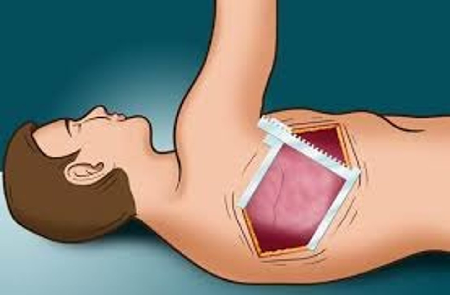
thoracotomy
incision into the chest cavity
tonsillectomy
excision of the tonsils
tracheoplasty
surgical repair of the trachea
tracheostomy
creation of an artificial opening into the trachea
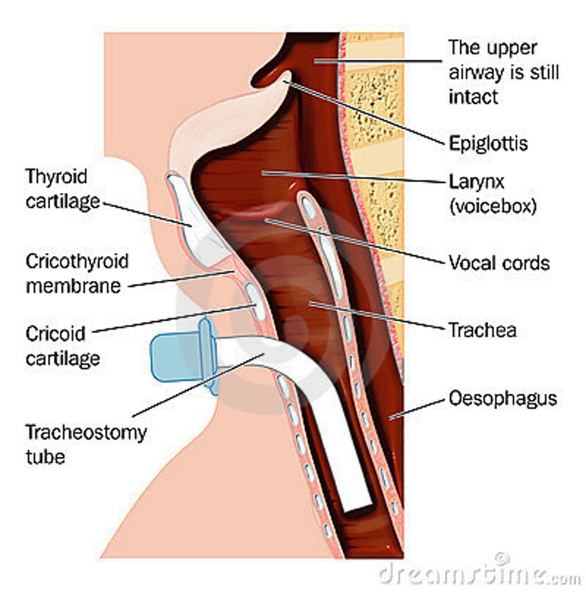
tracheotomy
incision into the trachea (Fig. 5.12)
bronchoscope
instrument used for visual examination of the bronchi
bronchoscopy
visual examination of the bronchi
endoscope
instrument used for visual examination within (a hollow organ or body cavity). (Endoscopes are used for surgical procedures as well as for viewing.)
endoscopic
pertaining to visual examination within (a hollow organ or body cavity) (used to describe the practice of performing surgeries that use endoscopes)
endoscopy
visual examination within (a hollow organ or body cavity)
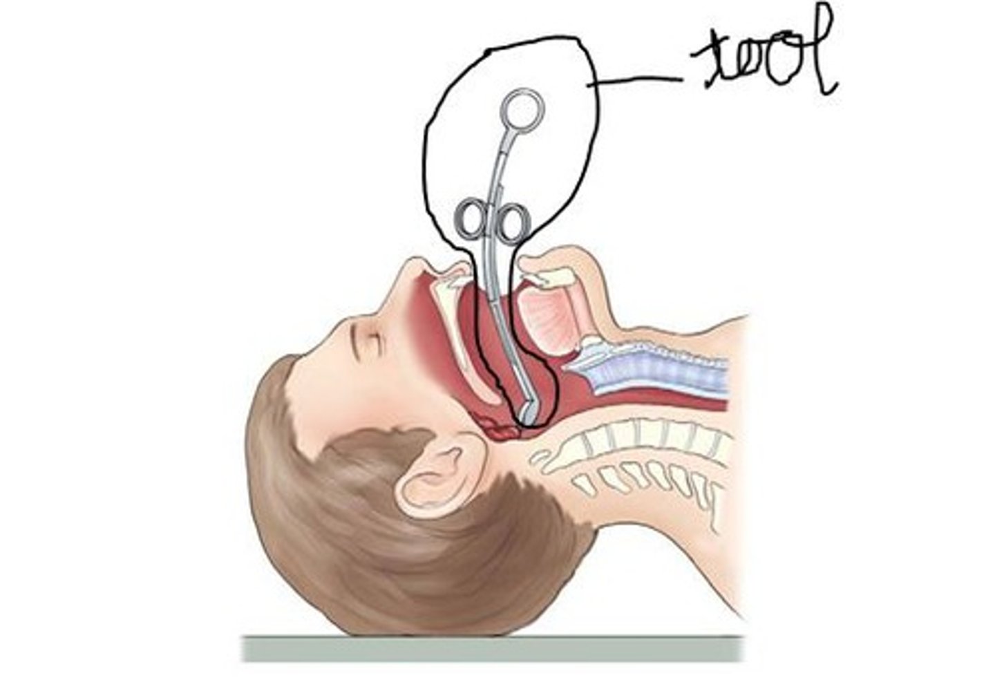
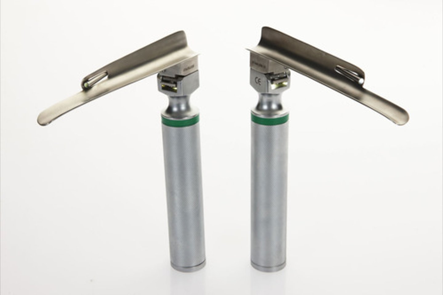
laryngoscope
instrument used for visual examination of the larynx
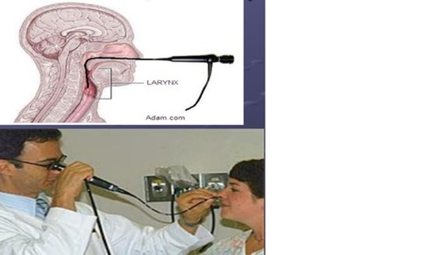
laryngoscopy
visual examination of the larynx
thoracoscope
instrument used for visual examination of the chest cavity (VAT)
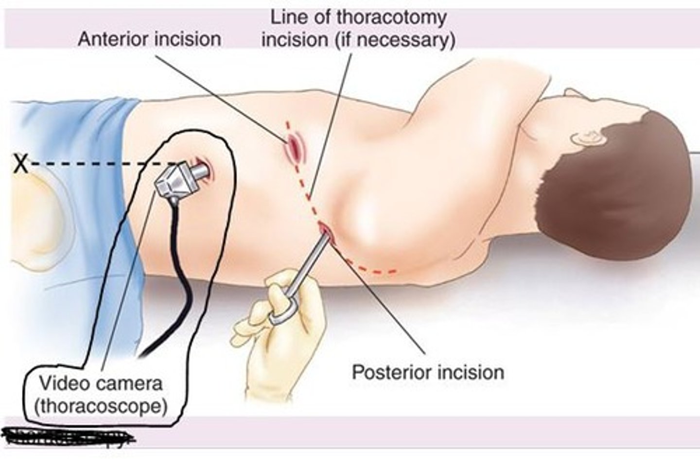
thoracoscopy
visual examination of the chest cavity
radiograph
record of x-rays
radiography
process of recording x-rays
sonogram
record of sound waves after they bounce off organs in the body
sonography
process of recording sound
tomography
process of recording slices (anatomical cross section)