Blood Supply
1/67
There's no tags or description
Looks like no tags are added yet.
Name | Mastery | Learn | Test | Matching | Spaced | Call with Kai |
|---|
No analytics yet
Send a link to your students to track their progress
68 Terms

Structure #2
Middle Cerebral Artery

Structure #4
Posterior communicating artery

Structure #1
Internal Carotid artery
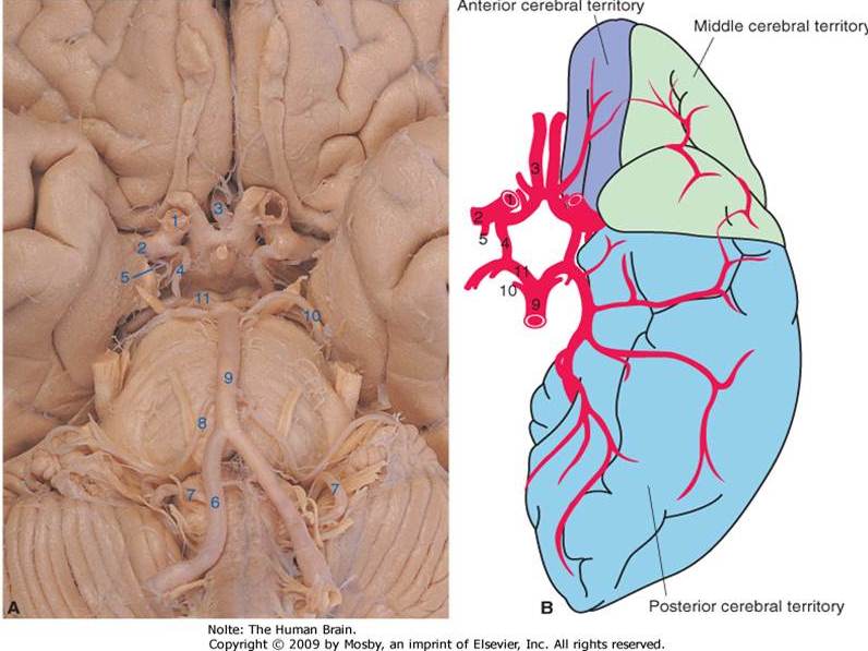
Structure #3
Anterior Cerebral Artery

Structure #6
Vertebral Artery

Structure #9
Basilar artery

Structure #1
Posterior Cerebral Artery

Structure #4
Anterior Inferior Cerebellar artery

Structure #6
Vertebral Artery

Structure #8
Anterior Spinal Artery

Structure #7
Posterior Inferior cerebellar artery
Structure #9
Basilar artery

Structure #5
Labyrinthine (internal auditory) artery

Structure #3
Pontine arteries

Structure #2
Superior cerebellar artery
Stroke is
5th leading cause of death & 1st leading cause of disability
Types of Stroke
Ischemic & Hemorrhagic
Ischemic stroke
most common (85%) and is BLOCKAGE in an artery
Hemorrhagic stroke
rupture of an artery (aneurysm)
Types of ischemic stroke
Embolic + Thrombotic
Infarct
area of dead cells/ necrotic tissue
ischemia
restriction in blood supply
Anoxia
without oxygen
Hypoxia
decreased oxygen
lesion
injury
Carotid endarterectomy (CEA)
A surgical procedure to remove plaque from the carotid arteries to improve blood flow and reduce stroke risk.
2 major arterial systems
Anterior circulation and Posterior circulation.
Anterior Circulation
Internal Carotid Arteries (80%) of the brain and supply most of the cerebral hemispheres
Posterior Circulation
Vertebral arteries + basilar artery (20%) of brain and supply to BS, SC, Diencephalon, etc
Structures of the ICA
8 total, Internal Carotid has 3 branches: Ophthalmic Artery, Anterior Choroidal Artery, Posterior Communicating Artery. 2 bifurcates: Anterior Cerebral Artery (ACA) with Anterior communicating artery & Middle Cerebral Artery (MCA) with lenticulostriate arteries
(ICA) Ophthalmic
Travels along with optic nerve to orbit to supply eye; when blocked causes BLINDNESS
(ICA) Anterior Choroidal Artery
Supplies several structures, fairly COMMOn site of stroke leads to deficits in functions
(ICA) Posterior Communicating Artery
Connects ICA to the posterior cerebral artery
(ICA) bifurcation Anterior Cerebral Artery (ACA)
runs medially and enters the longitudinal fissure and supplies tops + medial parts of frontal+parietal
(ICA) bifurcation Middle Cerebral Artery (MCA)
Larger than ACA, runs lateral while supplying the insula and is most common site of occlusion + stroke
(ICA) Anterior communicating artery
only ONE of these and most common site for aneurysms (20-25%) that may cause visual deficits
(ICA) Lenticulostriate Arteries
supply portions of internal capsule and basal nuclei, leads to symptoms out of proportion to the size
ACA Brain Area Affected: Primary motor cortex (medial precentral gyrus)
Contralateral hemiplegia (worse in legs)
ACA Brain Area Affected: Primary somatosensory
Contralateral hemiparatheisa (worse in legs)
ACA Brain Area Affected: Premotor area
Apraxia
ACA Brain Area Affected: Basal Nuclei
Planning and timing of movement + movement habits/patterns
ACA Brain Area Affected: Ventromedial prefrontal cortex + limbic system
Cognitive impairment + affective/personality changes
MCA Brain Area Affected: Primary motor cortex (lateral precentral gyrus)
contralateral hemiplegia (worse in arm, trunk, face)
MCA Brain Area Affected: Primary somatosensory cortex (lateral postcentral gyrus)
Contralateral somatosensory loss (worse in the arm, trunk, face)
MCA Brain Area Affected: Deep + superficial visual pathway
Visual + visual perceptual impairments
MCA Left hemisphere damage
Broca’s/Wernicke’s area (aphasia), Posterior multimodal association cortex (ideational apraxia), frontal lobe, limbic system (emotional lability, depression)
MCA Right hemisphere damage
Posterior association area (perceptual deficits), Frontal lobe (impulsive, poor judgement), Limbic system (euphoria, well-being)
Venous Drainage of Brain
Network of deep + superficial veins→ Dural sinuses→ Right & Left Internal Jugular Veins (IJVs)
Cerebral Arterial Circle (Circle of Willis)
Anastomosis connection that surrounds the optic chiasm + pituitary gland
Circle of Willis connects
Internal carotid + Vertebral-Basilar systems
Purpose of the Circle of Willis
ensure blood flow to brain when systems are compromised
“Textbook” Circle of Willis
Less than ½ population
Vertebral-Basilar System Structures
10, Vertebral Artery includes: Anterior spinla artery,Posterior spinal artery, Posterior inferior cerebellar artery (PICA), Basilar Artery includes: Anterior Inferior Cerebellar artery (AICA) Labyrinthine artery, Pontine arteries, Superior Cerebellar Artery, Basilar artery bifurcates at Posterior Cerebral Artery
Vertebral Artery (VA)
Arises from subclavian artery, ascends in transverse foramina of the cervical vertebra
(VA) Anterior Spinal Artery
1, supplies anterior 2/3 of spinal cord, damage leads to more motor systems
(VA) Posterior Spinal Artery
Supplies 1/3 posterior of spinal cord, damage leads to more sensory symptoms
(VA) Posterior Inferior Cerebellar Artery (PICA)
Supplies posterior portions of inferior cerebellum + lateral medulla, Damage to this artery results in Wallenbergs syndrome
Wallenberg’s syndrome
Damage to PICA, with dysphagia, hoarseness, vertigo + disquilibrium, nystagmus, uncontrollable hiccups
Basilar Artery (BA)
1 of these, begins inferiorly at the junction of the anterior pons + medulla, Damage leads to impaired blood flow
(BA) Anterior inferior cerebellar artery (AICA)
Supplies anterior portions of inferior cerebellum + inferior pons
(BA) Labyrinthine (internal auditory) Artery
Supplies inner ear, damage leads to vestibular symptoms and ipsilateral deafness
(BA) Pontine Arteries
smaller branches of basilar artery, supplies pons with blockage leads to Locked-In syndrome
(BA) Superior Cerebellar Artery
Branches at superior part of BA, Supplies superior cerebellum, interior midbrain, + superior pons
Cerebellar Arteries Occlusion/Hemorrhaging symptoms
incoordination, Ataxia, Intention tremors, Dizziness, Nystagmus, Balance problems, Dysphagia
Brainstem Arteries Occlusion/Hemorrhaging symptoms
Dysphagia, Nystagmus, Eye movement problems, Dizziness, Balance disorders, Coma, other CN symptoms
Posterior Cerebral Artery (PCA)
Curves around the midbrain, supplying the midbrain,, medial + inferior temporal + occipital lobes and parts of diencephalon
PCA Brain Area Affected (Occipital lobe, Temporal Lobe, Posterior association cortex)
Visual + visual-perceptual impairments
PCA Brain Area Affected (Midbrain)
SEE Above “Dysphagia, Nystagmus, Eye movement problems, Dizziness, Balance disorders, Coma, other CN symptoms”