Staphylococci & Other Catalase-Positive, Gram-Positive Cocci
1/30
Earn XP
Description and Tags
Micrococcus and CoNS
Name | Mastery | Learn | Test | Matching | Spaced | Call with Kai |
|---|
No analytics yet
Send a link to your students to track their progress
31 Terms
How are infections caused by gram-positive cocci spread? What type of infection does GPC cause?
through direct contact with individuals or objects
pyogenic infection - accumulation of WBCs, bacteria, and fluid at the site of infection
genus Micrococcus
GPC in tetrads or pairs
colonizes the skin, but rarely causes infection
can cause skin infection in immunocompromised individuals
How is Micrococcus identified in the lab? (colony morphology, catalase, oxidase)
produces pigmented, circular, smooth, convex colonies
M. luteus is bright yellow in color
catalase positive
oxidase positive
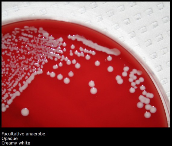
general Staphylococcus lab identification (oxygen requirements, catalase, motility, morphology)
facultative anaerobes
catalase positive
can be isolated on MSA
non-motile
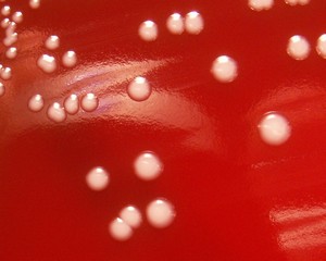
colony morphology
blood agar: raised, convex, creamy colonies
CNA: white, cream, or yellow pigment
Where can Staphylococcus sp. be found?
animals, the environment, and human skin and mucous membranes
can be a part of normal flora on the skin, nares, axillae, inguinal, and perianal areas
important human pathogen
What test is used to differentiate S. aureus from other species of Staphylococcus?
coagulase test
S. aureus is positive
all other Staphylococci are negative (coagulase-negative staphylococci, CoNS)
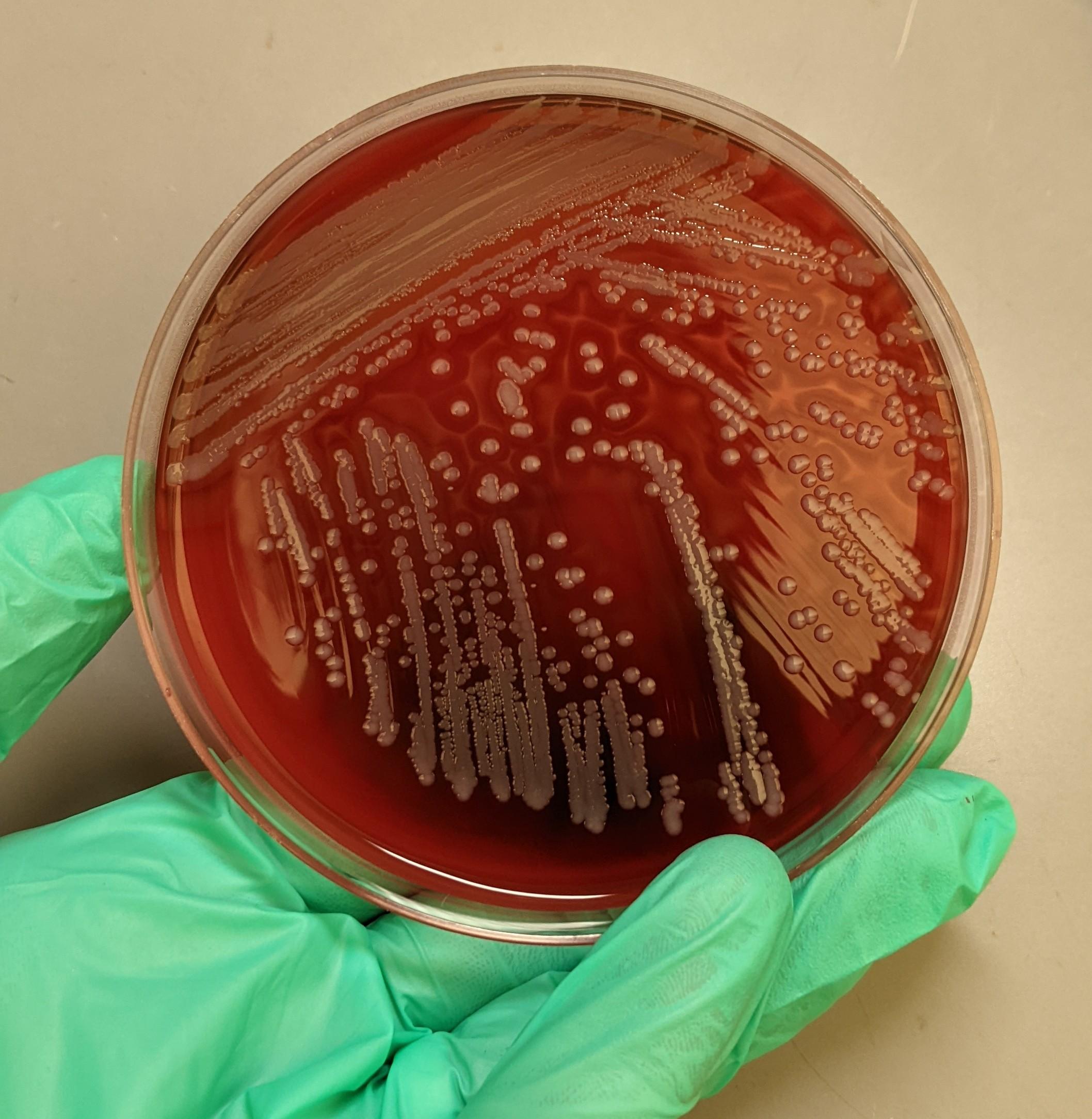
Colony morphology of S. aureus on SBA
white-yellow, raised, or creamy colonies with a smooth surface
medium to large
narrow zone of beta hemolysis
What is the most reliable test to ID S. aureus?
Coagulase test
most strains of S. aureus produce bound coagulase (aka clumping factor)
detected with the slide coagulase test
latex agglutination tests can also detect coagulase
Difference between slide and tube coagulase tests
slide coagulase test
bacterial colony is mixed with rabbit plasma
in a positive reaction, agglutination occurs (indicating presence of bound coagulase)
tube coagulase test
performed after negative slide tests to determine if free coagulase is present
organism is incubated with rabbit plasma at 37 C and then observed for a clot (indicates free coagulase)
false-negatives may occur due to fibrinolysin, which can break up the clot (test needs to be read periodically)
S. aureus is DNase:
positive
agar contains DNA and dye
S. aureus produces DNase, which breaks down DNA, allowing for a color change in the media
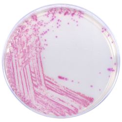
S. aureus can be isolated on chromogenic agar. What is this agar, and what does S. aureus look like when grown on it?
agar with chromogens (an artificial substrate) added
some organisms have enzymes that utilize the chromogens and form colored colonies
S. aureus: deep pink-fuchsia on HARDYchrome agar
S. aureus is a common HAI and can also cause CAIs. What type of infections does this organism cause?
skin and soft tissue infections
boils, folliculitis, bullous impetigo, scalded skin syndrome, acne, and acute or post-op wound infections
usual specimens: wounds, sputum, blood, urine, abscesses
List the 4 virulence factors of S. aureus
Protein A
binds to antibody and inhibits complement activation
Capsules
Peptidoglycan and teichoic acids
helps with attachment to mucus membranes and withstanding environmental stress
Penicillin-binding protein 2
alters how penicillin-type molecules bind to the bacterium
What 3 exotoxins does S. aureus release to increase its virulence?
hemolysin
lyses host RBCs
clumping factor/staphylocoagulase
aka coagulase, induces fibrin formation, fibrin binds to neutrophils, inhibiting phagocytosis
staphylokinase
dissolves fibrin clots
What enterotoxins does S. aureus produce?
hemolysin
heat-stable proteins
A, B, C, and D are associated with foodborne illness
B and C are associated with enterocolitis
superantigens
A class of antigens that cause non-specific activation of T-cells, leading to an exaggerated immune response
they are produced by certain bacteria, including S. aureus
What superantigens does S. aureus release?
enterotoxins
toxic-shock syndrome toxin 1 (TSST-1)
promotes cytokine release and progression of TSS
risk factors of TSS: menstrual products, childbirth, miscarriage, surgical wounds, skin infection, retained foreign objects
Describe the following S. aureus exotoxins: lipase, exfoliative toxins A and B, hyaluronidase, and leukocidin and Panton-Valentine Leukocidin
lipase
hydrolyzes lipids in the skin, allowing staph to colonize and cause infection
exfoliative toxins A and B
proteolytic, hydrolyzes tissues, responsible for scalded skin syndrome
hyaluronidase
lyses hyaluronic acid in CT, helping spread infection
leukocidin and Panton-Valentine Leukocidin (PVL)
enzymes toxic to neutrophils and macrophages, inhibiting phagocytosis
List some S. aureus infections
prosthetic joint infections
forms biofilms around implants
pneumonia (opportunistic)
bone infections
osteomyelitis
toxic shock syndrome
antibiotic resistance patterns of S. aureus
can produce beta-lactamase, causing resistance to the penicillins and other beta-lactams
such as methicillin and vancomycin
can also be resistant to clindamycin
How is MRSA detected in the lab?
cefoxitin disk screen test
chromogenic agars
latex agglutination
molecular methods
detects mecA and mecC genes
What are the 3 strains of S. aureus that are susceptible and resistant to vancomycin?
vancomycin-susceptible S. aureus (MIC of < 2 mg/mL)
vancomycin-intermediate S. aureus (VISA) (MIC of 4-8)
vancomycin-resistant S. aureus (VRSA) (MIC of 16+)
S. aureus clindamycin resistance
resistance may only be detected if the S. aureus is also exposed to erythromycin
inducible-clindamycin reistance (ICR)
ICR of S. aureus can be detected by a D test
lawn of bacteria is grown on Mueller-Hinton agar
clindamycin disk and erythromycin disk are placed next to each other
What are the Coagulase-Negative Staphylococci?
S. epidermidis, S. saprophyticus, S. lugdunensis, S. hominis, and S. haemolyticus
once considered contaminants and normal flora, but are now opportunistic pathogens
elderly, infants, chronically ill, those with prosthetics
they can cause blood infections, prosthetic biofilms, UTIs, and endocarditis
general lab identification of coagulase-negative staphylococcus
GPC in clusters
catalase positive
coagulase negative
DNase negative
grows on MSA, but does not ferment mannitol
How is S. epidermidis identified in the lab?
colony morphology
medium colonies that are white-gray, creamy, raised, and gamma-hemolytic
coagulase-negative, catalase-positive
DNase negative
novobiocin susceptible
What infections does S. saprophyticus cause, and how is it identified in the lab?
contaminant unless found in urine
causes UTIs (cystitis, pyelonephritis, catheter-associated UTIs)
can induce UTIs with low colony counts
lab ID
coagulase negative
DNase negative
resistant to novobiocin
grows on MSA with variable fermentation
S. lugdunensis
opportunistic pathogen that causes aggressive infections with a high mortality rate
associated with catheter-associated bacteremia, prosthetic joint infections, shunt infections, endocarditis, and UTIs
positive PYR and ODC
CoNS species: S. haemolyticus, S. hominis, and S. intermedius
S. haemolyticus
BETA hemolytic, associated with endocarditis and bacteremia
S. hominis
rare cause of septicemia in immunocompromised hosts
S. intermedius
common veterinary pathogen - dog bite wounds
S. schleiferi subsp. coagulans and S. warneri
S. schleiferi
veterinary pathogen
endocarditis and septicemia in humans
S. warneri
human and animal pathogen
can cause spontaneous abortions, UTIs, meningitis, or endocarditis
penicillin binding protein (PBP)
A set of proteins found in bacterial cell membranes that play a crucial role in building the cell wall, making them key targets for beta-lactam antibiotics
PBP2 and PBP2a
allowing cross-linking of peptidoglycan layers
targets for beta-lactam binding