Dysrhythmias, Heart Structures, & EKGs
1/25
There's no tags or description
Looks like no tags are added yet.
Name | Mastery | Learn | Test | Matching | Spaced | Call with Kai |
|---|
No analytics yet
Send a link to your students to track their progress
26 Terms
How does blood flow in the heart?
Deoxygenated Blood Enters (Right Heart):
Oxygen-poor blood returns from the body through the superior and inferior vena cava, entering the RA.
Right Ventricle to Lungs: Blood flows from the right atrium into the RV through the tricuspid valve. The right ventricle pumps the blood through the pulmonary valve into the pulmonary artery, taking it to the lungs to receive oxygen.
Oxygenated Blood Enters (Left Heart): Oxygen-rich blood returns from the lungs through the pulmonary veins and enters the LA.
Left Ventricle to Body: Blood flows from the left atrium into the LV through the mitral valve. The left ventricle (the strongest chamber) pumps this blood through the aortic valve into the aorta.
Distribution: The aorta supplies the oxygen-rich blood to the rest of the body.
What is the sinoatrial (SA) node?
The pacemaker of the heart. Beats 60-100bpm normally. The signal flows from the SA node —> AV node —> bundle of his —> bundle branches —> purkinje fibers
What is the atrioventricular (AV) node?
The backup pacemaker. Beats at 40-60bpm.
What is conductivity?
The ability to pass electrical impulse from cell to cell
What is contractility?
strength of contraction
What is the refractory period?
Recovery period after stimulation during which cardiac cells cannot respond to stimulation; resting state!!
What is the positioning for a 5 lead?
From R to L “Snow over grass” = white over green
From R to L “Smoke over fire” = black over red.
The RA (white) and LA (black) go above heart
The RL (green) and LL (red) go below the heart.
The V goes in the middle to the side of the sternum.
How many seconds are in a “big” box on ECG graph paper?
0.2 seconds
How many seconds are in a “small” box on ECG graph paper?
0.04 seconds

What is the P wave?
Shows the atria contracting (depolarization) and sending blood to the ventricles
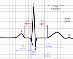

What is the P-R interval?
Beginning of the P wave to the beginning of the QRS complex. It represents the duration of time it takes for an electrical impulse to travel from the sinoatrial (SA) node, through the atria, through the atrioventricular (AV) node, and into the ventricles.
Should be 0.12 to 0.2seconds (3-5 small boxes)

What is the QRS complex?
Ventricles contracting (depolarizing) and atria repolarizing (relaxing; not seen).
Should be <0.12s (<3 small boxes)

What is the T wave?
Ventricles repolarizing (relaxing)


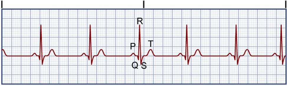
What rhythm is this?
Normal Sinus Rhythm
-Regular rhythm and rate (SA node fires 60-100 bpm)
-PR interval is within 0.12-0.2 (3-5 small boxes)
-QRS complex is <0.12s (<3 small boxes)
-P waves are paired with a QRS

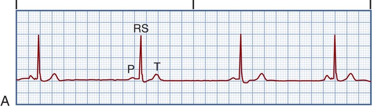
What rhythm is this?
Sinus Bradycardia
-Regular rhythm, but rate is <60bpm
-PR interval is within 0.12-0.2 (3-5 small boxes)
-QRS complex is <0.12s (<3 small boxes)
-P waves are paired with a QRS

What is the treatment for this rhythm?
Sinus Bradycardia
If symptomatic, give atropine (anticholinergic drug) which increases the HR. AEs include anticholinergic effects. If ineffective, use pacing or dopamine/epinephrine.

What are the symptoms of an atropine overdose?
“Hot as a hare” — Increased temperature (decreased sweating)
“Mad as a Hatter” — confusion, delirium
“Red as a beet” —- flushing
“Dry as a bone” —- decreased secretions, thirst
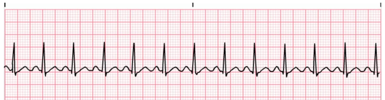
What is this rhythm?
Sinus tachycardia
-Regular rhythm, but rate is >100bpm
-PR interval is within 0.12-0.2 (3-5 small boxes)
-QRS complex is <0.12s (<3 small boxes)
-P waves are paired with a QRS


What is the treatment for this rhythm?
Sinus tachycardia
If symptomatic, treat cause.
Stable pts:
1) Vagal maneuver to drop HR
2) Beta blockers (metoprolol)
Unstable pts: cardioversion


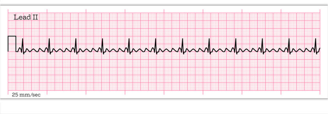
What is this rhythm?
Supraventricular tachycardia (SVT)
-Regular rhythm but rate is 150-220 bpm
-P wave is hidden or abnormal

What is the treatment for this rhythm?
Supraventricular tachycardia (SVT)
Vagal maneuver
IV adenosine given rapidly over 1-2 second via a stop c0ck as close to the heart as possible followed by a 20mL NS flush. Monitor for brief asystole, flushing, and dizziness.
For chronic SVT: B-adrenergic blocker (sotalol), calcium channel blocker (diltiazem), and cardioversion


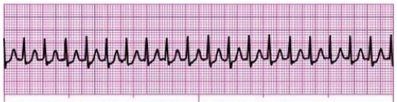
What is this rhythm?
Atrial flutter
-Rhythm is regular/irregular and rate is 220-430 bpm
-Many P waves in a sawtooth shaped flutter
What is the treatment for this rhythm?
Atrial Flutter
-Ablation of the singular ectopic foci
-CCB and beta blockers
-Cardioversion
-Amiodarone: antidysrhythmic and potassium channel blocker that delays repolarization (increases resting period).

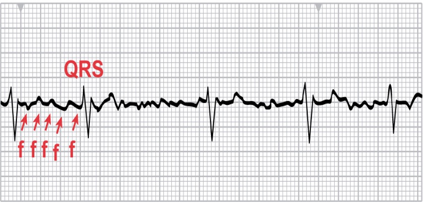
What is this rhythm?
Atrial fibrillation
-Irregularly irregular — distance between QRS is irregular with no identifiable pattern
-Rate is 350-650 bpm
-Hallmark fibrillatory waves
-No P waves visible

What is the treatment for this rhythm?
Atrial fibrillation
Cardioversion with amiodarone
Ablation
For chronic A-fib, lower HR with: digoxin, calcium channel blockers, β-adrenergic blockers. Use enoxaparin + warfarin for stroke prevention

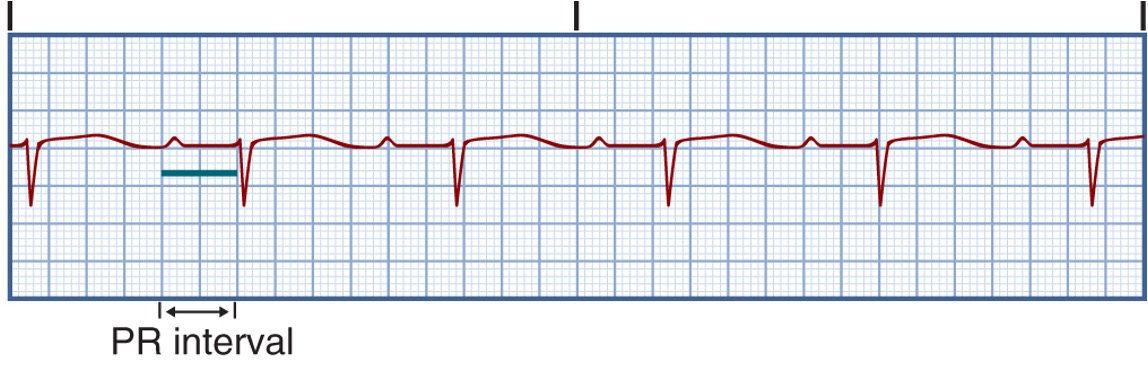
What is this rhythm?
1st degree AV heart block
-PR interval is >0.12s (> 5 small boxes) when normal PR interval is 0.12-0.2 (3-5 small boxes)
