S26 Anatomy 3-2 Renal Anatomy
1/44
Earn XP
Description and Tags
Renal Anatomy
Name | Mastery | Learn | Test | Matching | Spaced | Call with Kai |
|---|
No analytics yet
Send a link to your students to track their progress
45 Terms
The kidneys are located between which vertebrae?
T11/12 and L3
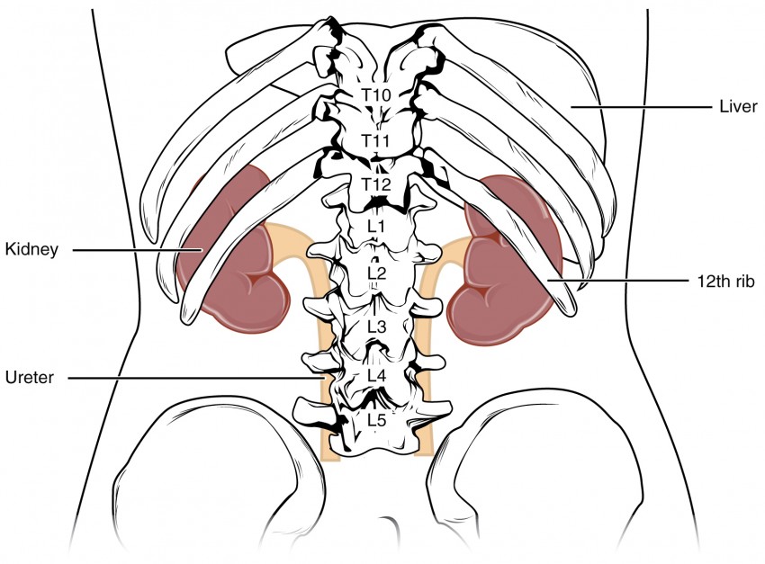
The right kidney is anterior to which rib?
Rib 12
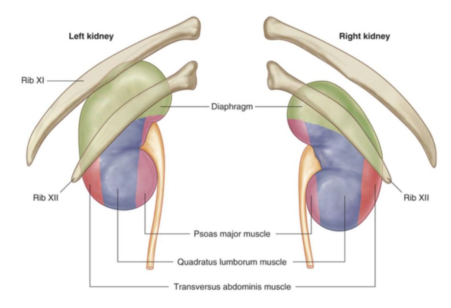
The left kidney is anterior to which ribs?
Rib 11 and 12

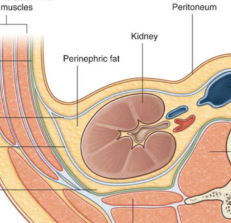
What type of fat surrounds all structures in the renal sinus?
Perinephric fat
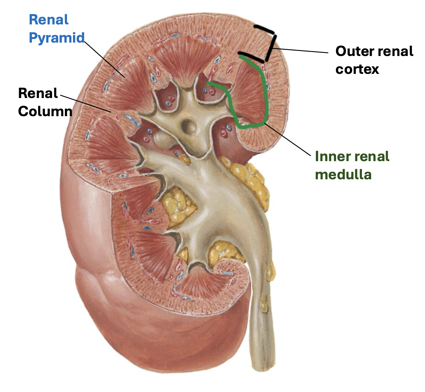
What does the renal cortex contain?
Renal corpuscles, proximal/distal tubules, and peritubular capillaries
What does the renal medulla contain?
Henle's loop, vasa recta, and collecting ducts for all nephrons
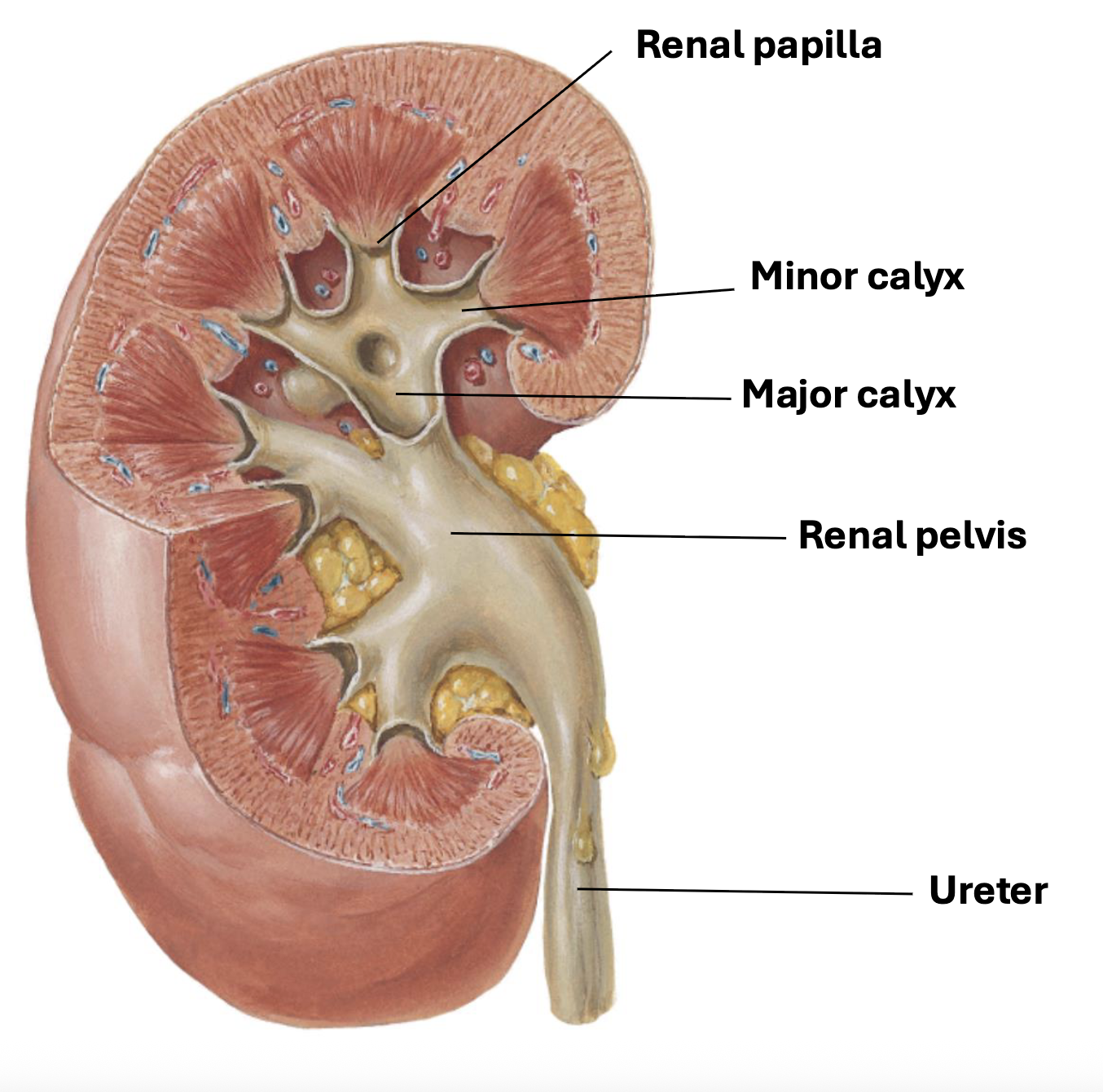
What does the renal pyramid contain?
Openings of the papillary duct draining the renal tubules
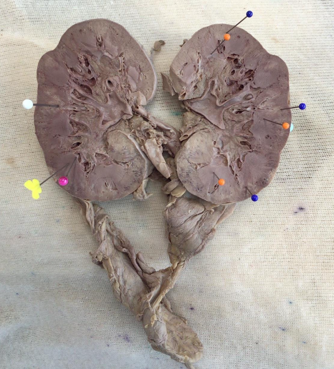
Blue Pins: Renal Cortex
Orange Pins: Renal Medulla
Yellow Butterfly: Renal Pyramids
Pink Pin: Renal Papilla
White Pin: Renal Column
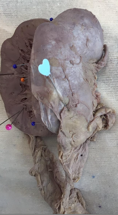
Blue heart: Fibrous Capsule
Describe the flow of urine from minor calices to the bladder.
Minor calices → Major calices → Renal pelvis → Ureter → Bladder +3

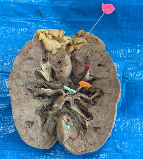
Red Pin: Minor Calyx
Orange Pin: Major Calyx
Teal Heart: Renal Pelvis
Blue Pin: Ureter
Pink Bird: Suprarenal Gland
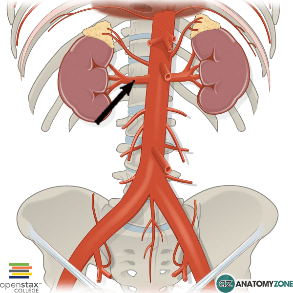
Renal arteries arise at what vertebral level?
Between L1 and L2
What arteries do renal arteries give off and what do they supply?
Segmental arteries (five renal segments), Interlobar (adjacent sides of neighboring renal lobes), and Cortical radiate arteries (bring blood to glomeruli) S.IL.C
Green Bird: IVC
Blue Star: Abdominal Aorta
Red Moon: Left Renal Vein
Pink Heart: Left Renal Artery
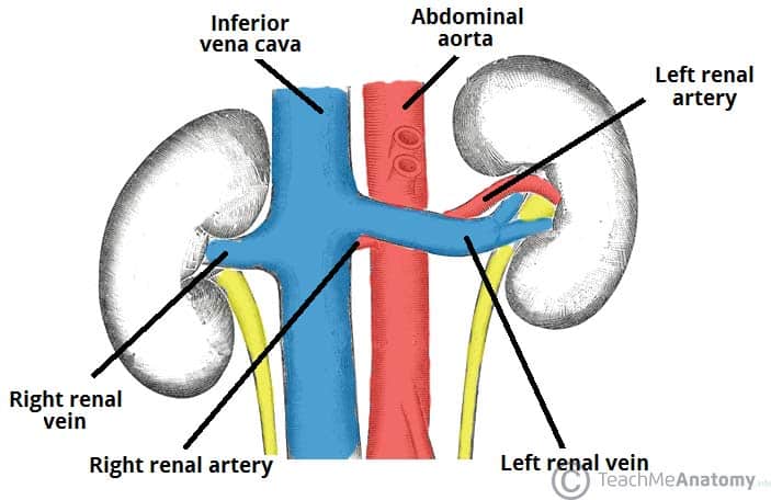
The right renal artery passes posterior to what anatomical structure?
Inferior Vena Cava (IVC)
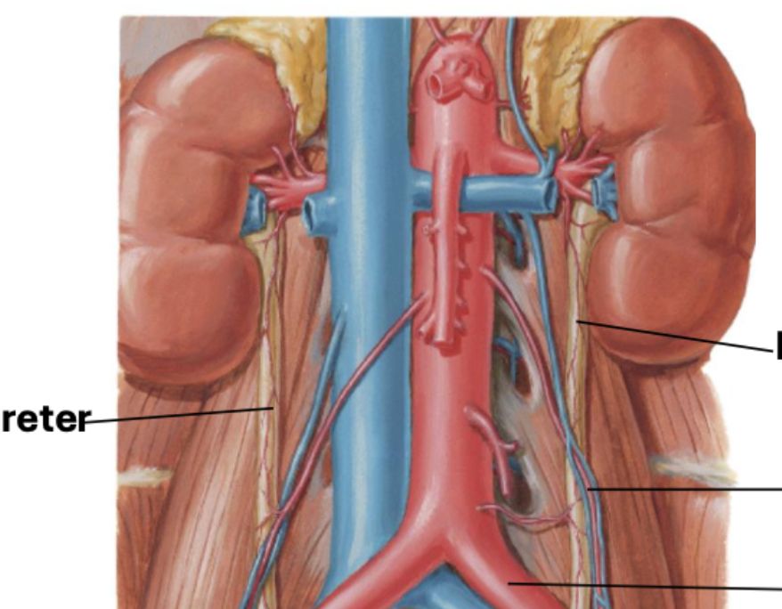
The left renal vein crosses between what two anatomical structures?
Anterior to the abdominal aorta and posterior to the superior mesenteric artery

Superior vesical arteries supply the?
Bladder
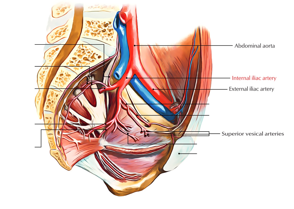
Pink Star: Common Iliac Artery
Blue Star: Internal Iliac Artery
Orange Pin: Superior Vesical Artery
Blue Button: Detrusor muscle
Yellow Pin: Internal Urethral Orifice
The bladder is mostly composed of which muscle?
Detrusor muscle +1
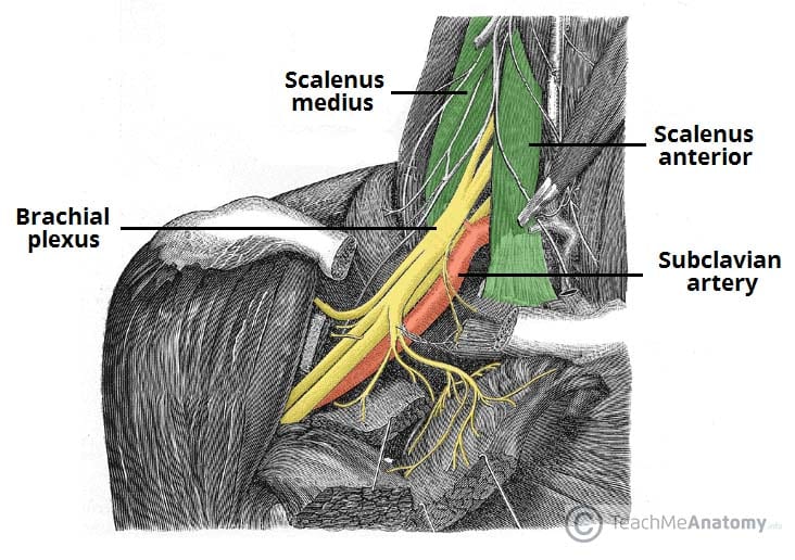
The roots of the brachial plexus exit between what muscles in the neck?
Anterior and middle scalene muscles
When may the upper roots of the brachial plexus be damaged?
With excessive rotation of the head to the opposite side
When may the lower roots of the brachial plexus be damaged?
With extreme abduction of the arms
The cords of the brachial plexus are named for their location to which artery?
Axillary artery
Where are the divisions of the brachial plexus located in the upper limb?
They begin at the lateral border of the 1st rib and posterior to the clavicle
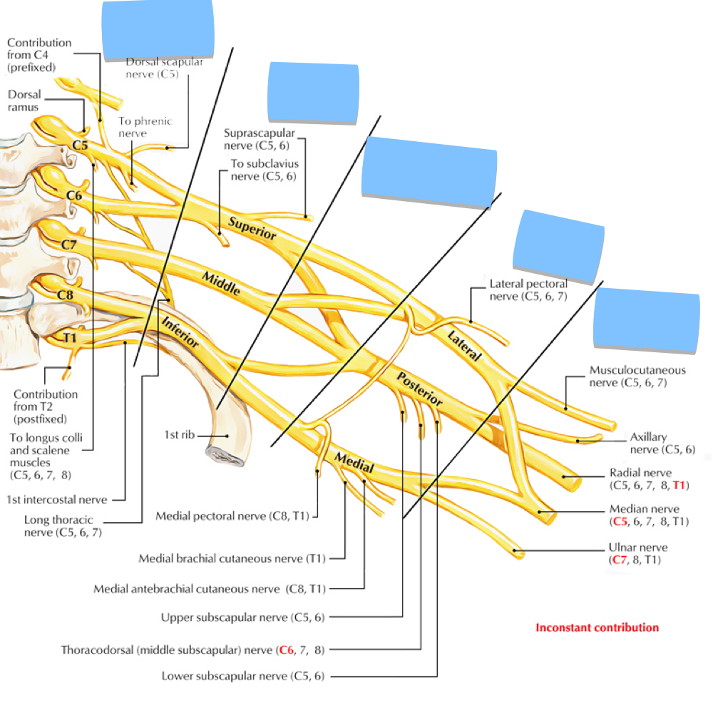
Rugby Teams Dont Cover Bruises
Roots, Trunks, Divisions, Cords, Branches

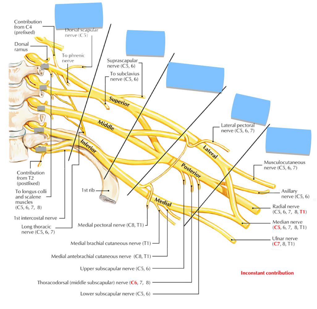
Name the Roots

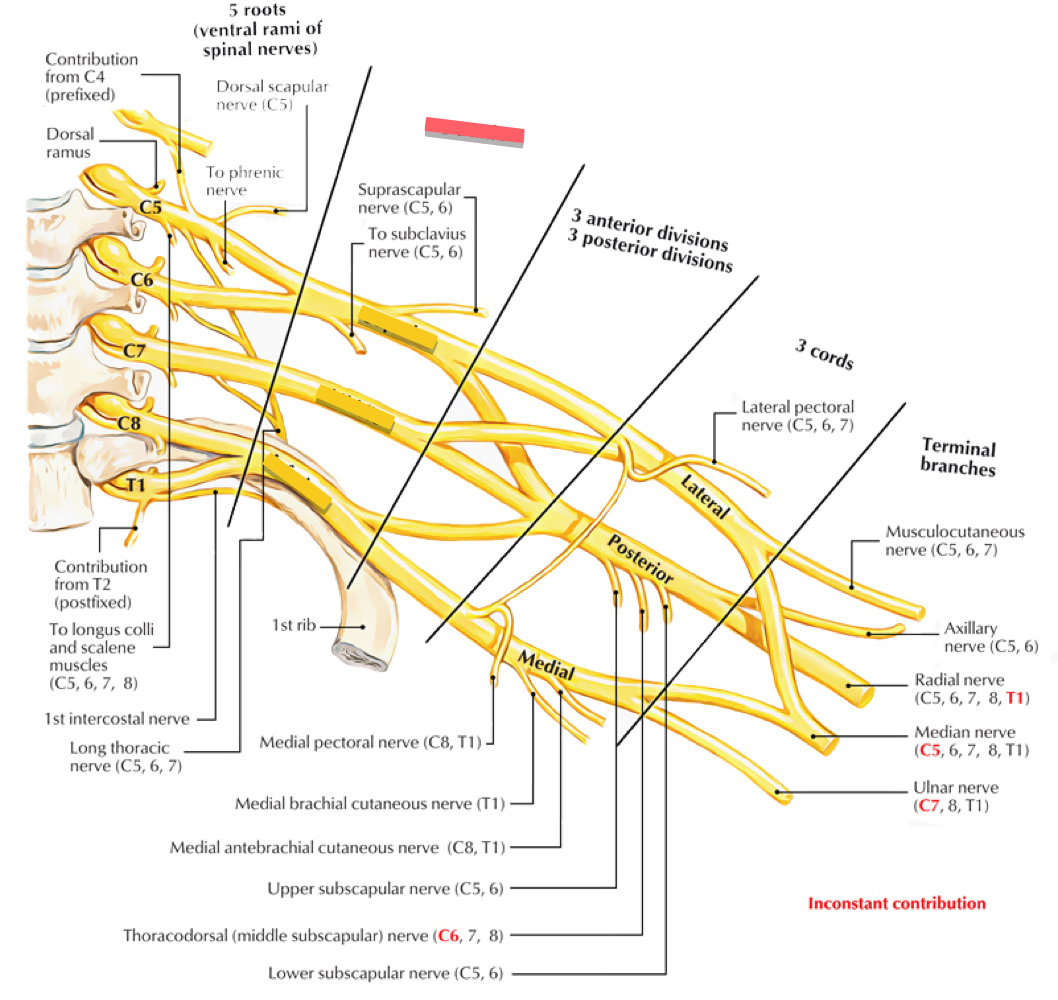
Trunks of the Brachial Plexus
Trunks: Superior, Middle, and Inferior
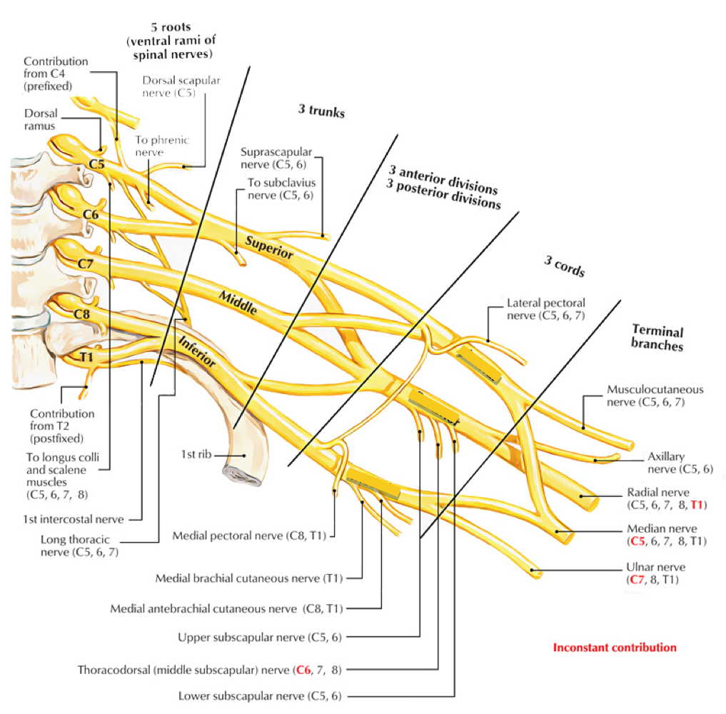
Cords of the Brachial Plexus
Lateral, Posterior, Medial
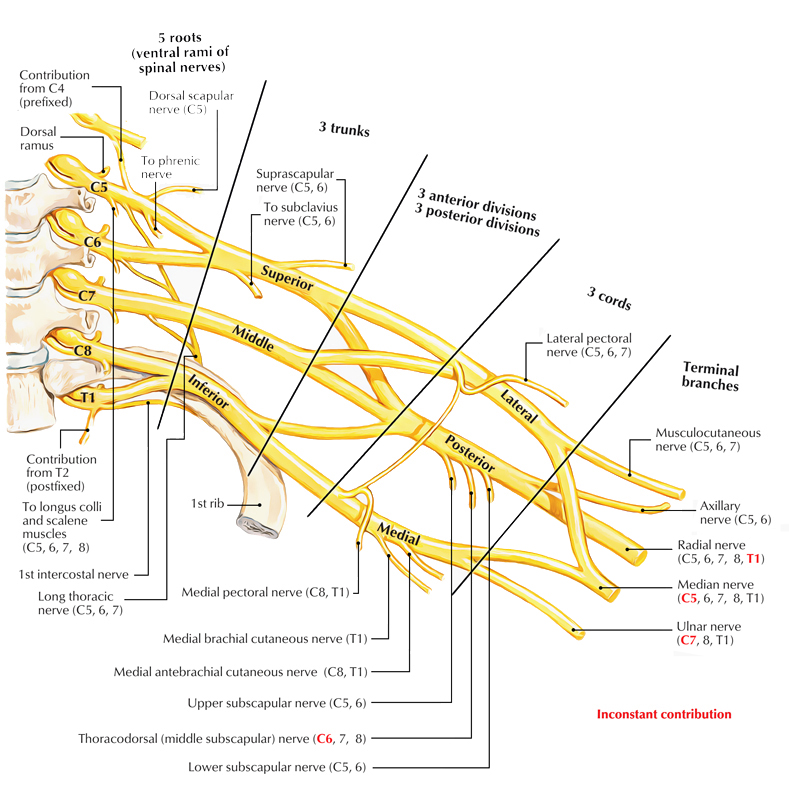
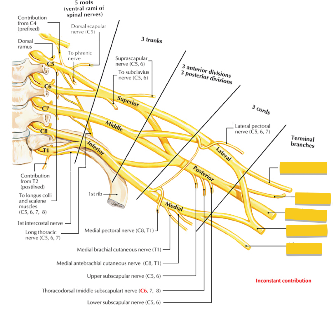
Terminal Branches of the Brachial Plexus
From Medial to Lateral:
Ulnar nerve, Median Nerve, Radial Nerve, Axillary Nerve, Musculocutaneous Nerve

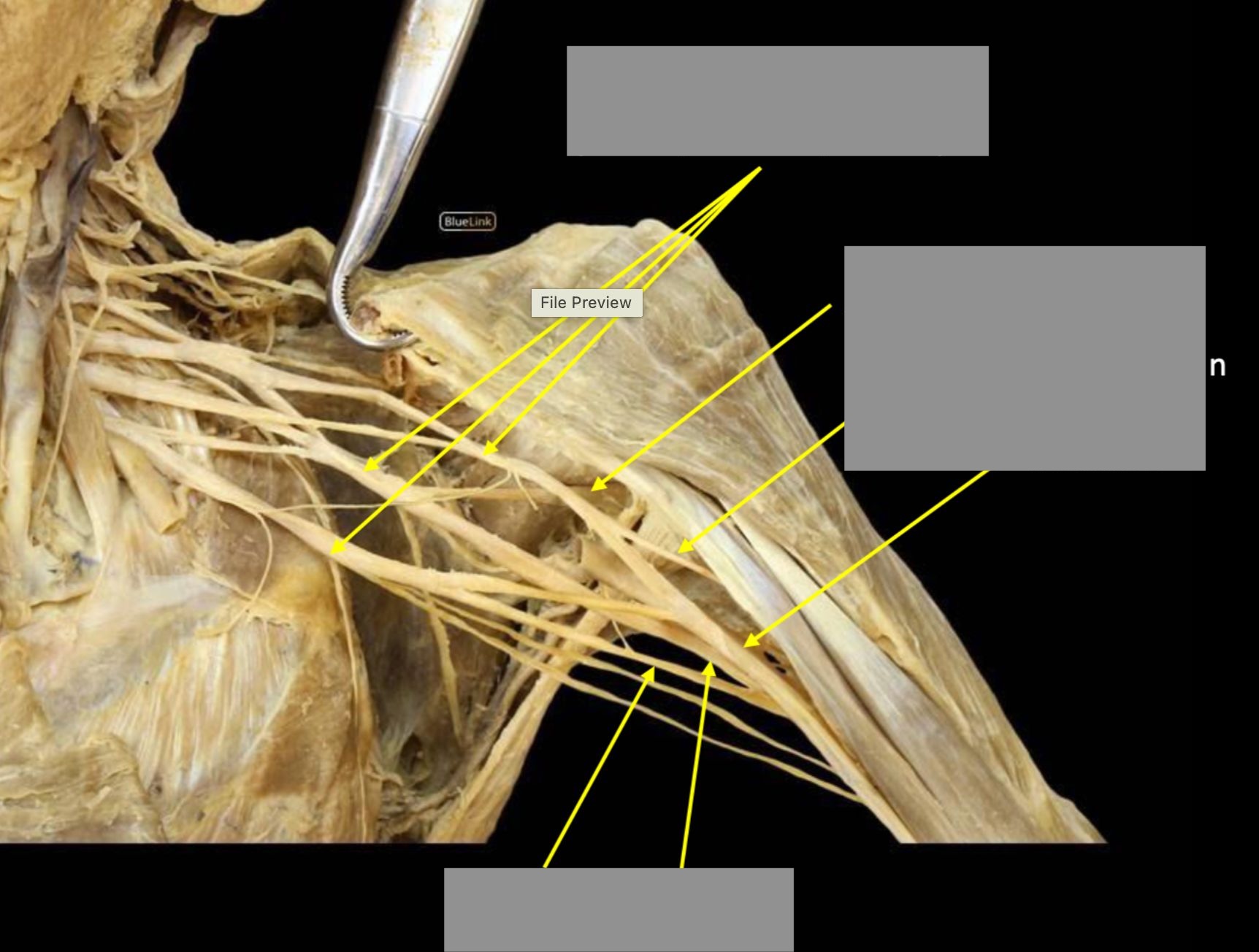
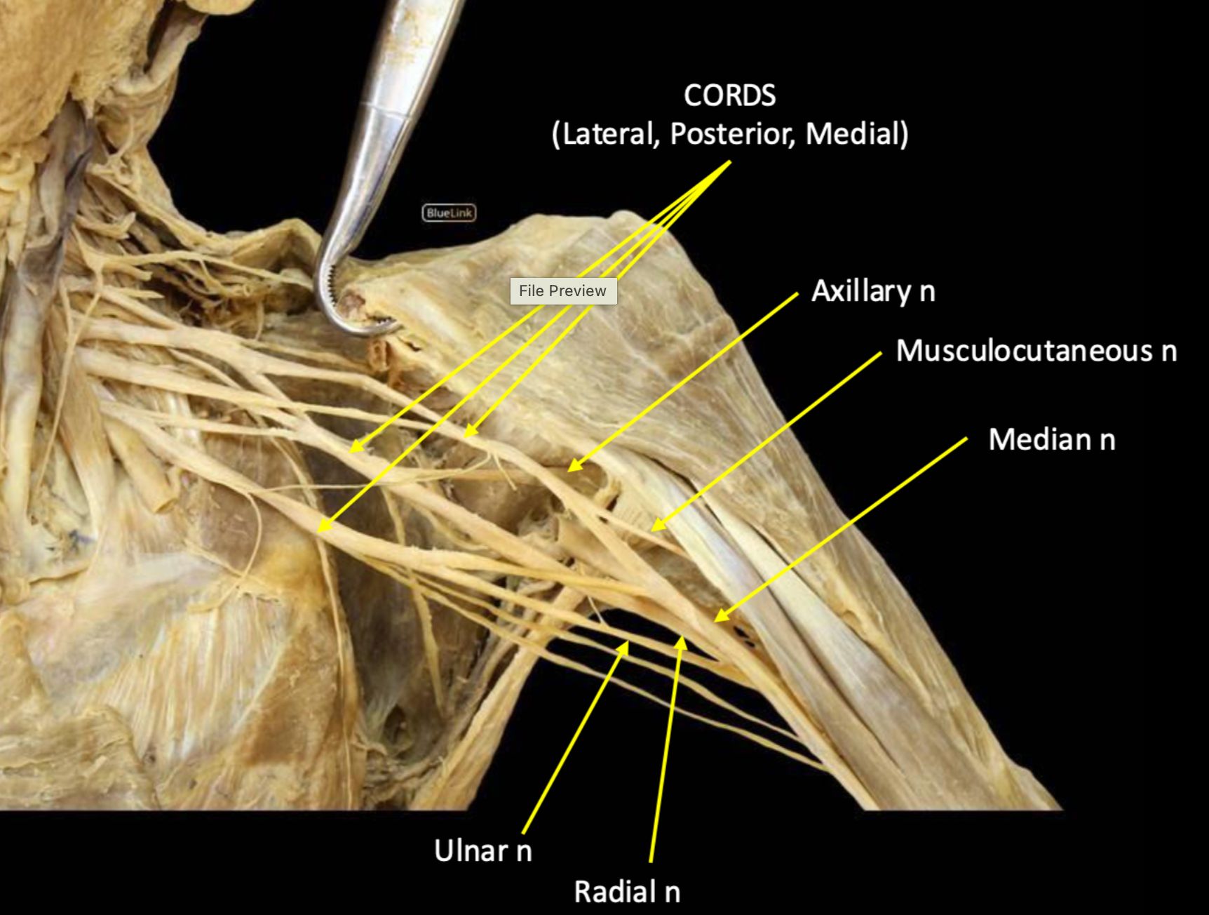
What does the musculocutaneous nerve innervate?
Anterior arm and skin of the lateral forearm
What does the axillary nerve innervate?
Deltoid and teres minor muscles
What does the radial nerve innervate?
Posterior arm and forearm
What do the median and ulnar nerves innervate?
Anterior forearm and palm
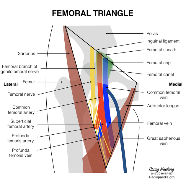
The femoral sheath is composed of the inferior prolongations of what fascia?
Transversalis, iliopsoas, and pectineal fasciae (TIP)
What is found in the femoral canal?
Loose connective tissue, fat, and lymph
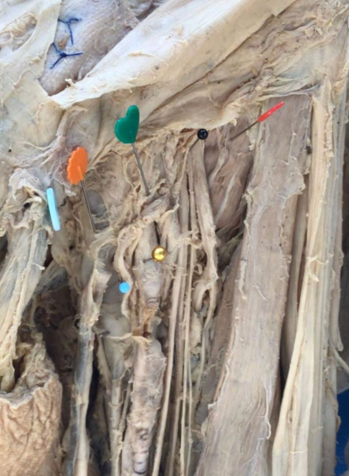
red - iliopsoas muscle
black - femoral nerve
green - lateral compartment
gold - femoral artery
orange - intermediate compartment
small blue - femoral vein
blue button - medial compartment

What are the boundaries of the femoral triangle?
Inguinal ligament, sartorius, and adductor longus

What are the contents of the femoral triangle from lateral to medial?
NAVL (femoral Nerve, femoral Artery, femoral Vein, Lymphatics)
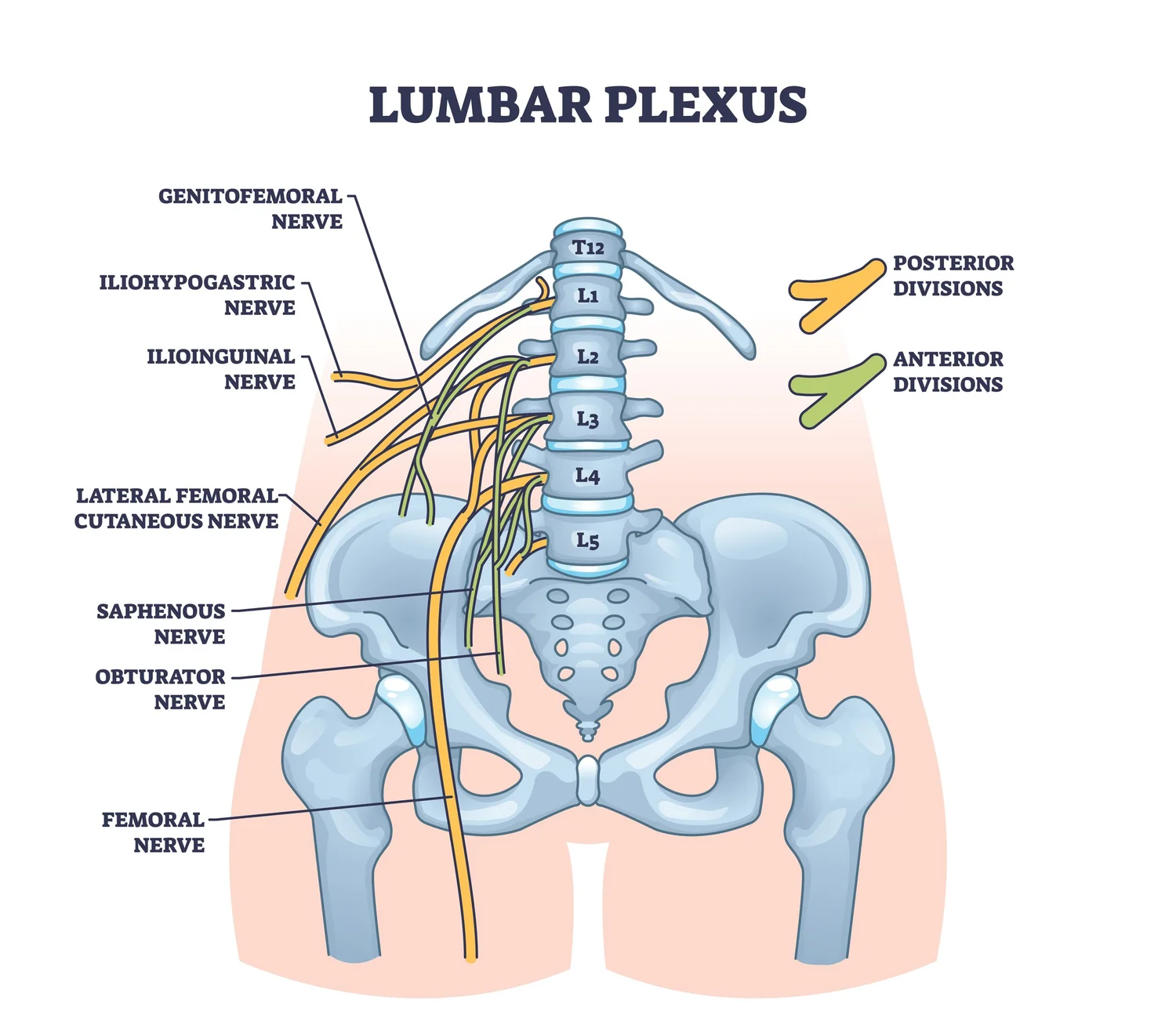
From which spinal nerve levels does the femoral nerve arise?
L2, L3, and L4
Is the femoral nerve in the fascial sheath?
No, it lies posterior and lateral to the femoral artery
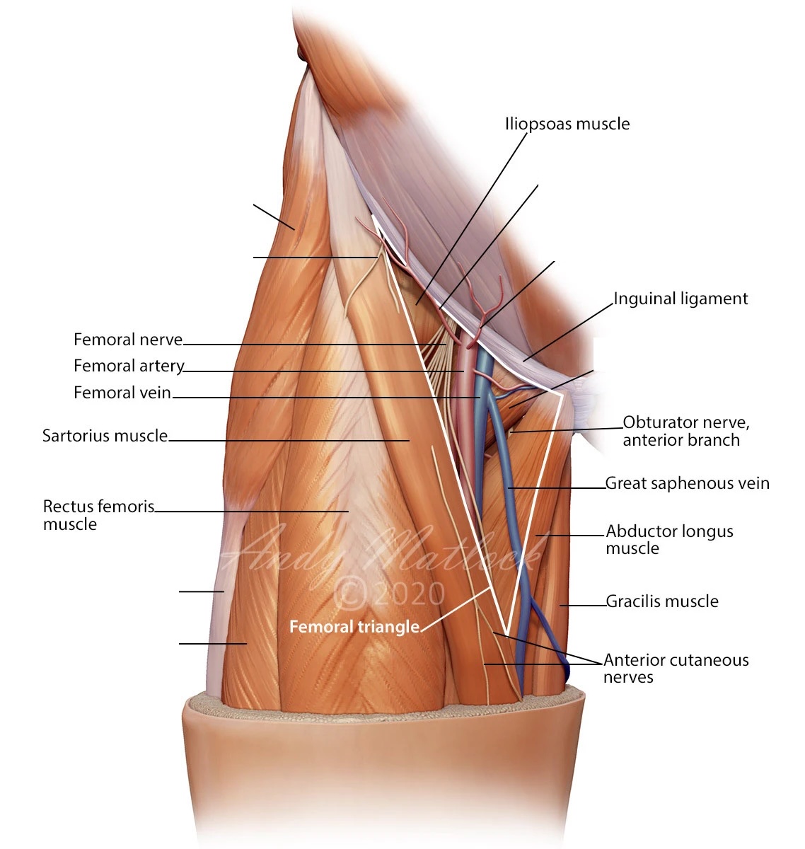
The femoral nerve is usually how many centimeters lateral to the femoral artery?
1 to 2 cm
The adductor canal extends from where to where?
From the apex of the femoral triangle to the adductor hiatus
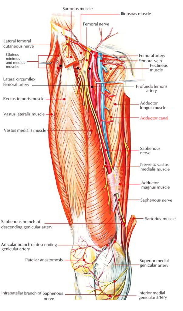
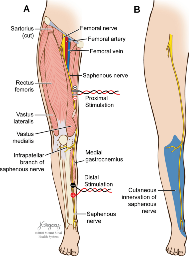
What skin does the saphenous nerve supply on the lower limb?
Skin over the medial, anteromedial, and posteromedial aspects from the knee to the foot
Describe the path of the saphenous nerve.
Terminal branch of femoral nerve; leaves femoral canal in the triangle, descends in adductor canal deep to sartorius with the superficial femoral artery