The Muscular System
1/89
There's no tags or description
Looks like no tags are added yet.
Name | Mastery | Learn | Test | Matching | Spaced | Call with Kai |
|---|
No analytics yet
Send a link to your students to track their progress
90 Terms
What are the 4 functions of the muscular system?
Movement
Maintenance of posture
Stabilization of joints (keeps your bones together)
Generation of heat
What are the 4 properties of muscle tissue?
Excitability
Extensibility
Elasticity
Contractility
What is the excitability property of muscle tissue?
Stimulating a muscle to do something; the ability to respond to a stimulus
What is the extensibility property of muscle tissue?
The ability to stretch without damage
What is the elasticity property of muscle tissue?
When you stretch muscle out of contract it, they come back to their original shape (recoils)
What is the contractility property of muscle tissue?
The ability to shorten
What are the 3 types of muscle tissue?
Skeletal muscle
Cardiac muscle
Smooth muscle
What is the location of skeletal muscle?
Attached to the skeleton
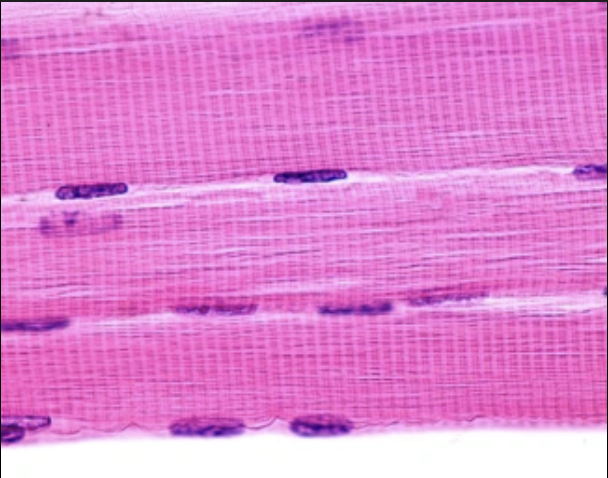
What is the structure of skeletal muscle?
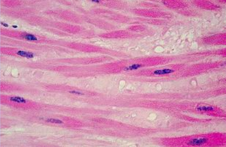
Long, thin cells (fibers) that are striated & multinucleated (nuclei are pushed off to the side)
What are the functions of skeletal muscle (3)?
Posture
Movement
Stabilizes joints
How is skeletal muscle stimulated?
Voluntarily controlled via the somatic nervous system
Skeletal Muscle
What is the location of cardiac muscle?
Wall of the heart
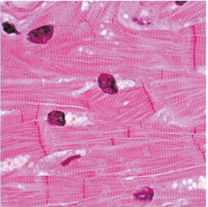
What is the structure of cardiac muscle?
Cells (myocytes) have some striations and branching, with 1-2 nuclei
Cells are connected by intercalated discs (gap junctions/desmosomes), which allow the cells to communicate & resist being pulled apart from contraction
What is the function of cardiac muscle?
Pushes the blood around throughout circulatory system
How is cardiac muscle stimulated (2)?
Involuntary, self-exciting
Autorhythmic, but can be controlled by the autonomic nervous system
Cardiac Muscle
What is the location of smooth muscle?
Walls of hollow organs (ex. small intestine, blood vessel walls)
What is the structure of smooth muscle?
Tapered (stretched-out football-shaped) with no striations & 1 nucleus
What is the function of smooth muscle?
Moves fluids
How is smooth muscle stimulated?
Involuntary, stimulated by autonomic nervous system, but could also be stimulated by hormones
Smooth Muscle
Are skeletal muscles organs?
What 2 structures are they made up of?
Yes
Skeletal muscle cells (fibers) & connective tissues
What are the 6 structures found within the gross anatomy of skeletal muscle?
Muscle fiber
Endomysium
Fascicle
Permysium
Whole muscle
Epimysium
Muscle fiber
Muscle cell
Endomysium
Very thin bit of connective tissue that surrounds each muscle fiber
Fascicle
1 bundle of muscle fibers
Perimysium
Connective tissues that surrounds a fascicle
Whole muscle
1 bundle of fascicles
Epimysium
Dense irregular connective tissue covering the entire muscle
What is an origin?
The attachment of a muscle (usually a bone) that does not move

What is the insertion?
The attachment of a muscle (usually a bone) that does move (gets pulled)
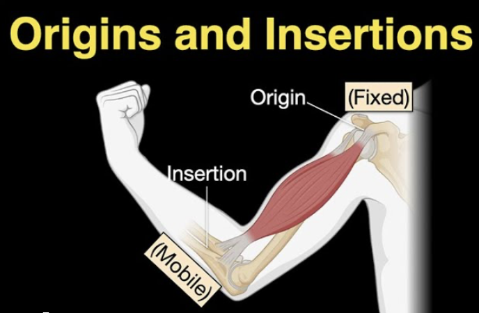
What is an action?
The movement when the muscle shortens
What is an innervation?
The name of the nerve that goes to a muscle
What are the 2 types of attachments?
Direct
Indirect
What is a direct attachment?
Periosteum of bone attaches to epimysium of muscle
What is an indirect attachment?
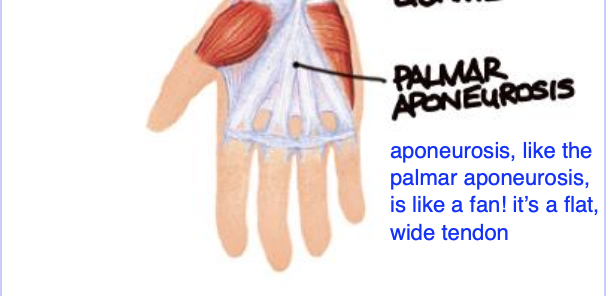
Attachment is made via tendon or aponeurosis
What is aponeurosis?
A specialized tendon that’s wide & flat
Is the respiratory diaphragm a skeletal muscle?
How do you know (2)?
Yes
It’s striated & you can voluntarily control your breathing
What is the sequence of gross muscle to microscopic?
Whole muscle → fascicle → muscle fiber (cell) → myofibrils → myofilaments
Sarcolemma
Plasma membrane of a muscle fiber (NOT the same thing as the endomysium)
Sarcoplasm
Cytoplasm of a muscle cell (fiber)
Myofibrils
Tubes within the muscle fibers that are responsible for muscle contraction
Transverse tubules (T-tubules)
Pieces of the sarcolemma that extend into the muscle cell (fiber)
Sarcoplasmic Reticulum
Smooth endoplasmic reticulum of the muscle cell (fiber) that releases calcium during contraction
Triad
Sandwich consisting of 1 sarcoplasmic reticulum (bread), a t-tubule (filling), and another 1 sarcoplasmic reticulum (bread)
Terminal cisternae
The end of the sarcoplasmic reticulum
What are myofibrils composed of?
Myofilaments
Name the 2 myofilaments
Actin
Myosin
What is actin (thick or thin filament)?
Thin myofilament
What is myosin (thick or thin filament)?
Thick myofilament
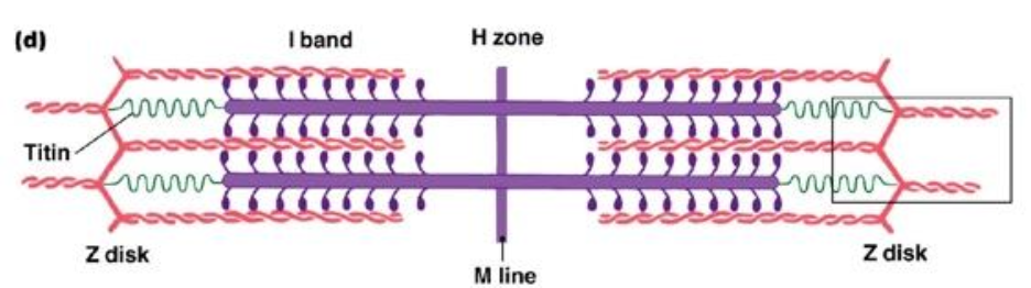
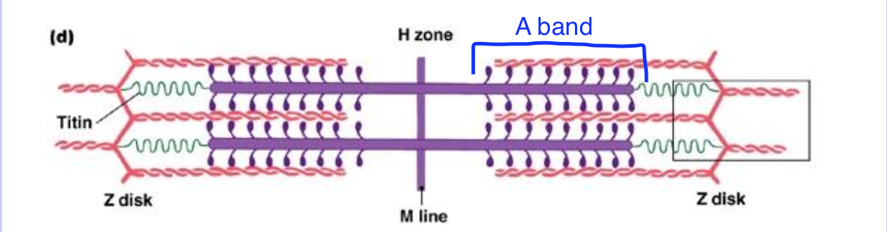
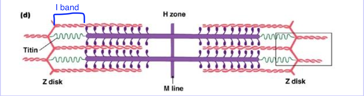
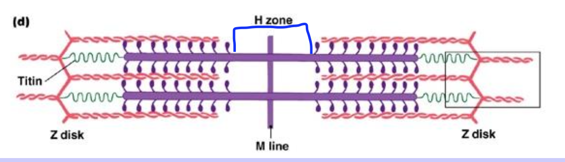
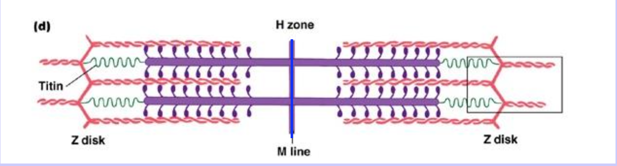
Sarcomere
The functional/contractile unit of the muscle
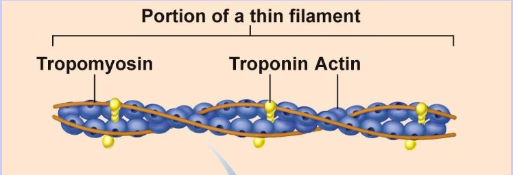
What are the 3 structures grouped within/alongside actin?
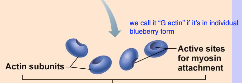
G actin
Troponin
Tropomyosin
G actin
A single actin subunit (1 blueberry)

F actin
Actin subunits when they’re in filament form
Troponin
“Clips on a rope”; Keeps tropomyosin in place, then moves it out of the way once calcium arrives
Tropomyosin
Rope that covers the active sites for myosin to bind during rest
What are the 3 important regions on the myosin head?
ATPase
Actin binding site
Hinge region
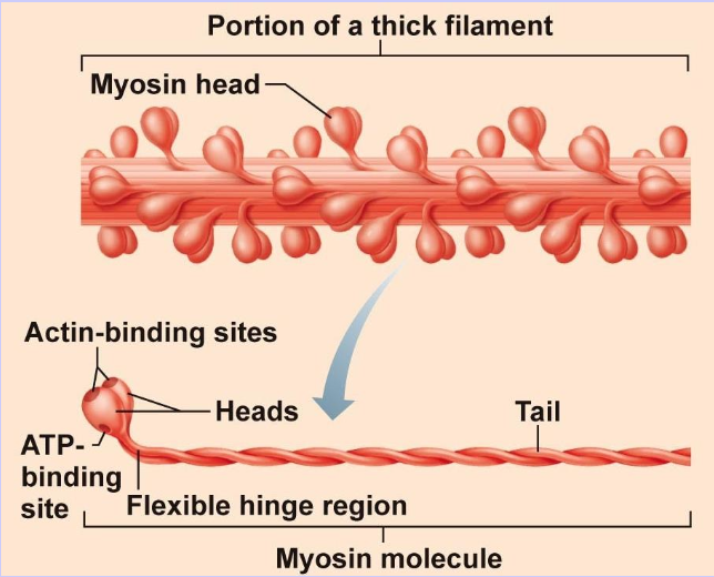
ATPase on myosin head
Enzyme to break down ATP during muscle contraction
Actin binding site on myosin head
Myosin heads fit into actin active sites
Hinge region of myosin head
Moveable neck of the myosin head
What is the elastic filament?
Titin

What is the function of titin in the sarcomere?
Holds myosin in place at each end of the sarcomere
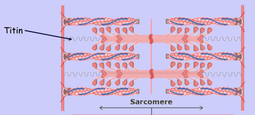
Name all of the bands, zones, & lines within the sarcomere (5)
Z disc
A band
I band
H zone
M line
Z discs
Zig-zag, Z-looking ends of the sarcomere
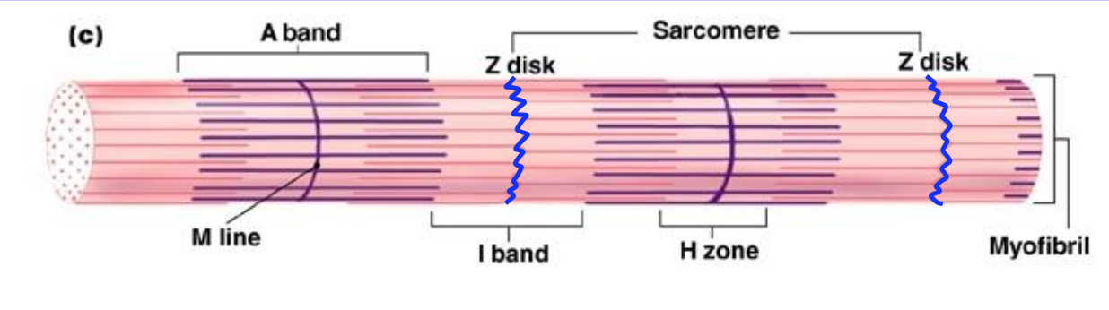
A band
Dark band under microscope; Contains both actin & myosin in the sarcomere
I band
Light under microscope; Contains actin only in the sarcomere
H zone
Region of the sarcomere that contains only myosin; Goes away during contraction (because of shortening!)
M line
Mid-line of the sarcomere
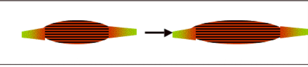
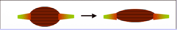
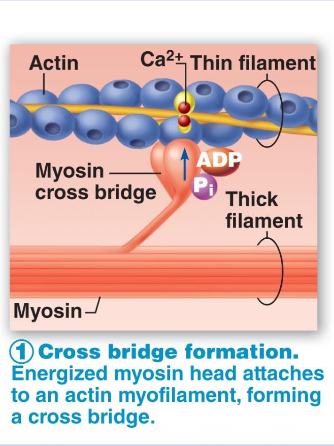
Sliding filament theory
In the presence of calcium, myosin heads can bind to actin to form a cross bridge
ATP breakdown causes the myosin heads to cock (hinge of myosin head moves), creating “power stroke,” then unbinds so the process can begin again
Actin & myosin don’t actually shorten. Instead, they slide against each other, pulling the Z discs closer
Cross Bridge Formation - Step 1 (3)
Calcium binds to troponin
Troponin moves tropomyosin out of the way
Myosin heads bind to actin
The Power Stroke - Step 2 (2)
Myosin head moves, pulling actin
Myosin releases ADP & phosphate (Pi)
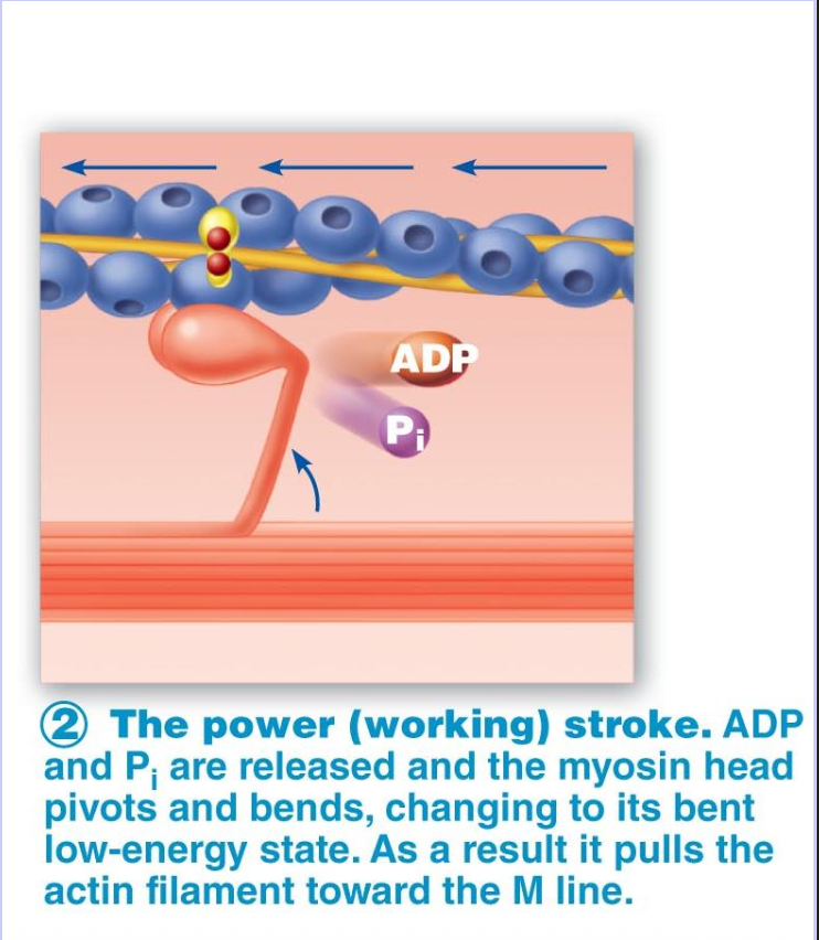
In Step 2 - The Power Stroke, what happens if ATP is not present?
Myosin head will stay stuck to actin (rigor)
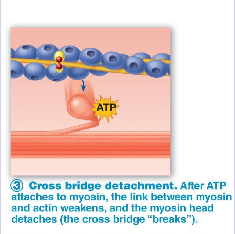
Cross Bridge Detachment - Step 3 (2)
ATP binds to myosin head
Myosin detaches from actin
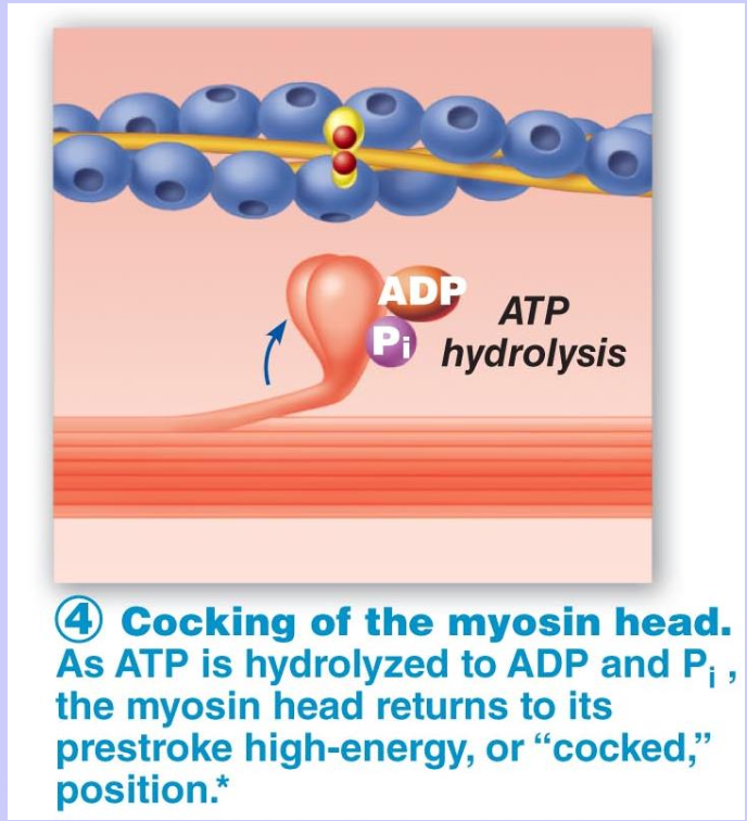
Resetting the System - Step 4
Myosin hydrolyzes the ATP molecule (breaks ATP into ADP & Pi)
Myosin head “cocks” in preparation for another cycle
Diffusion
The movement of solutes from one side of a semipermeable membrane to the other
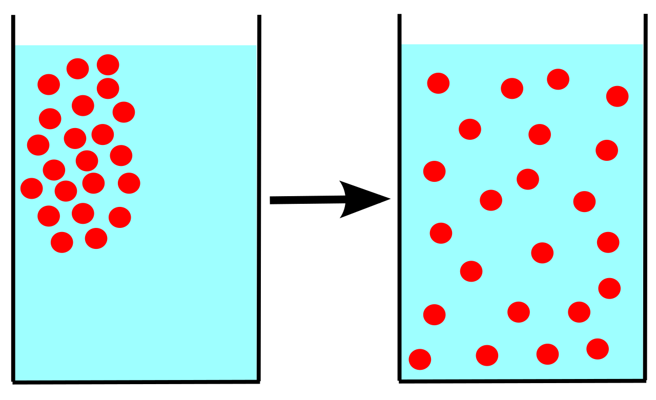
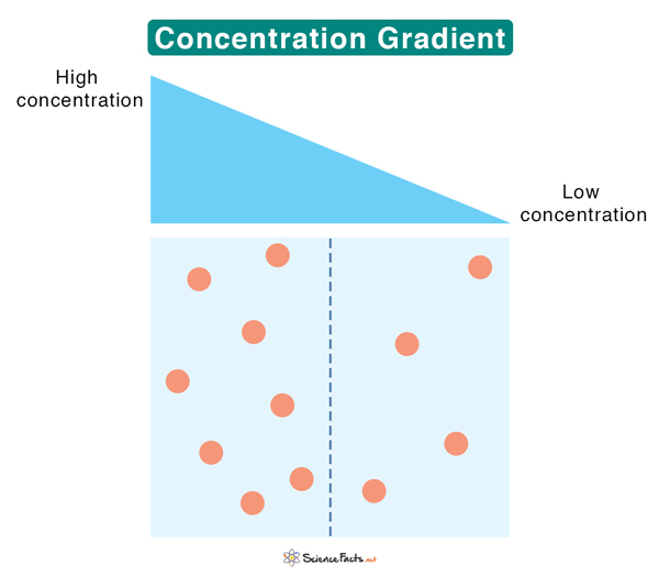
Gradient
The difference in concentration of molecules between one side of the semipermeable membrane and the other
Passive transport (Facilitated diffusion)
Molecules diffuse through the membrane easily from an area of high concentration to an area of low concentration; no energy required!
Active transport
Molecules move from low concentration to high concentration; requires energy
What is kinetic energy?
Energy that can do something that is release when positive & negative charges get together
What is potential energy?
Stored energy when we keep positive & negative charges separate
Potential/Potential Difference
The difference in charge between one place and another
Current
The flow of electrical charge from one place to another
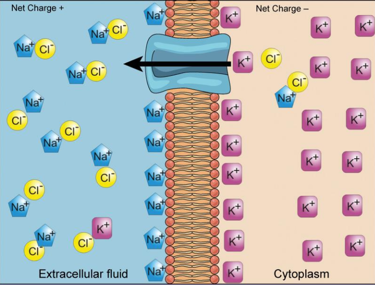
Resting Membrane Potential
The difference in voltage between the inside and outside of the cell; Determined by ions
Membrane Permeability
The capacity of a membrane to have things pass through it; Depends on the number of open ion channels present for a specific ion (the more channels, the more permeable the membrane)
Electrical Gradient
If the net charge on the outside of the cell is more positive than the inside, an ion like potassium (K+) will be pulled in by the electrical gradient because the inside of the cell where it currently sits is more negative (opposites attract)
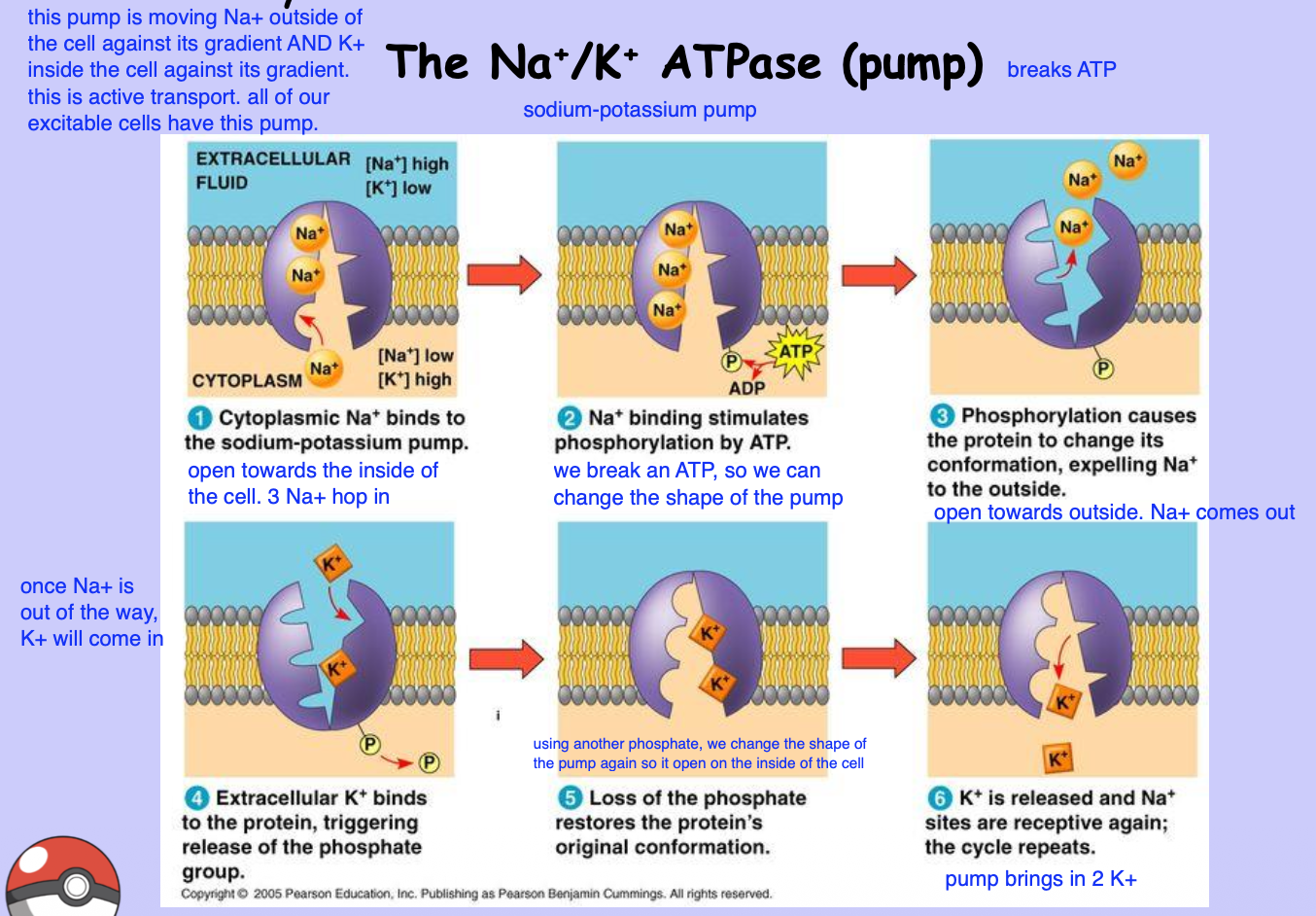
6 Steps in the Sodium-Potassium Pump (Na+/K+ ATPase Pump)
Sodium-potassium pump is open towards the inside of the cell. 3 Na+ hop in
An ATP is broken in order to change the shape of the pump
Pump flips and opens towards the outside of the cell. Na+ exits the pump
Once Na+ comes out, K+ will come into the pump from the outside of the cell
Using another phosphate, we change the shape of the pump again so it’s open on the inside of the cell once more
Pump brings in K+
Polarized Membrane
Membrane is at resting membrane potential of -70mV
Depolarized Membrane
Membrane is getting closer to 0 mV
Hyperpolarized Membrane
Membrane becomes more negative