Oral Pathology Final (only bone stuff)
1/23
There's no tags or description
Looks like no tags are added yet.
Name | Mastery | Learn | Test | Matching | Spaced | Call with Kai |
|---|
No analytics yet
Send a link to your students to track their progress
24 Terms
-Ameloblastic fibro-odontoma
-Calcifying odontogenic cyst
-Central cemento-ossifying fibroma
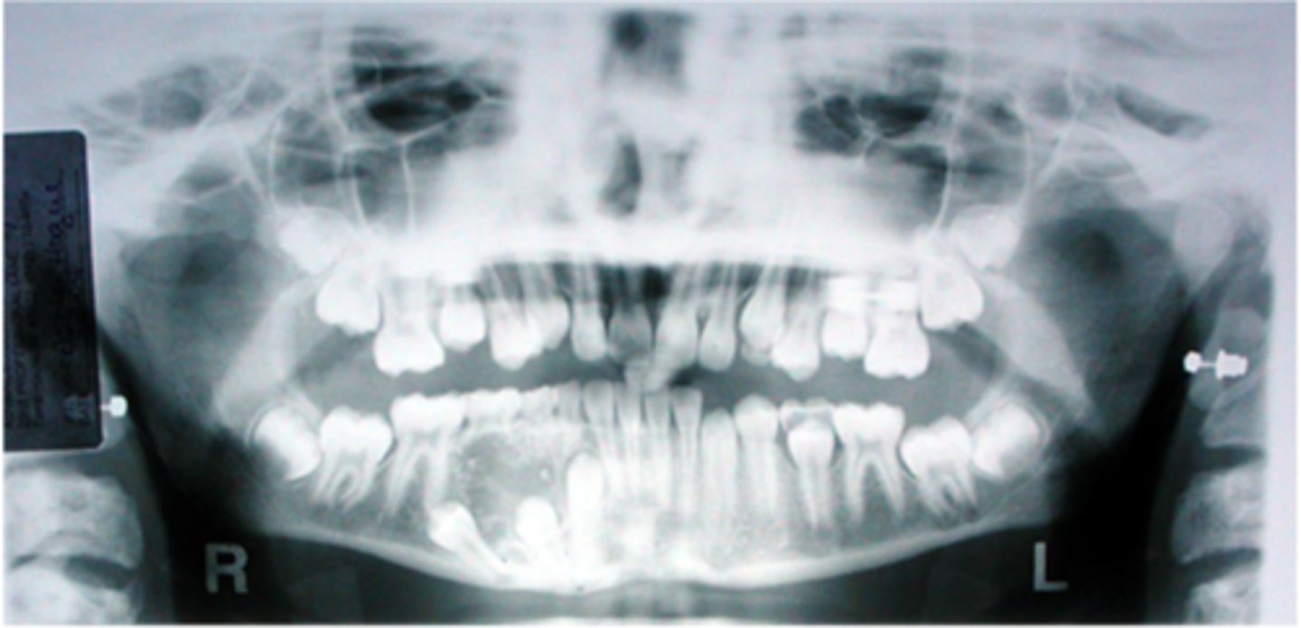
This 11-year-old female presented with an asymptomatic swelling of the mandible. A panoramic radiograph was taken.
What are three diagnoses that you would include in the differential
diagnosis?
C
This 11-year-old female presented with an asymptomatic swelling of the mandible. A panoramic radiograph was taken.
What would be appropriate diagnostic procedures?
A. Enucleation
B. Excision
C. Aspiration
D. Curettage
E. Resection
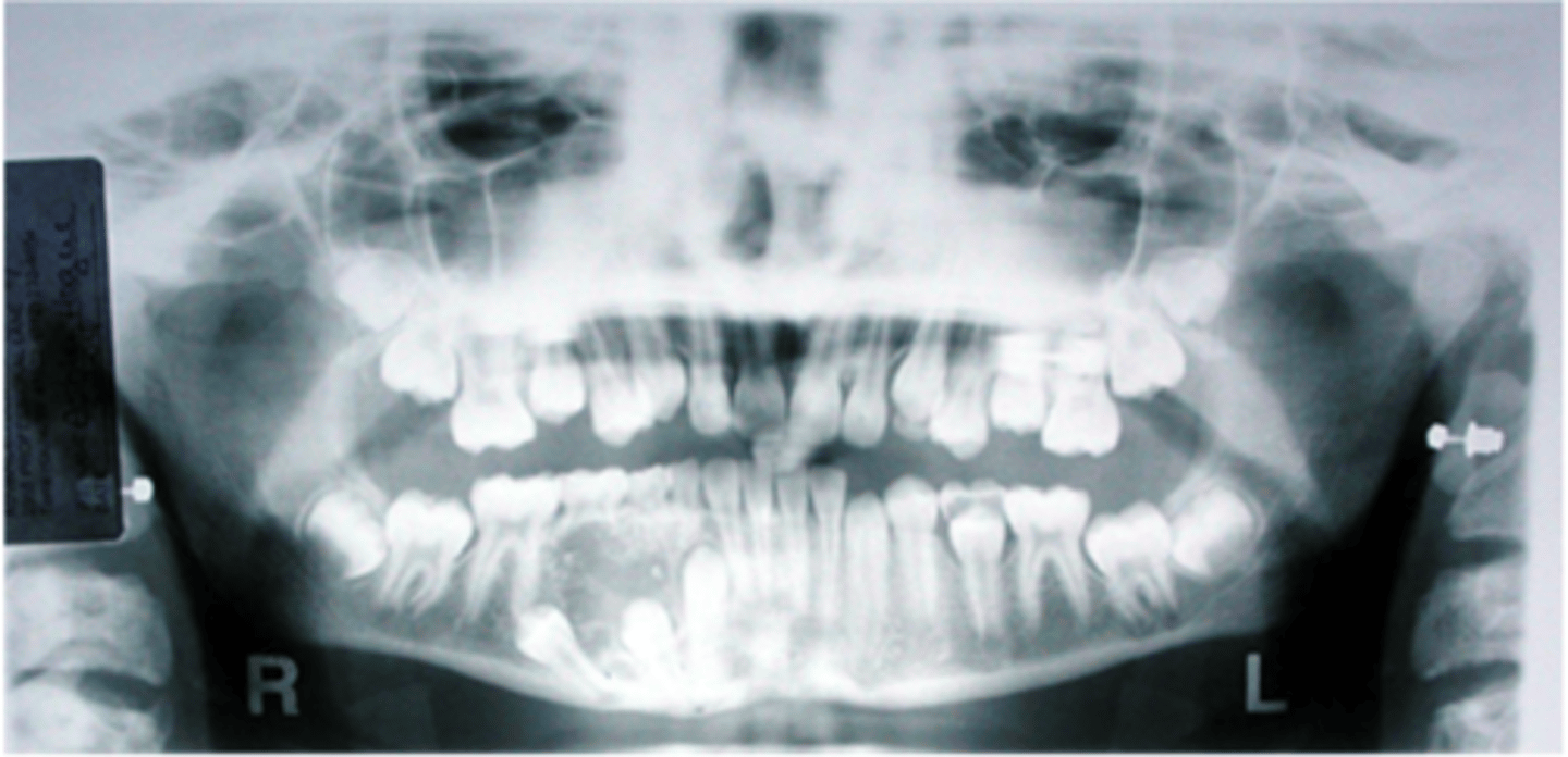
Central giant cell granuloma
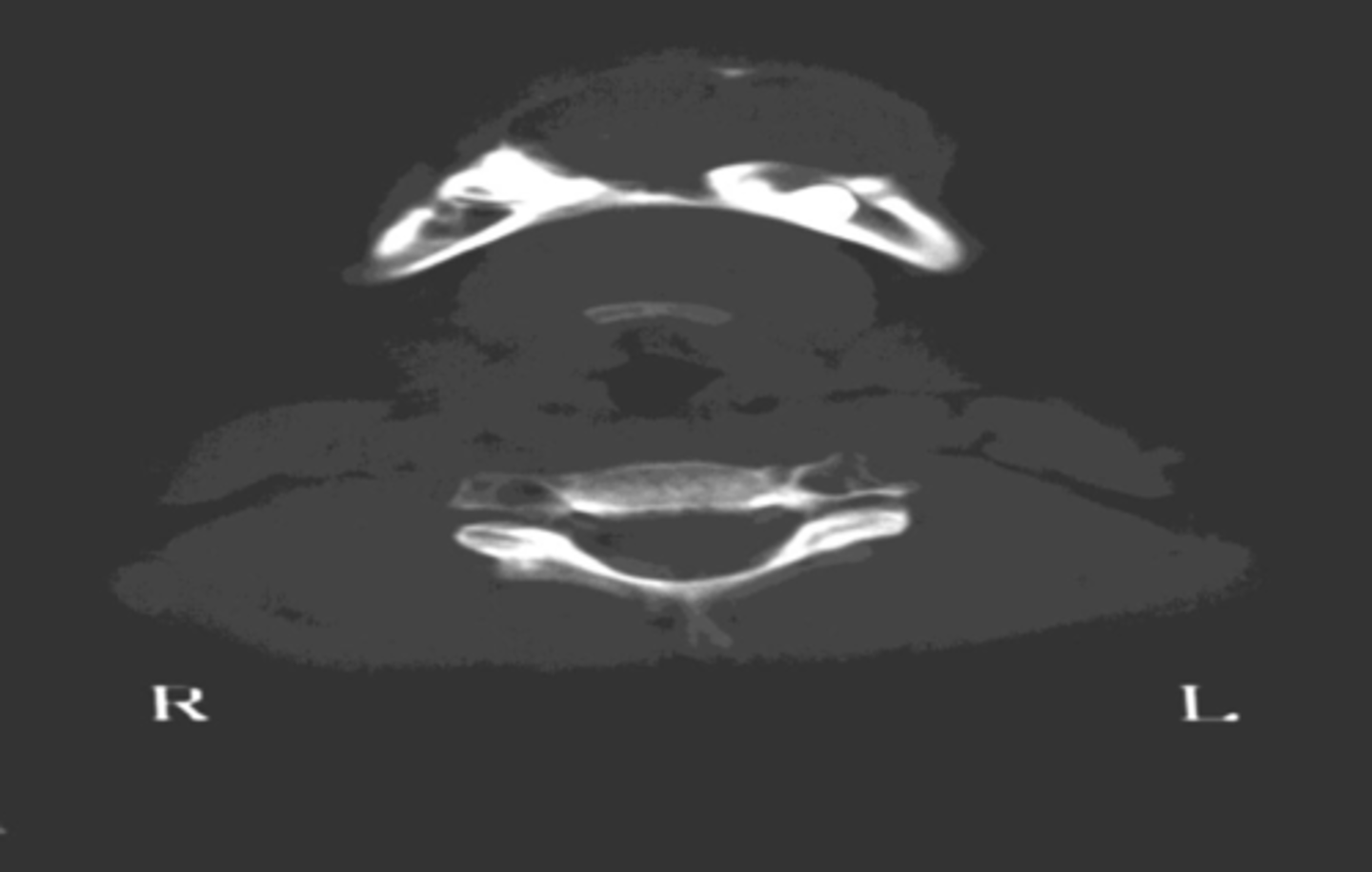
This 12-year-old female presented to your office with a painless enlargement of her anterior mandible. Computed tomography was utilized to obtain images.
What would be the most likely diagnosis in this case?

Dark red blood
This 12-year-old female presented to your office with a painless enlargement of her anterior mandible. Computed tomography was utilized to obtain images.
If you were to aspirate this lesion, what would you expect to obtain?
Hyperparathyroidism
This 12-year-old female presented to your office with a painless enlargement of her anterior mandible. Computed tomography was utilized to obtain images.
For what systemic disease would you evaluate the patient?

Question the patient relative to parathyroid or kidney disease and obtain three serum calcium levels over a week's time.
This 12-year-old female presented to your office with a painless enlargement of her anterior mandible. Computed tomography was utilized to obtain images.
How would you go about doing this?

-Dentigerous cyst
-Odontogenic keratocyst
-Conventional
ameloblastoma
This 52-year-old male was noted to have this presentation upon routine radiographic evaluation. He was asymptomatic.
What are three entities that you would include in a differential
diagnosis?
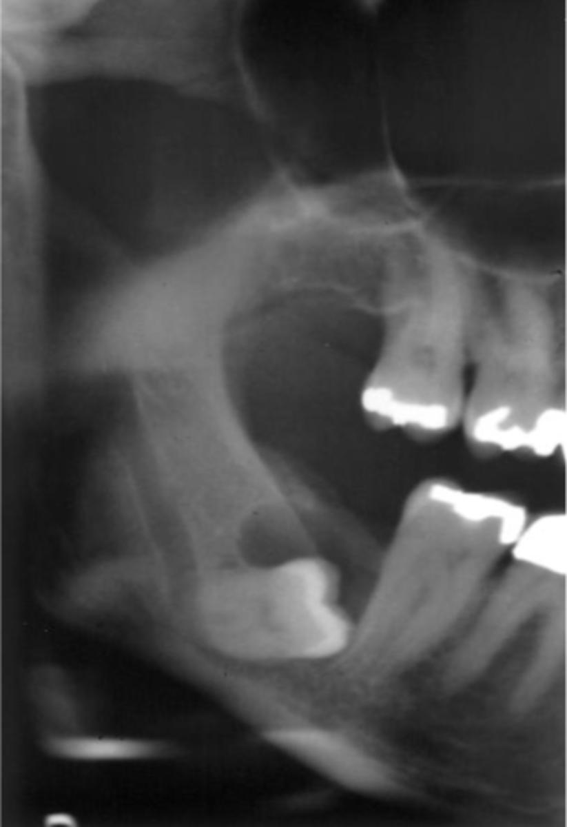
-Ameloblastoma
-Mucoepidermoid carcinoma
-Squamous cell carcinoma
This 52-year-old male was noted to have this presentation upon routine radiographic evaluation. He was asymptomatic.
What are three neoplasms that could arise in such a setting, provided
this originally was an odontogenic cyst?

-Caries (irreversible pulpitis, periapical inflammatory disease)
-Fibrous dysplasia
This 14-year-old female came to the emergency clinic complaining of pain and swelling in her maxilla. The swelling was firm to the touch.
What are the two conditions present in this radiograph?

Pulp test the tooth and ask the patient if they have any other bony lesions or discolorations of her skin.
This 14-year-old female came to the emergency clinic complaining of pain and swelling in her maxilla. The swelling was firm to the touch.
How would you evaluate the patient?
-Primary malignancy of bone (Malignant lymphoma)
-Metastatic disease
-Odontogenic carinoma, multiple myeloma
This 77-year-old male presented with a complaint of a loose tooth, swelling of the mandible and tingling of his lower lip. A panoramic radiograph was taken.
What are three diagnoses that you would include in a differential diagnosis?
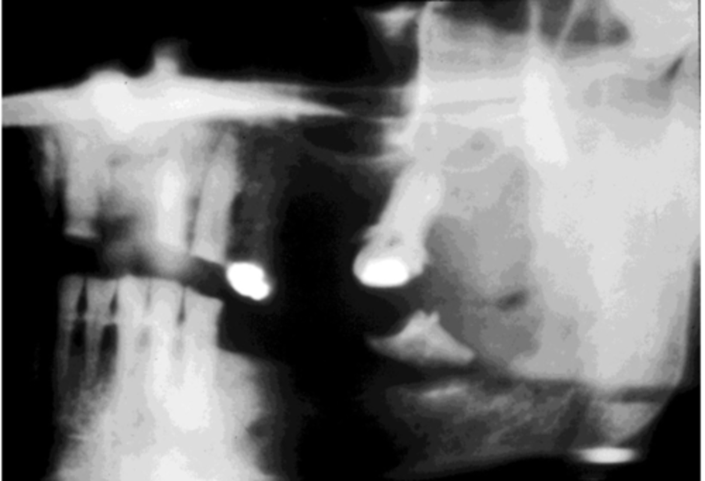
Because the diagnosis is adenocarcinoma, that indicates that this is metastatic disease and that the patient should be comprehensively evaluated and questioned as to knowledge of malignancy elsewhere in the body.
This 77-year-old male presented with a complaint of a loose tooth, swelling of the mandible and tingling of his lower lip. A panoramic radiograph was taken.
A diagnostic procedure is performed and a diagnosis of adenocarcinoma is rendered. What does that tell you?

-central giant cell granuloma
-ameloblastoma
What is your differential diagnosis for a multilocular expansile radiolucency in the anterior mandible that crosses the midline of a 24 y.o male? (2)
multiple myeloma
This was the radiographic finding (pathological fracture, punched out erosion-skull) of a 60 y.o black male with macroglossia.
What is your top differential?
amyloidosis
This was the radiographic finding (pathological fracture, punched out erosion-skull) of a 60 y.o black male with macroglossia.
What would you expect the tongue biopsy to reveal?
bisphosphonate-related osteonecrosis of the jaw (BRONJ)
This 52 y.o WF has a history of breast CA.
What would be your top differential for this clinico-radiographic presentation? (This is not a metastasis)
Diagnosis = focal cemento-osseous dysplasia
Race = white caucasian
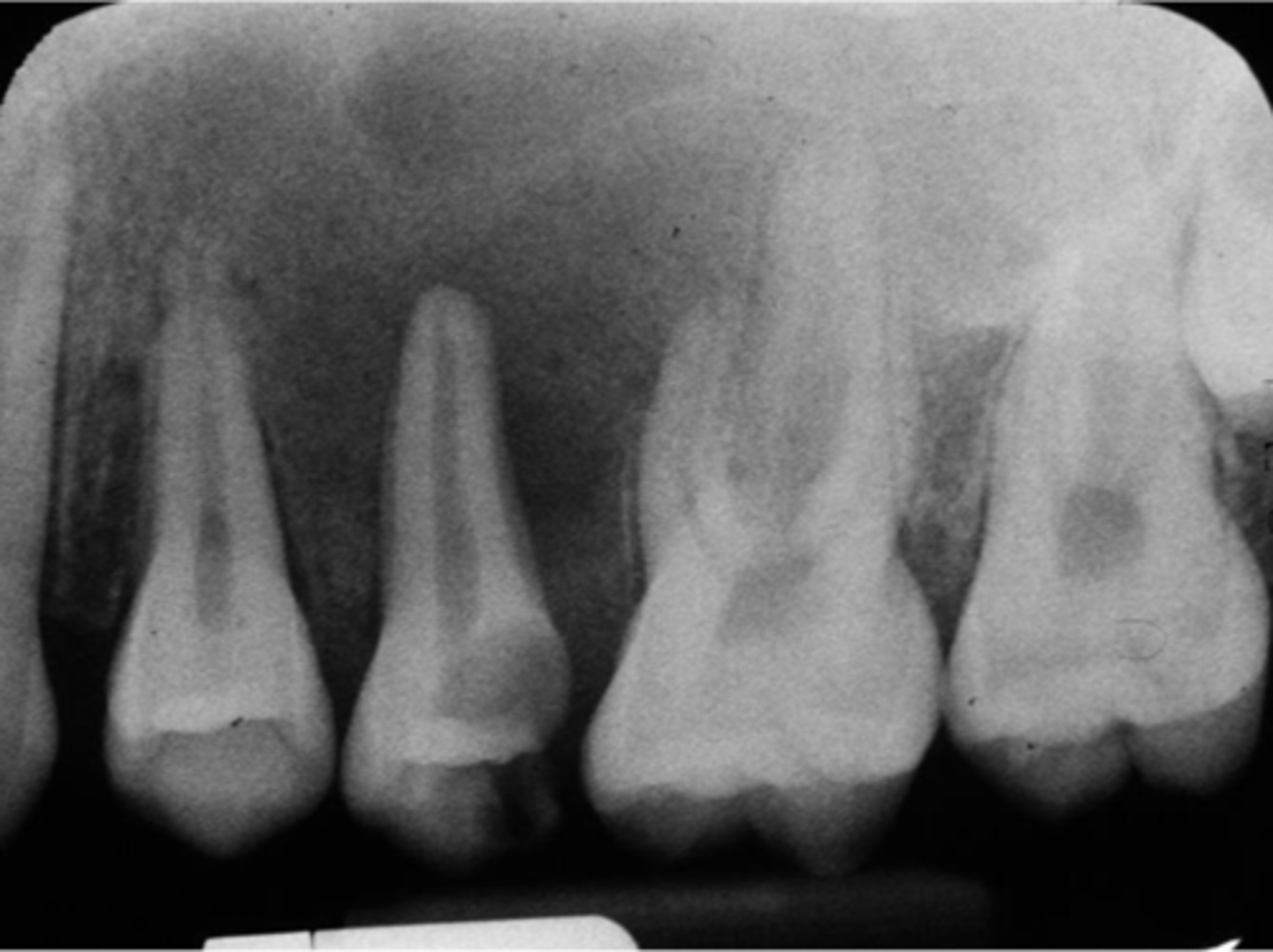
This radio-opacity/lucency was associated with the apex of mandibular 1st molar tooth.
The diagnosis is ________ and the race of the patient is ____________.
periapical cemento-osseous dysplasia
The diagnosis if it is in association with apices of the lower anterior teeth in a black female ________
Diagnosis = florid cemento-osseous dysplasia
Race = Black
The diagnosis and race if it involves the entire mandible_________.
Paget's disease of bone
-hypercementosis
What is the condition associated with "cotton wool" appearance? And name the most common radiographic dental finding.
Ground glass
Fibrous dysplasia has a _____________ __________ radiographic appearance
Osteosarcoma
Most common primary malignancy of bone is _____________.
Female; anterior mandible
CGCG - is most common in ________ patients (sex) and _________________ location.
do nothing - no treatment necessary.
The best treatment for "cemento-osseous dysplasia" is ______________.