Cell functions (membrane proteins, organelles, etc.) Unit 2
1/23
There's no tags or description
Looks like no tags are added yet.
Name | Mastery | Learn | Test | Matching | Spaced | Call with Kai |
|---|
No analytics yet
Send a link to your students to track their progress
24 Terms
What is selective/semi permeability
Has control over what can enter or leave the cell.
Molecules that are not permeable through membrane
Large, charged molecules cannot enter through the cell membrane via simple diffusion. They can enter through other means, though.
Eg. ions have a charge - the hydrophobic tails do not like them
Molecules that are permeable through membrane
Small, uncharged/non-polar molecules can enter through the cell membrane via simple diffusion.
Eg. oxygen/CO2
Also, non-polar steroids
Simple diffusion
Passive movement of particles from a high concentration to a low concentration until equilibrium (same concentration reaches at both sides)
Facilitated diffusion
Passive flow of molecules that are too big/polar to diffuse through the membrane - instead uses a channel protein to diffuse from high to low concentrations. Eg. ion channels, glucose channels, aquaporins. Different channels needed for different molecules.
Active transportation
Movement of particles from areas of low to high concentration - going against concentration gradient requires energy.
What makes the membrane selectively permable
Controls what enters/leaves the cell with integral protein manufacturing, or no manufacturing. If it does not want glucose to enter the cell, it won’t produce a glucose channel. If it wants less water to enter the cell, it will manufacture less aquaporins. The nature of the phospholipid bilayer’s hydrophobic tails will always repel the polar molecules and the bilayer will repel molecules that are too large since the membrane is packed.
What are glycoproteins
Glycoproteins are typically integral membrane proteins in the with a carbohydrate chain extending outside and work to join cells together/serve as recognition
What are glycolipids
Glycolipids are lipids that span in the hydrophobic tails and have a little carbohydrate chain extending outside that serves as recognition in the immune system.
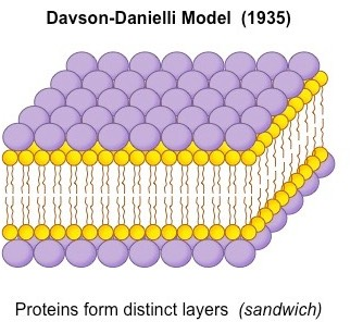
What was the Davson Danielli model?
The model first made for what they thought the cell membrane looked like. Using the new electron microscope, they saw the dark areas and light areas of the membrane, and assumed proteins. Thus, they believed the structure was protein-lipid-protein sandwich. This was proved wrong
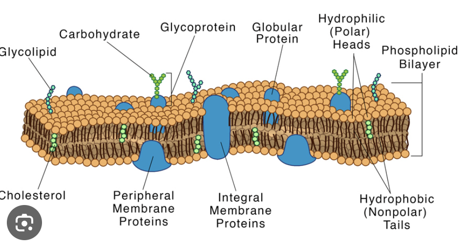
What was the Singer Nicolson model?
After using freeze fracture (a method that froze the membrane, then cracked it open) they found instead of a smooth model, a model with bumps. This proved the Davson Danielli model wrong, since it’s clear that proteins were embedded into the membrane. They developed the fluid mosaic model which shows the phospholipid bilayer with proteins in between, and the proteins can move through the membrane as the membrane isn’t rigid. The membrane contains: phospholipids, channel proteins, pump proteins, cholesterol, glycolipids, glycoproteins, enzymes, etc.
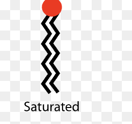
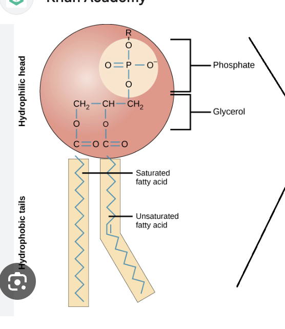
Saturated fatty acid
Makes membrane less fluid as fatty acid has no double bond thus no kink, so can pack more tightly together. Optimal in warmer environments when less fluidity needs to be maintained even through heat (which can cause the membrane to be more fluid).
Unsaturated fatty acid
Makes membrane more fluid as fatty acid has a double bond thus a kink, so can pack less tightly together. Optimal in colder environments when the fluidity needs to be maintained even through cold temperatures (which can cause the membrane to firm up).
Role of cholesterol
Acts to regulate membrane fluidity and flexibility (and thus permeability) in ANIMAL CELLS. It is non polar & hydrophobic, so it is in the tails. It can help in preventing membrane rigidness when temperatures are very low, and prevents membranes being too fluid when temperatures are very high. Important for not too fluid to ensure it still is a barrier, and not too rigid to ensure endo/exocytosis is still possible and cell can move around.
Endocytosis
Engulfs materials from outside of the cell and moves them inside of the cell using the lipid bilayer. This requires ATP
There is material outside of cell that cell wants
Cell membrane indents, then pinches off to surround the material inside of the cell.
Now it is a vesicle.
Fluidity of membrane is required for this since a stiff membrane won’t bend or pinch off, but this is needed to get materials from outside of cell to inside of cell.
Exocytosis
Vesicle containing contents that cell wants to excrete (from rough ER ribosomes/golgi)
Fuses with the cell membrane
Releases the contents to the outside of the cell, now vesicle rejoins cell membrane
Growth in membrane
Vesicles from rough ER pinch off and fuse with the cell membrane, making it bigger
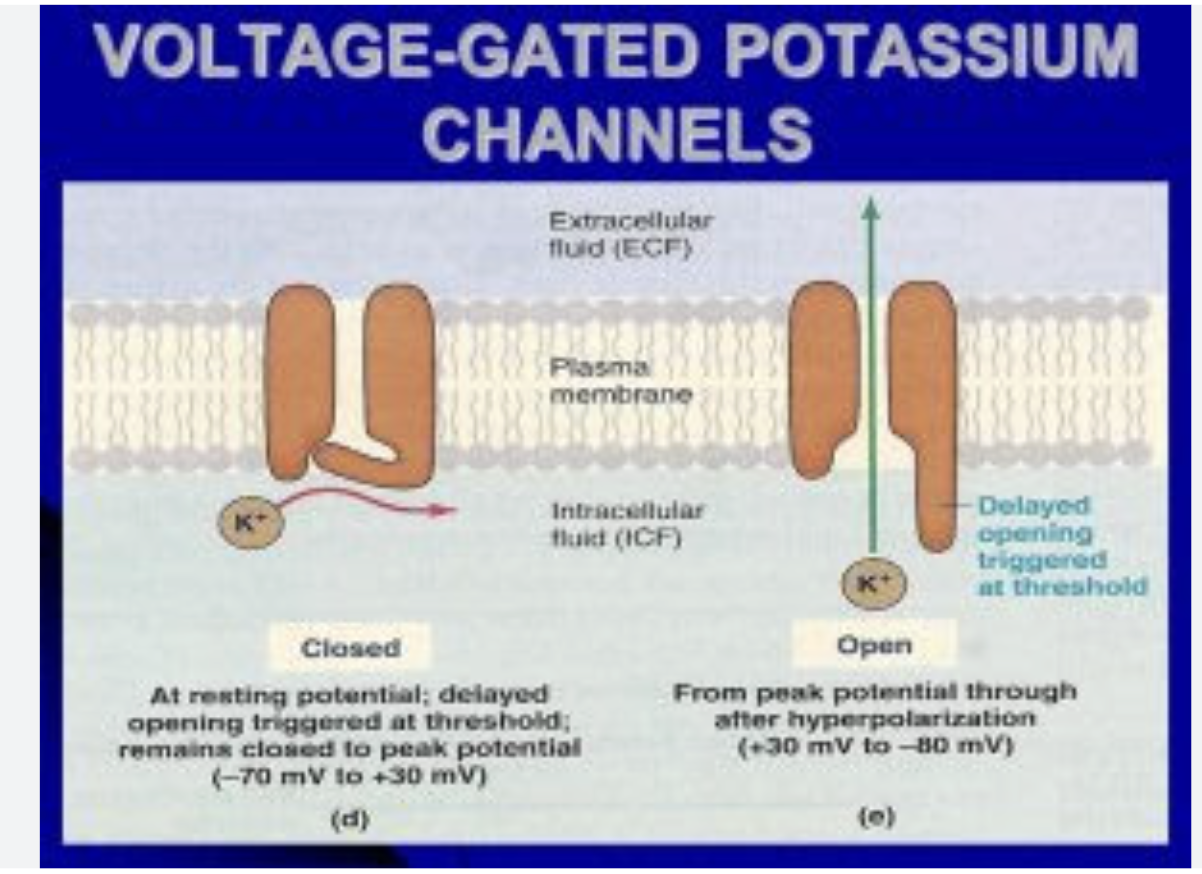
How does the voltage gated sodium & potassium ion channel work
This is an example of facilitated diffusion, passive transport via a channel in muscle & neuron cells which opens & closes in response to voltage changes.
Outside of the cell there is a large amount of Na+ and inside a small amount of Na+. Inside the cell there is a large amount of K+ and a small amount of K+ outside of the cell. Thus, the ions want to move to their lower concentration (K+ out, Na+ in).
The resting state of the cell membrane is -70mV.
A stimulus arrives and the membrane becomes slightly less negative at -55mV.
This change in volts leads to the Na+ channel opening, so a lot of Na+ rushes in to fix the concentration & reach equilibrium.
This depolarizes the membrane til about +30mV.
This change in voltage causes the K+ channel to open, and a lot of K+ ions rush out of the membrane. This repolarizes the membrane until about -80mV.
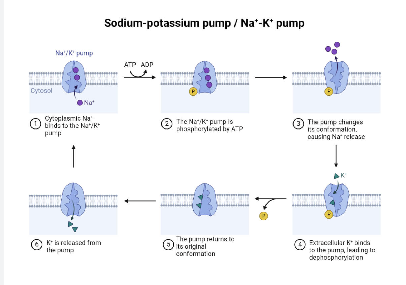
How does the sodium potassium pump work
This pump is an example of active transportation in exchange transportation with Na+ and K+ working against their concentration gradient in neuron and muscle cells.
1) 3 Na+ ions inside the cell attach to the pump
2) ATP → ADP + Pi, the Pi attaches to the pump & phosphorylates it
3) This causes the pump to change shape, forcing the Na+ to leave the cell into a higher concentration
4) Then, 2 K+ ions attach to the pump
5) The phosphate group detaches from the pump, dephosphorylating it and changing back to its original shape. This forces the 2 K+ ions into the cell into a higher concentration.
3 sodium out, 2 potassium in.
How does the nicotinic acetylcholine receptor protein work?
1) Acetylcholine is the message (neurotransmitter) from one neuron to the next. It will diffuse out of the sender neuron and bind to the acetylcholine receptor on the receiving neuron.
2) This binding causes the receptors shape to change and open to allow the flow of Na+ into the receiver cell. This is passive because Na+ is going from areas of high to low concentration.
3) This increase in Na+ depolarizes the membrane and cell and initiates a change.
4) Acetylcholine can detach and the receptor can change shape again and close
How does the sodium dependent glucose cotransporter work?
This is an example of indirect active transport. This is used in the kidneys and small intestines to ensure glucose is coming into cells.
1) The Na+ K+ pump pumps out sodium against its concentration gradient. It forces it to go from low to high.
2) Now, a lot of Na+ is outside of the cell waiting to come in & wanting to diffuse into a lower gradient. There is also a low concentration of glucose also outside of the cell, and a higher concentration inside of the cell. Glucose does not want to enter since this is against its concentration gradient.
3) Na+ binds to the cotransporter and so does glucose, changing the shape of the cotransporter and opening it.
4) Na+ flows through to a low concentration via simple facilitated diffusion down its concentration gradient.
5) This flow of a lot of Na+ releases energy and powers the glucose to go against its concentration gradient. The glucose has gone from low to high concentration.
Tight cell-cell junction
Tight junctions stick cells very close to one another and prevent any gaps. This ensures no materials can slip through them, but need to pass the membrane. Needed in stomach
Desmosomes/Anchoring cell-cell junction
Anchors cells to each other to secure them together and ensure no tearing apart when force is applied. Eg. epidermis skin
Gap cell-cell junction
A junction that allows the movement of small ions and molecules to pass directly between cells. Eg. in cardiac muscle/smooth muscle