Imaging - Advanced Imaging Techniques
1/102
There's no tags or description
Looks like no tags are added yet.
Name | Mastery | Learn | Test | Matching | Spaced | Call with Kai |
|---|
No analytics yet
Send a link to your students to track their progress
103 Terms
Computed tomography (CT)
creates imaged using multiple x-rays (ionizing radiation) in cross sectional (axial) slices, created by up to 1,000 projections from different angles
During rotation around the patient the x-ray source produces a narrow, fan shaped beam of x-ray that passes through a section of patient's body
How does a CT scanner work?
detectors in rows opposite the x-ray source
What registers the image in a CT scan?
Scout image
two-dimensional digital radiograph produced by the CT scanner that is used to localize the structures to be scanned
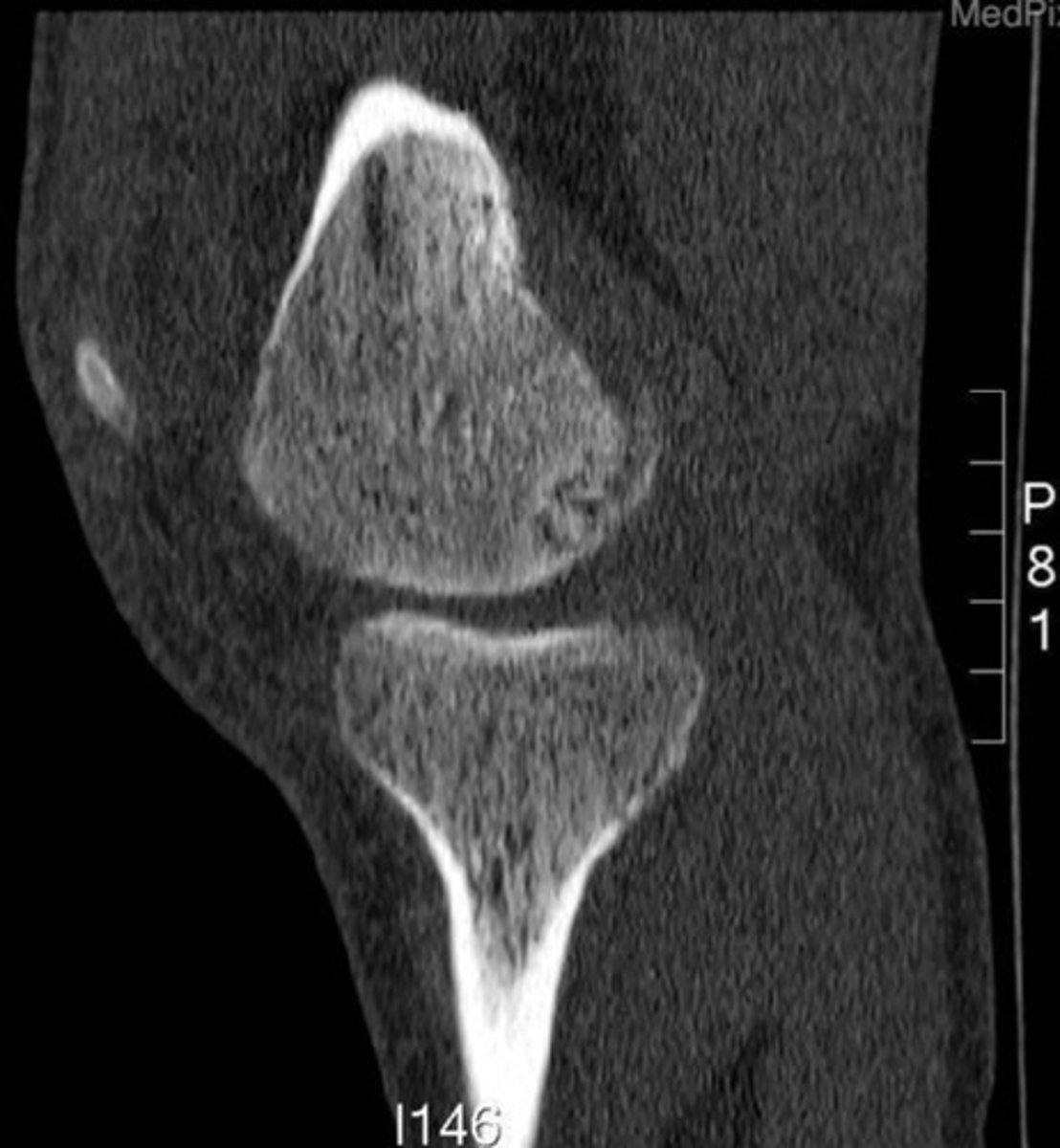
sagittal
What view of a CT is this?
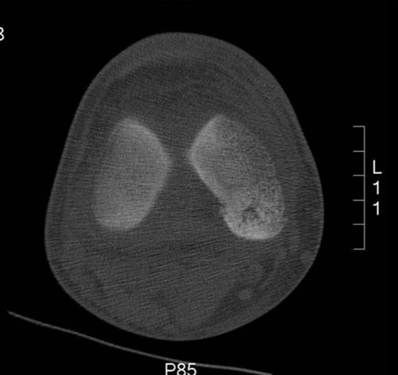
axial
What view of a CT is this?
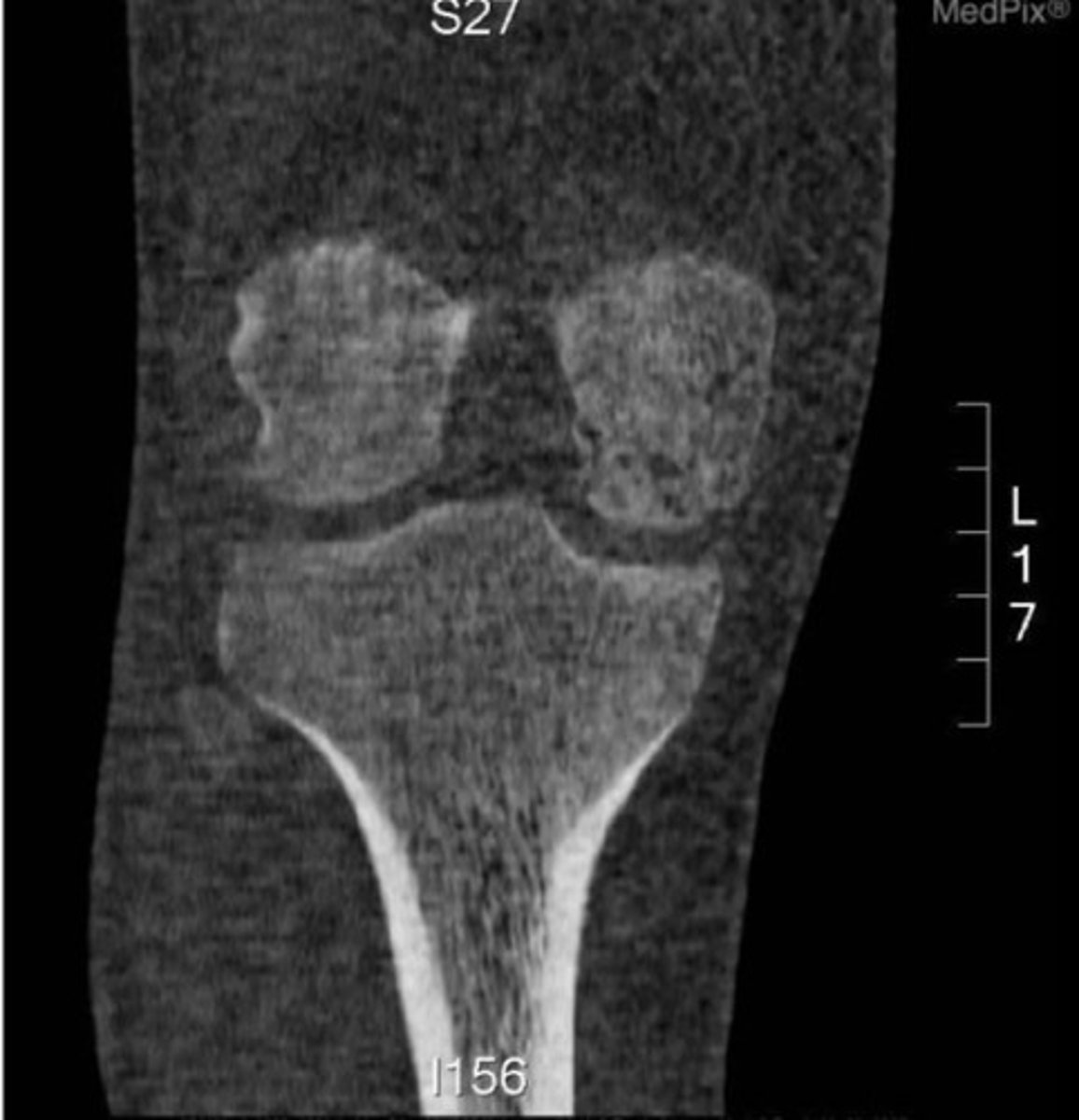
coronal
What view of a CT is this?
reflection of tissue radiodensity without superimposition of other tissues
What do you see on a CT image?
to reduce the range of radiodensities displayed and focus on a particular tissue
Why would you window a CT image?
anatomy (larger for larger joints)
What is slice thickness dependent on?
volume is smaller
What does a thinner slice mean in terms of image volume?
Three-Dimensional CT
Multiplanar reconstruction
CT myelogram
injection of contrast material into the spinal fluid
Cone beam CT
single volume of data means shorter scanner time, less radiation (used in dental practice)
subtle or complex fractures
degenerative changes
trauma causing injuries to bone and soft tissue
Spinal stenosis
Disc condition
loose bodies in a joint
What is CT best at imaging?
less time consuming
less expensive
What are benefits to using a CT scan?
radiation exposure
radiodensity
What are limitations of a CT?
Neuroimaging
Cardiac Imaging
Pulmonary imaging
What other medical specialties are CTs common in?
levels imaged
contiguous or interrupted slices
slice thickness
reforming/reconstructions
angulation of gantry
windows provided
use of contrast agent
What is reported in a CT report?
- signal generation based on the properties of magnetic resonance
- relaxation process
- signal detection
- encoding of spatial information
- reconstruction of the image from the signal
- manipulation of tissue-dependent contrast
What is the process to create an MRI image?
early signal decay
What does T1 weighted image capture?
late stage of signal decay
What does T2 weighted image capture?
Longitudinal relaxation (T1)
process for radiofrequency energy to be transferred back to the surroundings
Transverse Relaxation/Spin-Spin Relaxation (T2)
loss of transverse magnetization due to loss of phase coherence or order among the protons in the transverse plane
the time it takes for 63% of the original excited transverse magnetization to remain
What does T2 refer to?
the time it takes for the tissue to recover 63% of its original longitudinal magnetization
What does T1 refer to?
No
Do tissues show up the same on T1 and T2 images?
T1
T2
proton density
What is contrast in an image based on differences in?
time to repetition (TR)
time to echo (TE)
What are the 2 parameters most important for creating contrast in an MRI image?
time to repetition (TR)
time at which the signal is captured
time to echo (TE)
time at which the RF pulse is repeated to again displace protons
short TR and TE times
What is T1 imaging characterized by?
long TR and TE times
What is T2 imaging characterized by?
very low
What is 1?

intermediate
What is 2?

intermediate to low
What is 3?

high
What is 4?

low
What is 5?

intermediate
What is 6?

low
What is 7?

very low
What is 8?

low to intermediate
What is 9?

intermediate
What is 10?

intermediate
What is 11?

low
What is 12?

intermediate
What is 13?
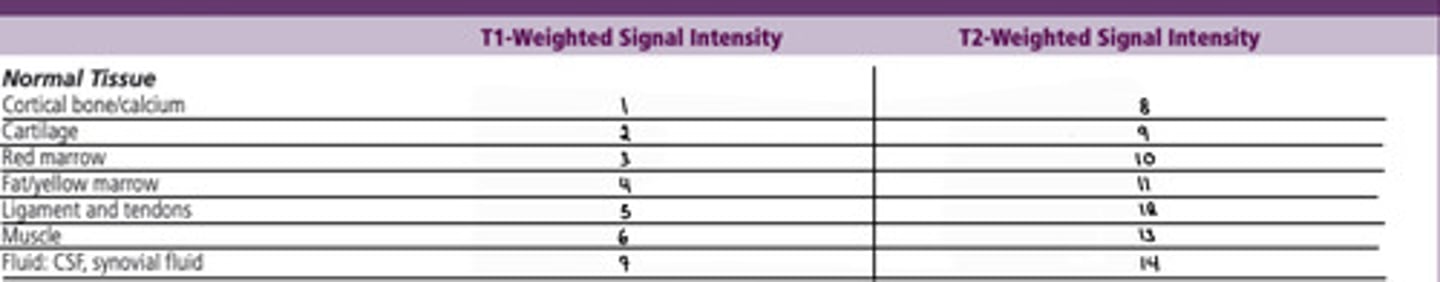
high
What is 14?

high
What signal intensity shows up brighter?
low
What signal intensity shows up darker?
greater ability to scan claustrophobic or obese patients
reduction of noise
possibility to perform tests or procedure during scan
What are advantages of an open MRI scanner?
lower field strength
lower signal to noise ratio
longer time
What are disadvantages of an open MRI scanner?
examine spine under WB conditions
can patients too large to fit in normal
scan pt who must stay in upright position
What are advantages of an upright MRI scanner?
longer time
possible image degradation
pt may be in painful position
What are disadvantages of an upright MRI scanner?
variations in bone marrow
soft tissue detail
disc herniations
neoplasms
What is MRI good at imaging?
Ferromagnetic surgical clips can be displaced
orthopedic hardware can cause distortion
no pacemakers
claustrophibia
What are disadvantaged to an MRI?
MR arthrography
gadolinium in iodine is injected into joint and created bright signal which allows the radiologist to see small defects in the capsule, articular surfaces, ligaments, or labra
MR myelography
study of the spinal canal and subarachnoid space using high resolution MRI with strong T2 weighting
greater contrast resolution
better or organs surrounded by bone
no radiation
less risk of missing disease processes
What are advantages of MRI over CT?
less expensive
greater availability
faster
less operative involved
thinner slice
not as affected by motion
easier with metal implants
What are advantages of CT over MRI?
higher resolution
lower cost
no known hazards
easy comparison
ability to follow structure
can modify area being examined
What are advantages to ultrasound over MRI?
small field of view
operator dependt
does not penetrate bone
does not cross air interfaces
harder on obese pts
What are limitations of an ultrasound?
pulser
transductor
What equpimnet is needed to complete an ultrasound?
reflection of sound wave
What is the ultrasound image based on?
echogenicity
difference in acoustic impedance of tissues at interface
smoothness of the reflecting interfaces
angle of reflection
What is the amount of reflection of sound waves during an ultrasound determined by?
different in acoustic impedance between the two tissues types
angle of incidence
What does refraction of a sound wave during an ultrasound depend on?
angle of incidence
the farther from the perpendicular, the greater the refraction
blood flow in an artery or vein
What does a doppler ultrasound measure?
sagittal slice
What does a longitudinal sonogram result in?
axial image
What does a transverse sonogram result in?
Hyperechoic structures
reflect more energy than other surrounding structures
Hypoechoic structures
reflect less energy than other surrounding structures
white/lighter
How do hyperechoic structures appear?
black/darker
How do hypoechoic structures appear?
Anechoic structures
do not reflect energy
bone
tendon/ligament
nerve
muscle
subcortical bone
What is the order of tissues from hyperechoic to hypoechoic?
hyperechoic relative to muscle
How does a tendon/ligament show up on an ultrasound?
hypoechoic line
How does a bursa show up on an ultrasound?
hypoechoic relative to fascia or tendons
How does a muscle show up on an ultrasound?
hypoechoic layer
How does hyaline cartilage show up on an ultrasound?
hyperechoic
How does fibrocartilage show up on an ultrasound?
hypoechoic to tendon, hyperechoic to muscle
How does a nerve show up on an ultrasound?
bright echo
How does a bone-soft tissue interface show up on an ultrasound?
hypo or anechoic
How does subcortical bone show up on an ultrasound?
hyperechoic, smooth, continuous
What is 1?

hyperechoic, distinct parallel fiber pattern
What is 2?

hyperechoic, with parallel fibrous hyperechoic bands
What is 3?

thin hypoechoic line
What is 4?

hypoechoic layer next to cortex
What is 5?

Hyperechoic, relative to muscle
What is 6?
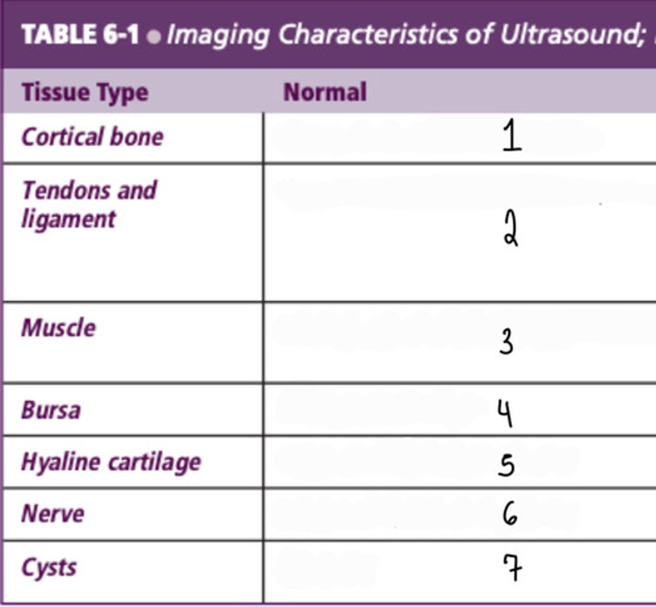
Anechoic
What is 7?

hyperechoic, smooth, continuous
What is the normal findings of cortical bone in an ultrasound?
hyperechoic;distinct parallel fiber pattern
What is the normal findings of tendons and ligaments in an ultrasound?
hypoechoic with parallel fibrous hyperechoic bands
What is the normal findings of muscle in an ultrasound?
thin hypoechoic line
What is the normal findings of bursa in an ultrasound?
hypoechoic layer next to cortex
What is the normal findings of hyaline cartilage in an ultrasound?
hyperechoic, relative to the muscle
What is the normal findings of nerves in an ultrasound?
anechoic
What is the normal findings of cysts in an ultrasound?
break in continuity, uneven surfaces
What are abnormal findings of cortical bone on an ultrasound?
strains: thickening of mixed echogenicity (hypoechoic if inflammation occurs or a hematoma) and disrupted fiber pattern
ruptures: disruption of structure, initially filled with hypoechoic hematoma and separation of ends
What are abnormal findings of tendons and ligaments on an ultrasound?
increased width. In later stages, hyperechoic thickining of the walls
What are abnormal findings of bursa on an ultrasound?