3.1.1 exchange surfaces
1/61
Earn XP
Description and Tags
All of exchange surfaces - revision
Name | Mastery | Learn | Test | Matching | Spaced | Call with Kai |
|---|
No analytics yet
Send a link to your students to track their progress
62 Terms
Need for exchange
Allows for diffusion across plasma membranes for materials such as oxygen, glucose and carbon dioxide and to remove excretory materials such as urea and heat with their environment
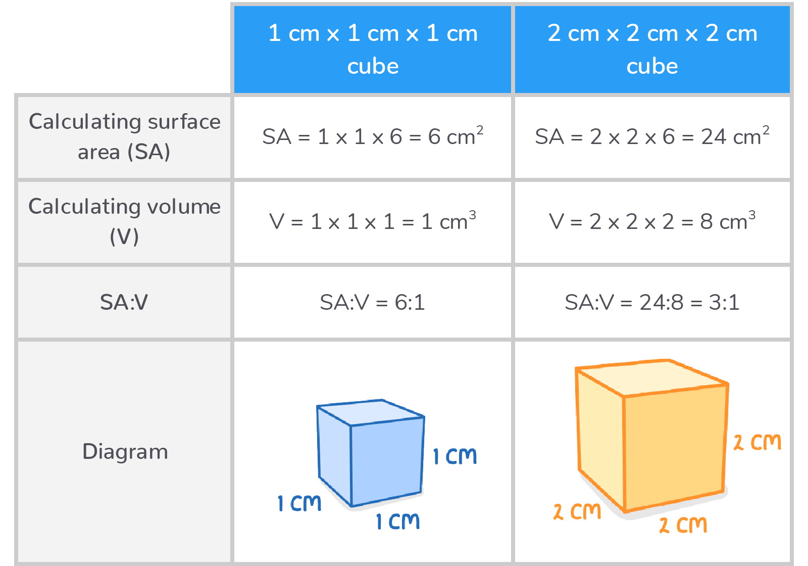
High SA:V ratio
Organisms with a high surface area to volume ratio have a larger surface area relative to their volume which means substances diffuse across a shorter distance through plasma membranes faster - smaller organisms
Low SA:V ratio
Organisms with a lower surface area to volume ratio have a smaller surface area relative to their volume so substances have to diffuse across a larger distance through plasma membranes so diffusion is slower - larger organisms
Calculating SA:V ratio
calculate surface area l x w x 6
calculate volume l x w x d
( for a cube )
Why do multicellular organisms require specialised exchange surfaces ? (give 3 reasons)
cells are not in direct contact with their external environment
diffusion distances between cells and their environment are larger
larger organisms have higher metabolic rates so they need more oxygen and glucose
How do single celled organisms exchange ?
diffusion occurs across the cell membrane directly from their environment
In single-celled organisms, substances diffuse directly across the cell membrane.
What are exchange surfaces ?
Specialised structures which allow for materials to be transported between cells and the surrounding environment
4 Key features of exchange surfaces
large surface area
thin cell walls
extensive / rich blood supply and/or ventilation
surrounded by selectively permeable membranes
purpose of large surface area
larger area in which substances can be exchanged
purpose of thin cell walls
minimise diffusion distance
purpose of extensive blood supply and/or ventilation
maintains a steep concentration gradient
purpose of selectively permeable membranes
controls what substances are exchanged
Why is the gas exchange system located inside the body ?
air is not dense enough to support these delicate structures
body would lose water and dry out
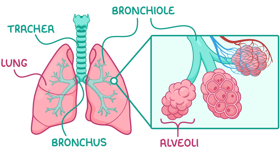
Pathway of air
Air (containing oxygen) enters the nose / mouth
Air travels down the trachea
Air travels into the bronchi (both) and branches into each bronchus (one)
Air travels into the bronchioles which lead to tiny air sacs - alveoli
Gas exchange
Surrounding the alveoli is an extensive network of capillaries
Oxygen travels from inside the alveoli into erythrocytes (RBC’s) containg haemoglobin
Oxygen is carried by erythrocytes to body cells where it is used for respiration which produces carbon dioxide as a waste product into the blood
From the blood in the capillaries carbon dioxide is exchanged into the alveoli and into the bronchioles, bronchus, trachea and then outside from the mouth/nose into the air
What is ciliated epithelium ?
tissue located throughout most of the airways
Structure of ciliated epithelium
Ciliated epithelial cells
Goblet cells
function of goblet cells
Secrete mucus which traps dust and microbes
function of cilia on ciliated epithelial cells
waft mucus up toward the mouth so it can be swallowed
Trachea - Structure and adaptations (4)
Rings of cartilage to prevent airway from collapsing (keeps it open)
Smooth muscle can constrict or relax to constrict or dilate the airway and control air flow
Elastic tissue containing elastic fibres which allows for stretching and recoiling
Lined with ciliated epithelial cells and goblet cells
Bronchi - Structure and adaptations(4)
Same as trachea
Reinforced with to keep airways open
Smooth muscle. can constrict or relax to constrict and dilate the airway to control air flow
Elastic tissue with elastic fibres which allows for stretching and recoiling
Lined with ciliated epithelial cells and goblet cells
Bronchioles - Structure and adaptations (4)
NO cartilage, can change shape
Smooth muscle constricts and relaxes to constrict and dilate airways to control air flow
Elastic tissue with elastic fibres which allows for stretching and recoiling
Simple squamous epithelium (only larger bronchioles have a ciliated epithelium)
How do the alveoli carry out gas exchange?
(brief)
Oxygen diffuses from the alveoli into the pulmonary capillary and and binds to haemoglobin in erythrocytes
Carbon dioxide disassociates from haemoglobin and diffuses from the blood into the alveoli
Adaptations of alveoli for gas exchange (8)
Wall consists of squamous epithelial cells
Large surface area
Dense network of capillaries
Ventilation of air
Collagen fibres
Elastic fibres
Partially permeable membrane
Moist inner surface
Purpose : wall consists of squamous epithelial cells
Allows for rapid diffusion
Purpose : Large surface area
Increases rate of gas exchange
Purpose : Dense network of capillaries
Brings blood closer to oxygen for more efficient gas exchange
Purpose : Ventilation of air
Maintains a steep concentration gradient
Purpose : Collagen fibres in alveoli
Strong collagen prevents alveoli from bursting and limits overstretching
Purpose : Elastic fibres
Allow stretching and recoiling
Purpose : Moist inner surface
Allows for gases to dissolve and lung surfactant helps alveoli remain inflated
Pulmonary blood vessels (3)
Pulmonary arteries
Pulmonary veins
Pulmonary capillaries
function of pulmonary artery
delivers deoxygenated blood away from the heart into the pulmonary capillaries
function of pulmonary vein
delivers deoxygenated blood from capillaries to the heart
function of pulmonary capillary
site of gas exchange between alveoli and blood
Adaptations of the pulmonary capillaries for gas exchange (5)
thin walls
erythrocytes pressed against capillary walls
large surface area
movement of blood
slow blood movement
Purpose : thin walls
shorter diffusion distance
Purpose : erythrocytes against capillary walls
reduces diffusion distance
Purpose : large surface area
increases diffusion speed
Purpose : movement of blood
maintains steep diffusion gradient
Purpose : slow blood movement
allows more time for diffusion
Muscles involved in ventilation
Diaphragm
External intercostal muscle
Internal intercostal muscle
What happens during inspiration ?
The external intercostal muscles contract while the internal intercostal muscles relax, moving the ribcage up and out.
The volume of the thoracic cavity increases.
The diaphragm contracts and flattens, further increasing the volume of the thoracic cavity.
The lung pressure decreases below atmospheric pressure.
Air flows into the lungs down the pressure gradient.
What happens during expiration ?
The external intercostal muscles relax, moving the ribcage down and in.
The volume of the thoracic cavity decreases.
The diaphragm relaxes and unflattens, further decreasing the volume of the thoracic cavity.
The lung pressure increases above atmospheric pressure.
Air is forced out of the lungs down the pressure gradient.
Main structures of the insect gas exchange system
Trachea: air-filled tubes branching throughout the organism's body.
Tracheoles: fine branches of tracheae that deliver gases to cells.
Spiracles: external openings of the tracheal system on the exoskeleton of insects.
Spiracles + adaptation
Spiracles are external openings along the abdomen and thorax
Opens to allow fresh air in
Closes to minimise water loss
Tracheae + adaptations
Air filled tubes that branch throughout insects body
Reinforced with chitin :
prevents tracheae from collapsing
stays open so air can pass through
Tracheoles + adaptation give 4 aspects
Smaller tubes that penetrate into body tissues
highly branched:
increases surface area for gas exchange
thin walls:
reduce distance gases need to diffuse
not reinforced with chitin
allows gases to exchange freely across lining
tracheal fluid
helps dissolve oxygen
easier to diffuse into body cells
Why do insets need gas exchange?
To deliver oxygen to cells - This allows aerobic respiration to occur to release energy for cellular processes.
To remove carbon dioxide from cells - The build up of carbon dioxide produced as a waste product of respiration reduces pH, which can denature enzymes.
Features that minimise water loss in insects
closed spiracles
exoskeleton covered with waterproof cuticle
generally small SA:V ratio
Gas exchange in insects
Air reached the end of tracheoles and oxygen dissolves into the tracheal fluid
Oxygen diffuses into surrounding body cells
Carbon dioxide released from body cells and diffuses into the tracheoles
Why and how are concentration gradients in insects maintained ?
Body cells are constantly respiring so oxygen concentration in body cells is lower than in tracheal fluid and concentration of carbon dioxide is higher in body cells than in tracheal fluid
fresh air rich in oxygen constantly supplied to tracheal system which keeps oxygen concentration in tracheal fluid high
air rich in carbon dioxide expelled from insects body via spiracles which keeps carbon dioxide concentration in the tracheal fluid low
Insect gas exchange has adapted to what ?
Gas exchange by maximizing gas exchange efficiency and minimizing water loss
Structure of an insect's gas exchange system
Insects have an open respiratory system comprised of tubular systems that transport air.
Adaptations of the insect gas exchange system
Tracheae reinforced with chitin spirals to prevent collapsing.
Multiple tracheae to increase surface area.
Tracheoles penetrate directly into tissues to reduce diffusion distance.
Thin walls to reduce gas diffusion distance.
Highly branched to maximize surface area.
Tracheal fluid allows oxygen to dissolve to aid diffusion and reduce water loss.
Spiracles open and close to control gas exchange and minimize water loss.
What is lactate accumulation?
Lactate accumulates during anaerobic respiration and reduces the water potential of tracheal fluid.
Water leaves the tracheoles through osmosis, exposing a higher surface area to air for gas exchange.
Why is it difficult to extract oxygen from water ?
water is more dense and more viscous so slower diffusion of oxygen
water has less oxygen content than air
bony fish are very active so they have higher demands for oxygen
Structure of gills
gills are covered by an operculum flap
gills consist of stacked filaments containing gill lamellae
Gill lamellae are surrounded by extensive blood vessels.
Adaptations of the gills for efficient gas exchange (5)
The lamellae provide a large surface area.
The lamellae membranes are thin to minimise diffusion distance.
The gills have a rich blood supply to maintain steep diffusion gradients.
The countercurrent flow of blood and water creates even steeper concentration gradients.
Overlapping filament tips increase resistance, slowing water flow over gills and allowing more time for gas exchange.
Explain the countercurrent flow system
Blood and water flow over the lamellae in opposite directions.
This means that oxygen-rich blood meets water that is at its most oxygen rich when it first moves across the gills, maximising diffusion of oxygen into the blood.
Oxygen-poor blood returning from body tissues meets oxygen-reduced water that has had most of its oxygen removed, still allowing diffusion of oxygen into the blood.
This maintains a steep concentration gradient across the entire gill.
Why is this more efficient than parallel flow?
Countercurrent flow systems allow for more efficient gas transfer as parallel flow reduces the concentration gradient so less oxygen is absorbed
Process of ventilation via the buccal cavity
Bony fish ventilate their gills by opening and closing their mouths, changing the volume of the buccal cavity:
When a fish opens its mouth, this increases the volume of the buccal cavity.
This decreases the pressure, which pulls water into the buccal cavity.
Water flows over the gills.
Water flows out through the operculum.
This drives unidirectional water flow for ventilation, providing freshly oxygenated water and removing carbon dioxide.