Gross Anatomy - Unit 1-5: Vertebral Canal, Spinal Cord, and Meninges
1/100
There's no tags or description
Looks like no tags are added yet.
Name | Mastery | Learn | Test | Matching | Spaced | Call with Kai |
|---|
No analytics yet
Send a link to your students to track their progress
101 Terms
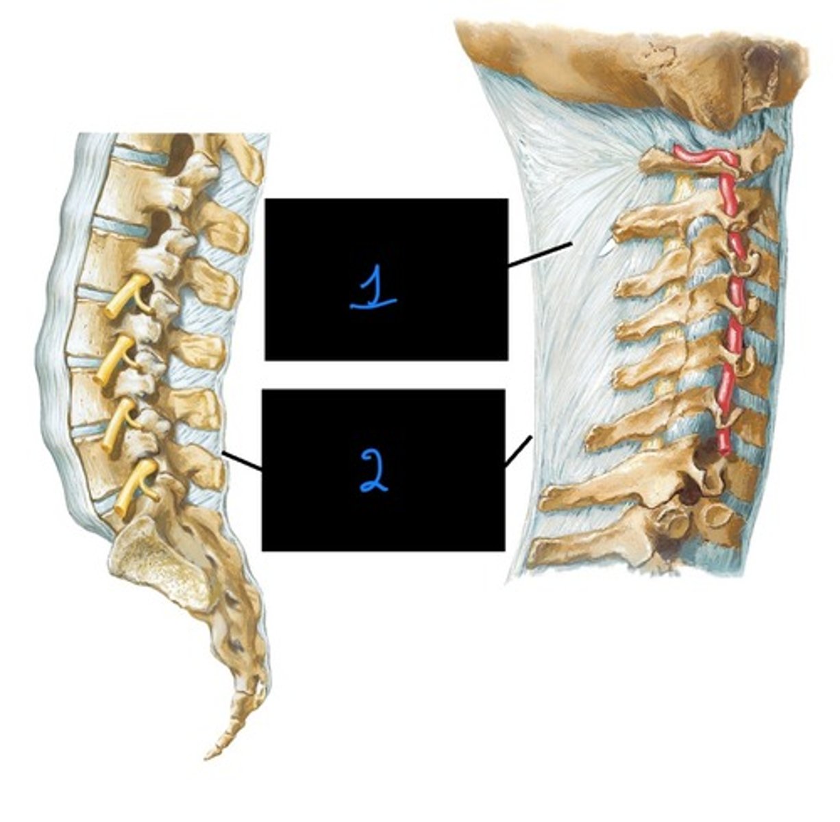
anterior longitudinal ligament
what vertebral ligament that limits vertebral extension?
ligamenta flava, posterior longitudinal ligament, supraspinous ligament, nuchal ligament
what vertebral ligaments that limits vertebral flexion?
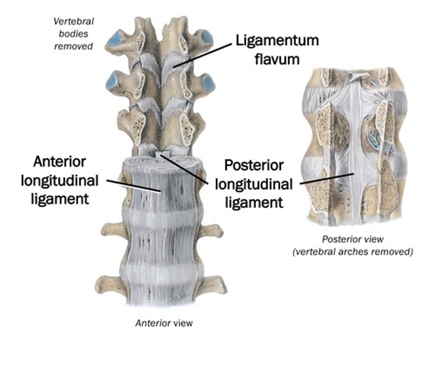
anterior longitudinal ligament
Ligament of the vertebral column that:
- Runs along and attaches to vertebral bodies and
- IVDs anteriorly
- Prevents hyperextension

posterior longitudinal ligament
Ligament of the vertebral column that:
- Runs within vertebral canal along vertebral bodies and IVDs posteriorly
- Mainly attaches to IVDs
- Limits hyperflexion (weakly)

ligamentum flavum
Ligament of the vertebral column that:
- Attaches to adjacent laminae
- Resists separation of laminae
- Helps to limit hyperflexion
anterior longitudinal ligament
what is image 1?

posterior longitudinal ligament
what is image 2?
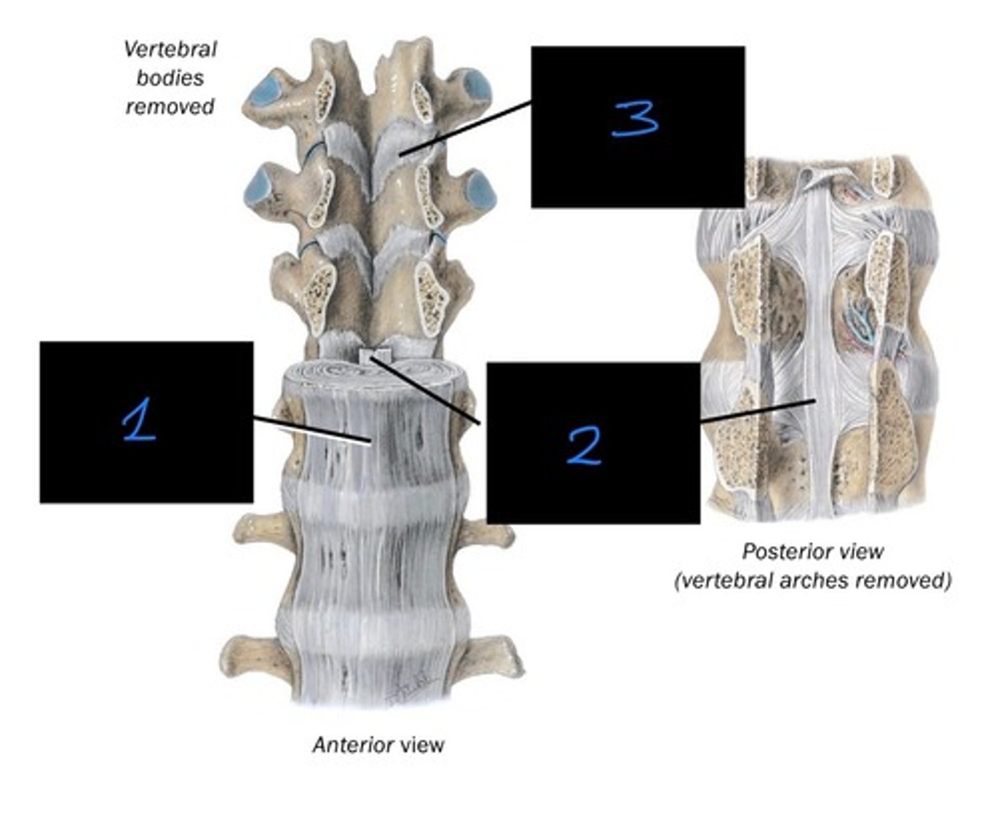
ligamentum flavum
what is image 3?

Supraspinous ligament
What ligament of the vertebral column:
- Runs along the tips of spinous processes from C7-sacrum
- Helps to limit hyperflexion

Nuchal Ligament
What ligament of the vertebral column:
- Starts at the external occipital protuberance through the C7 Spinous process
- Also C1 posterior tubercle and C2-C6 spinous processes -> from median fibrous septum between R & L muscles
- Attachment for trapezius, rhomboid minor, serratus posterior superior and splenius.
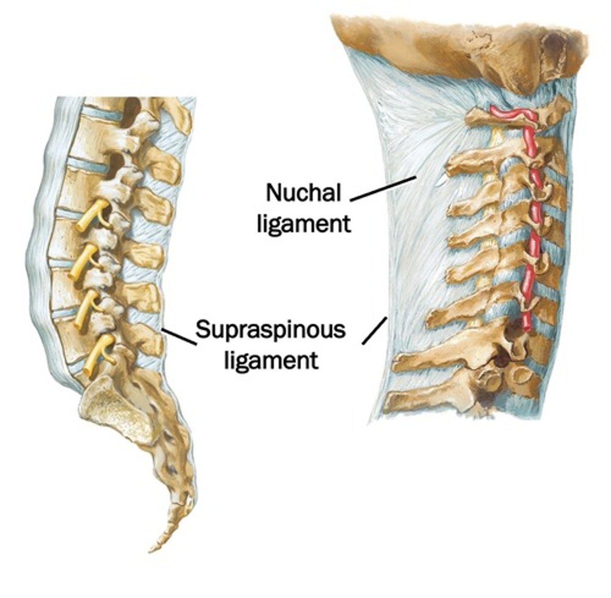
nuchal ligament
what is image 1?
supraspinous ligament
what is image 2?

whiplash
severe hypderextension of the neck (cervical region)
anterior longitudinal ligament
what ligament is stretched or torn when a person experiences whiplash?
intervertebral discs
These are:
- Attached to adjacent vertebral bodies
- Deformable (acts as a shock absorber)
fibrocartilaginous joint (symphysis)
nucleus pulposus
what is image 1?
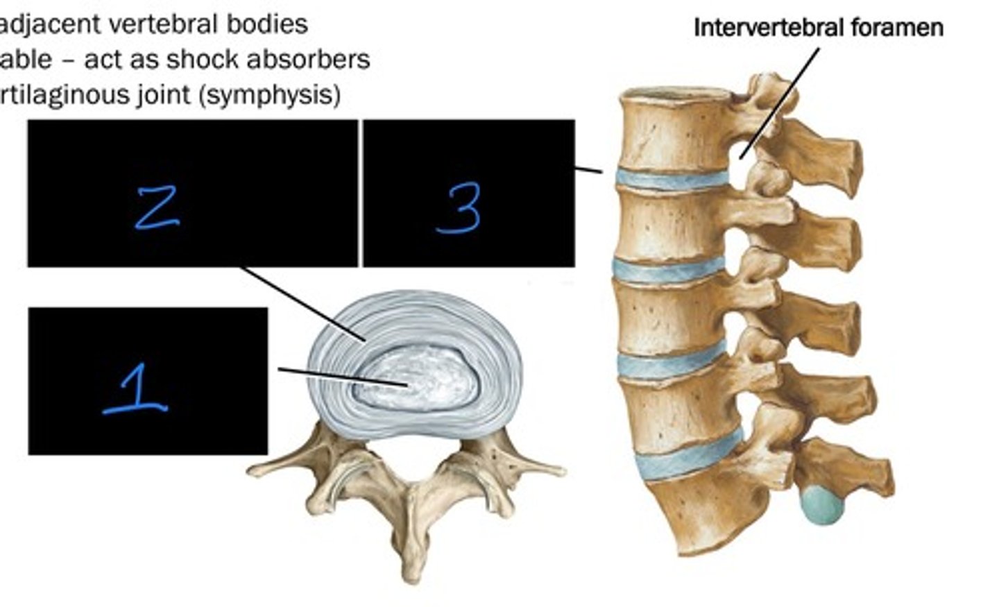
annulus fibrous (contains nucleus pulposus)
what is image 2?

intervertebral disc (IVD)
what is image 3?

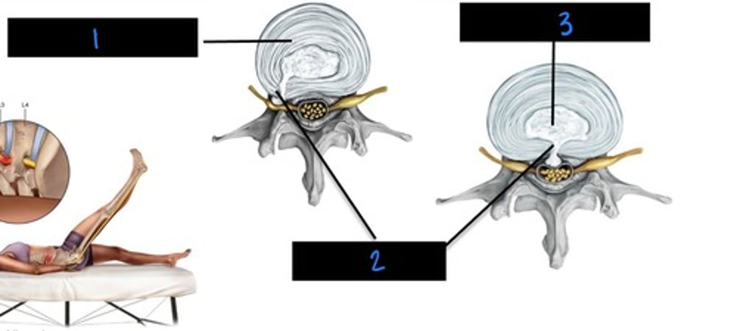
herniated dic
this occurs when the nucleus pulposus protrudes through annulus fibrousus. This causes it to put pressure on the nerve.
- Perception of pain in the dermatome supplied by the nerve.
annulus fibrosus
what is image 1?

herniation
what is image 2?

nucleus pulposus
what is image 3?
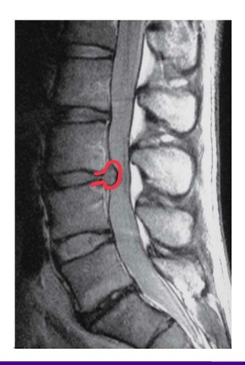
lumbar herniation
what injury is in this image?
laminectomy
surgical removal of the spinal process and laminae to relieve pressure on the spinal cord or nerve roots caused by a tumor, herniated dic, or bony hypertrophy.
spinal fusion
surgical stabilization after a spinal fracture, abnormal curvature, stenosis or due to herniated disc.
dura mater, arachnoid mater, pia mater
what are the three membranous layers around the spinal cord?
dura mater
this membranous layer around the spinal cord forms the dural sheath surrounding the spinal cord and part of cauda equina.
arachnoid mater
this membranous layer around the spinal cord is very thin, and has small filaments connecting it to the underlying pia mater
pia mater
this membranous layer around the spinal cord is the deepest layer adherent to the spinal cord.
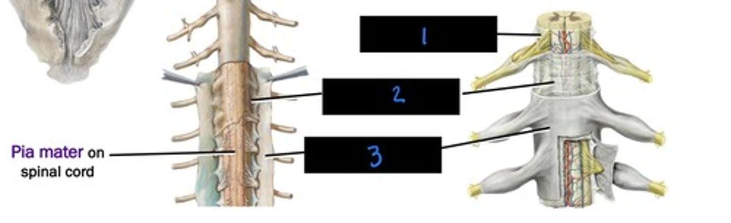
pia mater
what is image 1?

arachnoid mater
what is image 2?

dura mater
what is image 3?
Epidural space
the space within the vertebral canal, exteral to dural sheath, contains fat and internal venous plexus
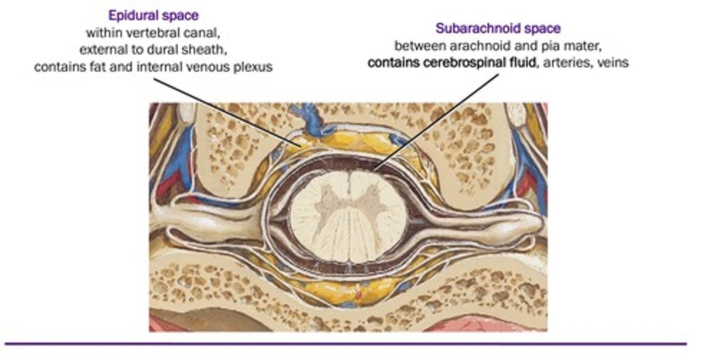
subarachnoid space
the space between arachnoid and pia mater, contains CSF, arteries, veins

epidural space
what is image 1?
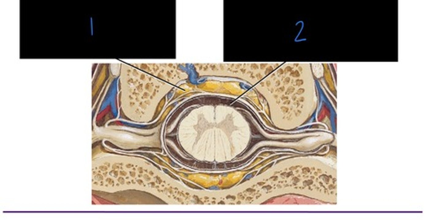
subarachnoid space
what is image 2?

dura mater
what is image 1?

arachnoid mater
what is image 2?

pia mater
what is image 3?

epidural space
what is image 4?
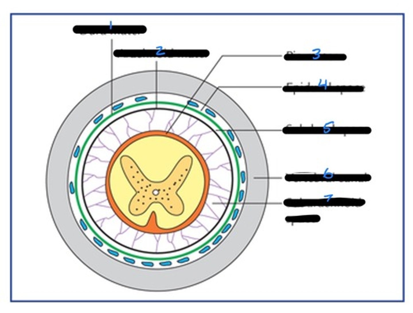
subdural space
what is image 5?

vertebral canal
what is image 6?

subarachnoid space
what is image 7?

L1/L2 IVD
at what point does the spinal cord end?
conus medullaris
this is inferior tapering at the end of the spinal cord.
cauda equina
the long spinal nerve roots that descend inferior to the termination of the spinal cord
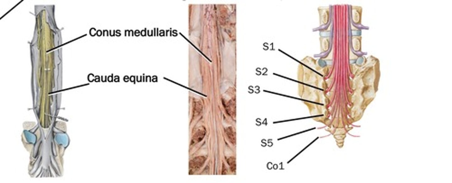
lumber cistern
the enlarged subarachnoid space caudal to the conus meduallaris containing CSF and cauda equina.

denticulate ligament
derivative of the pia mater:
- extending from the spinal cord.
- between the dorsal and ventral roots. R and L sides attach to dural sheath as (21) - small saw tooth-like processes.
filum terminale
derivative of the pia mater:
- spinal cord to the coccyx
- anchor spinal cord
filum terminale internum
Pia mater portion superior to the end of the dural sheath
filum terminale externum
dural sheath portion inferior to the dural sheath
denticulate ligament
what is image 1?
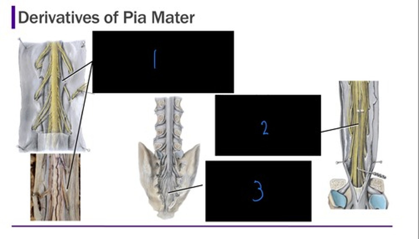
filum terminale internum
what is image 2?

filum terminale externum
what is image 3?

vertebral colum
what grows faster in development, vertebral column or spinal cord?
L2 or L3
at what vertebrae does the spinal cord end for infants?
C1
spinal cord segment C1 is parallel to which vertebrae?
T1
spinal cord segment T1 is parallel to which vertebrae?
T11
spinal cord segment L1 is parallel to which vertebrae?
L1
spinal cord segment S1 is parallel to which vertebrae?
spina bifida
this condition occurs when there is a deficit in vertebral arch of L5 or S1, failure to close in midline.
Often asymptomatic, may have heir tuft over spinous process
inferior to L1/L2 IVD
where on the vertebrae should a lumbar puncture be done?
subarachnoid space
where does a lumbar puncture need to go in order to collect CSF or inject anesthesia?
ligamentum flavum
an epidural injection only pierces what ligament in the vertebral canal?
visceral/autonomic and somatic
efferent neurons can be further categorized into what two divisions?
sympathetic and parasympathetic
two division of the visceral/autonomic efferent neurons
...
draw the typical spinal nerve
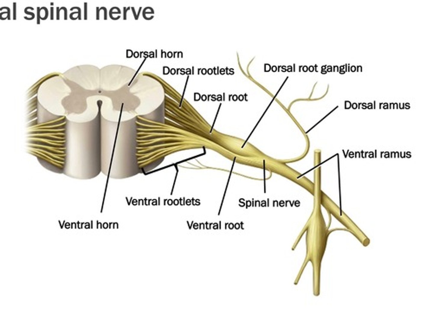
dorsal rami
these efferent fibers run to the epaxial muscles
- Sensation from overlying strip of the skin
ventral rami
these efferent fibers run to the hypaxial muscles
- Sensation from overlying skin
stenosis
narrowing of the vertebral canal or intervertebral foramen by bone or IV disc bulging.
- Leads to compression of spinal cord and/or exiting spinal nerves
- Often pain and discomfort when standing; numbness weakness and loss sensation in lower extremities; and loss of sensation or flexes in lower extremities
suboccipital nerve
what innervates the suboccipital muscles?
dorsal rami
what innervates all epaxial muscles (not including the suboccipital region
dorsal scapular nerve (C4-C5)
what innervates the levator scapulae (with branches from C3-C4), and rhomboids
thoracodorsal nerve (C6-C8)
what innervates the latissimus dorsi?
lower subscapular nerve (C5-C6)
what innervates the teres major, and the subscaularis?
Axillary Nerve (C5-C6)
what innervates the deltoid and teres minor?
suprascapulary nerve (C4-C6)
what innervates the supraspinatus and the infraspinatus?
T1-T5
what branches of the thoracic ventral rami innervates the serratus posterior superior?
T9-T12
What branches of the thoracic ventral rami innervates the serratus posterior inferior?
accessory nerve (CN XI)
what innervates the trapezius (with branches from C3-C4 ventral rami)
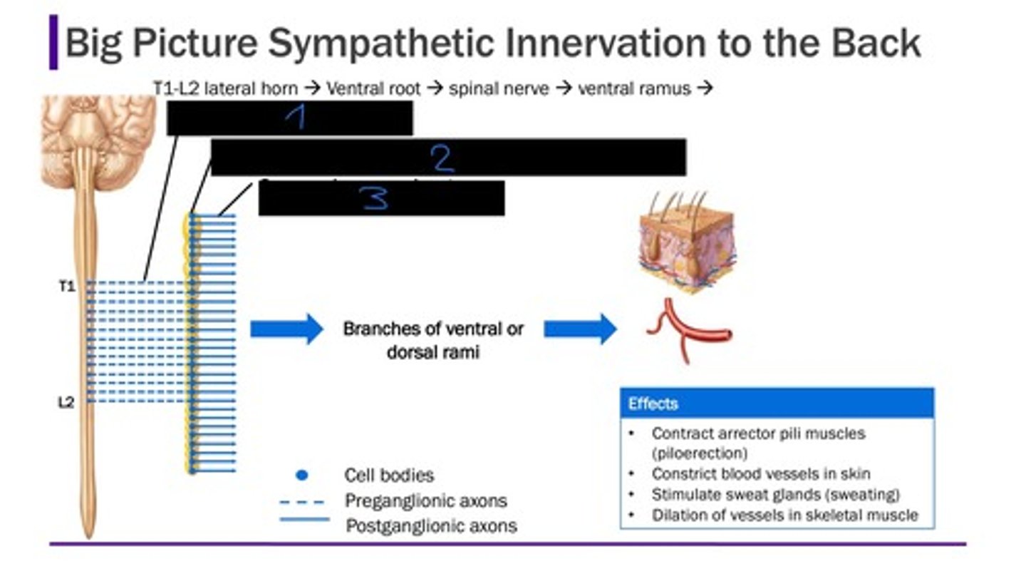
preganglionic neurons
The first neuron in a sympathetic reaction, located on the lateral horn of the spinal cord T1-L2
T1-L2
the preganlionic neurons are located where?
postganglionic neurons
the second neuron is paravertebral/sympathetic ganglia, prevertebral ganglia
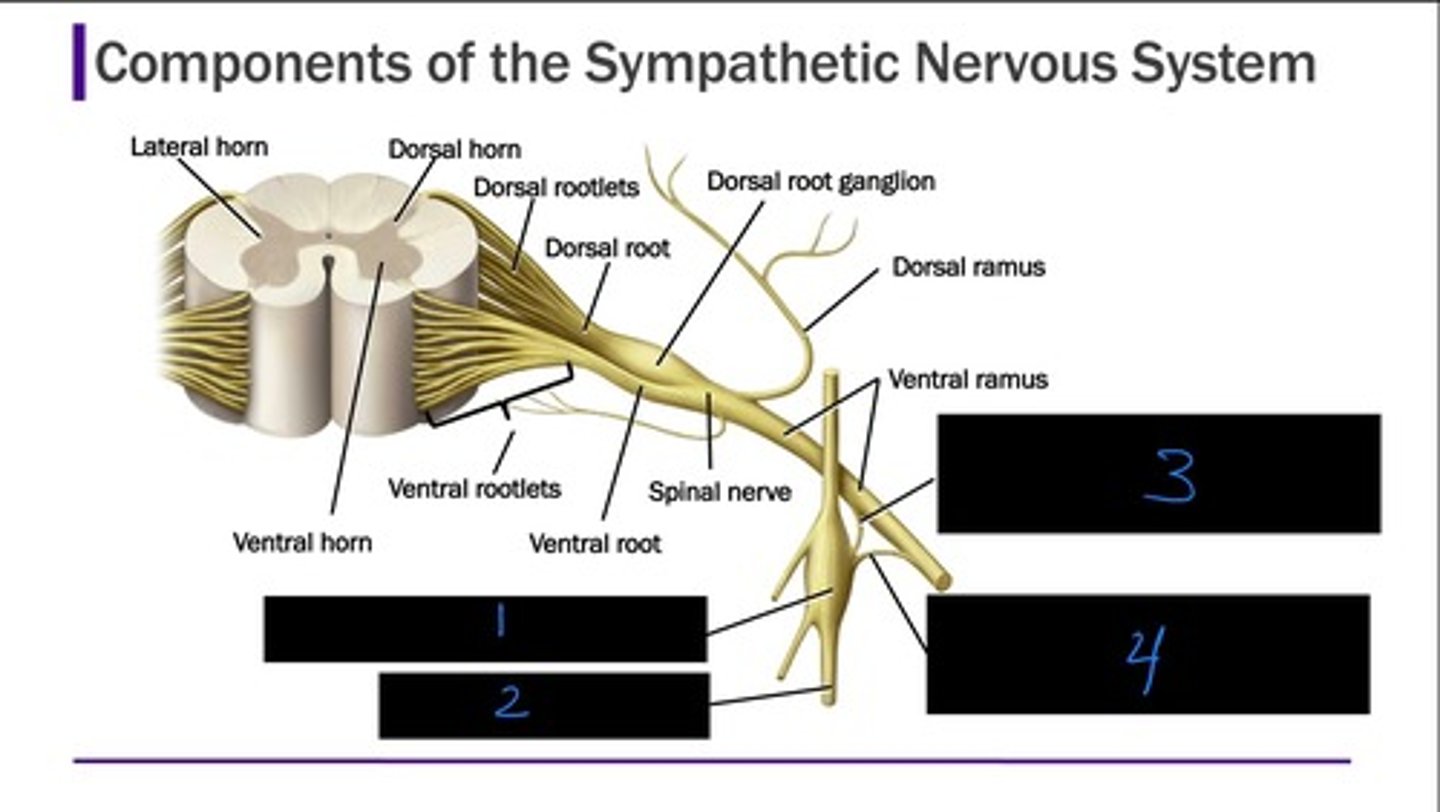
sympathetic ganglion
what is image 1?

sympathetic chain
what is image 2?

gray ramus communicans
what is image 3?

white ramus communicans
what is image 4?
3
how many cervical ganglia are there?
11-12
how many thoracic ganglia are there?
~4
how many lumbar ganglia are there?
~4
how many sacral ganglia are there?
1
how many coccygeal ganglia are there?
gray rami
does gray or white rami communicate at all levels?
T1-L2
white rami communicates to what vertebral levels?
sympathetic
the body wall only receives this type of innervation from the autonomic nervous system
Sympathetic preganglionic axons
Ventral root
Spinal Nerve
Ventral ramus
White ramus communicans
sympathetic chain ganglion
Stay at same level, ascend or descend.
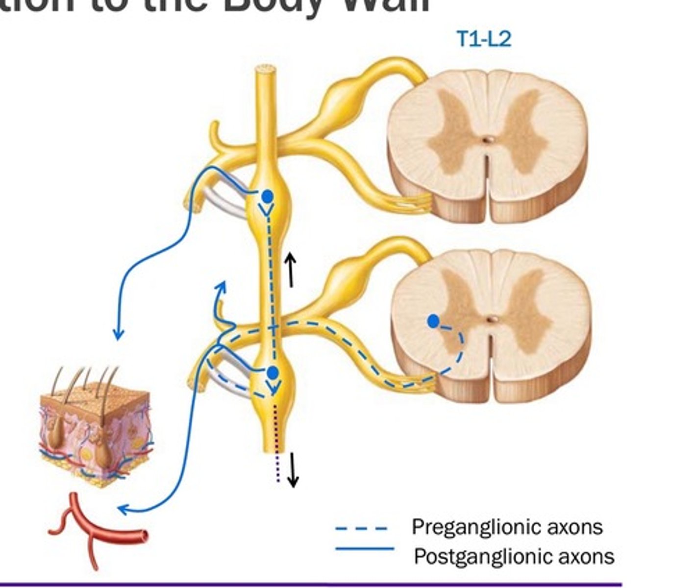
paravertebral ganglia
where are the postganglionic cell bodies located?
gray ramus communicans
where does the sympathetic chain exit to the ventral, to skin, blood vessels or arrector pili muscle in the skin
white rami communicans
what is image 1?

sympathetic chain and sympathetic ganglia
what is image 2?