FINALS RADLAB - LESSON PROPER
1/112
There's no tags or description
Looks like no tags are added yet.
Name | Mastery | Learn | Test | Matching | Spaced | Call with Kai |
|---|
No analytics yet
Send a link to your students to track their progress
113 Terms
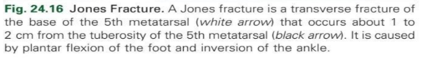
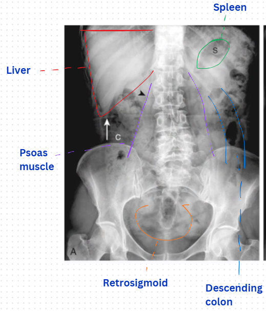
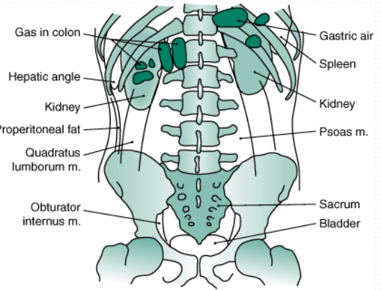
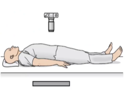
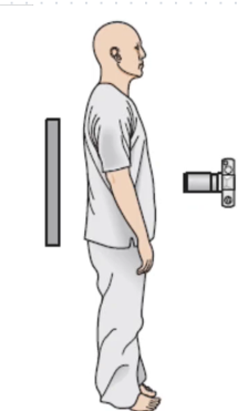
What view
Supine abdomen
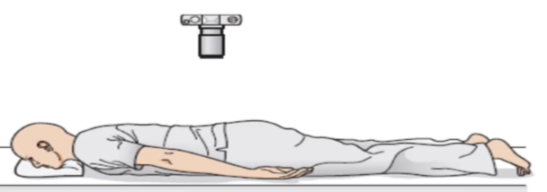
What view
Normal Prone abdomen
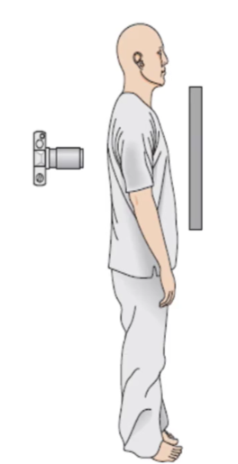
What view
Supine abdomen
What view
Prone view
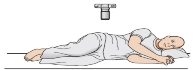
What view
Lateral Rectum view
substitute view for Prone view
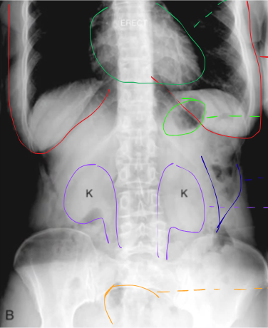
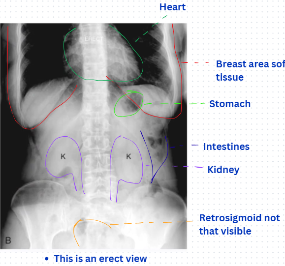
What view and what substitute view?
Upright abdomen
Substitute view: Left Lateral decubitus view
What view and what substitute view?
Upright chest
Substitute view: Supine view
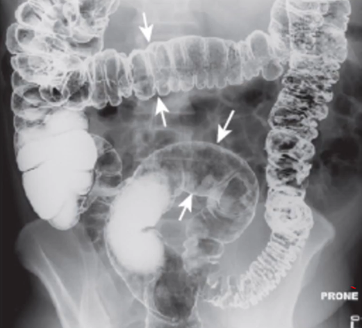
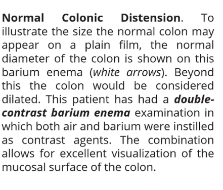
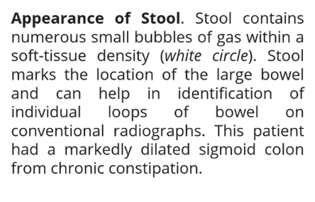
Describe the radiograph
Normal colonic distension
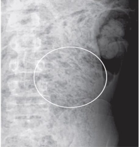
Describe the radiograph
Appearance of stool
Aerophagia
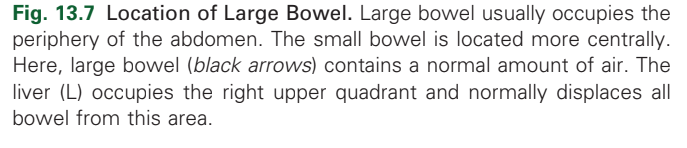
Large bowel with normal amounts of air
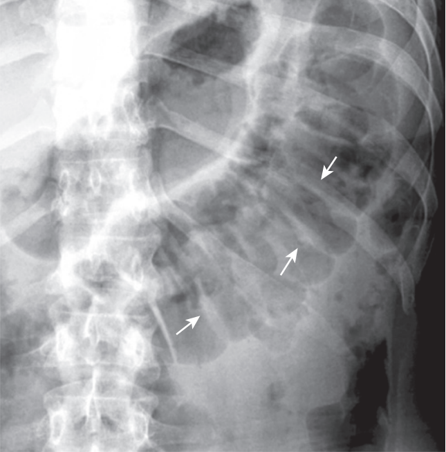
Normal Large Bowel Haustral Markings.
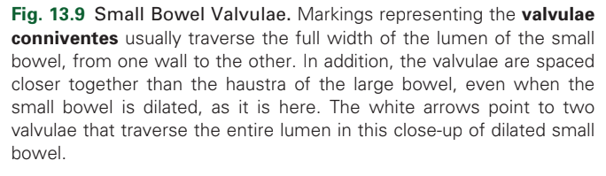
Small bowel valvulae
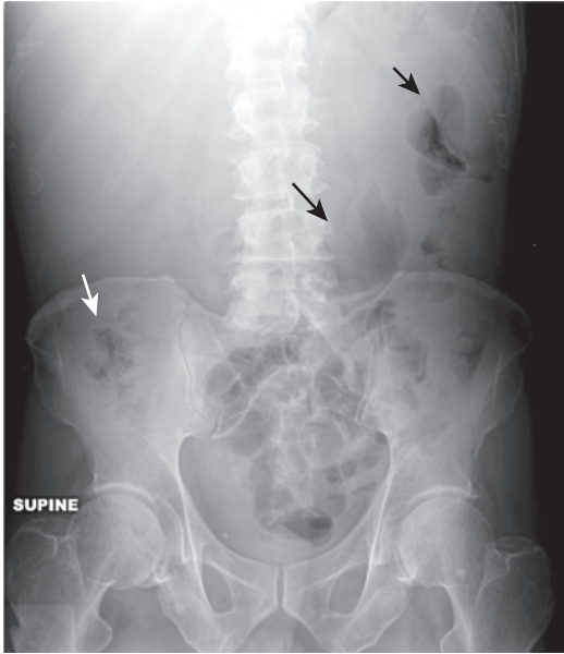
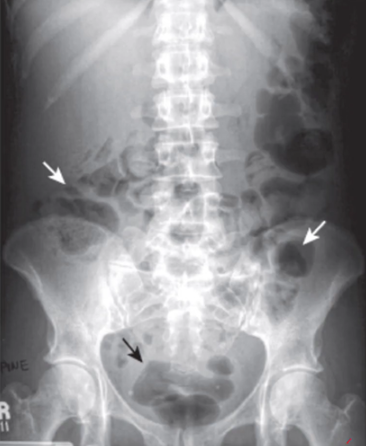
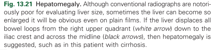
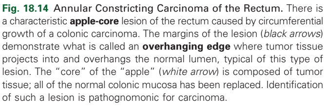
diagnosis
hepatomegaly
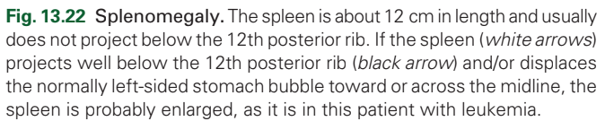
diagnosis
Splenomegaly
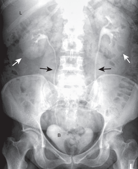
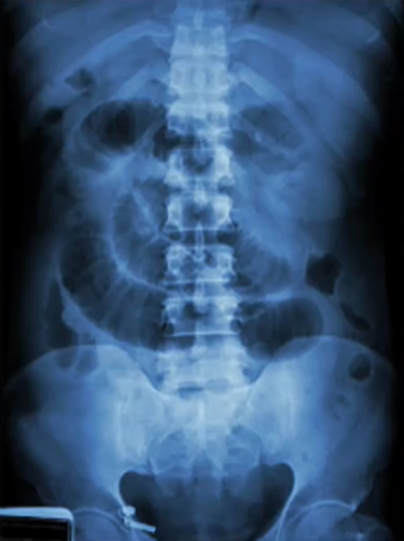
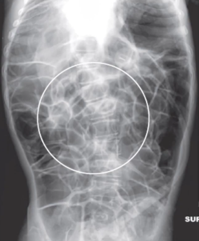
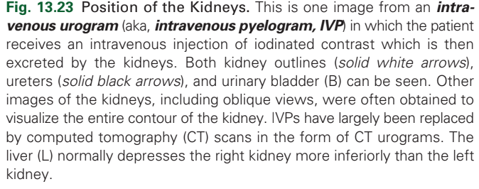
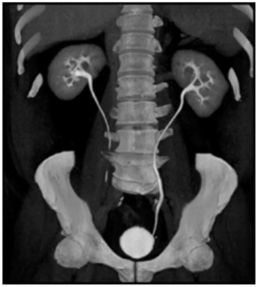
White arrows are the?
kidneys
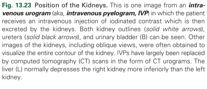
Black arrows are the?
Ureters
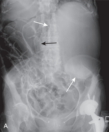
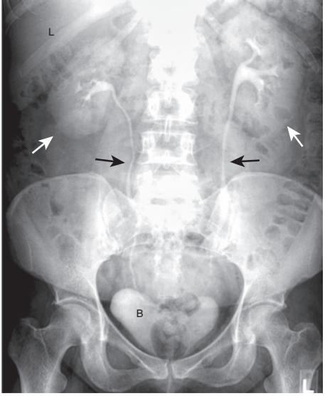
diagnosis
enlarged kidneys

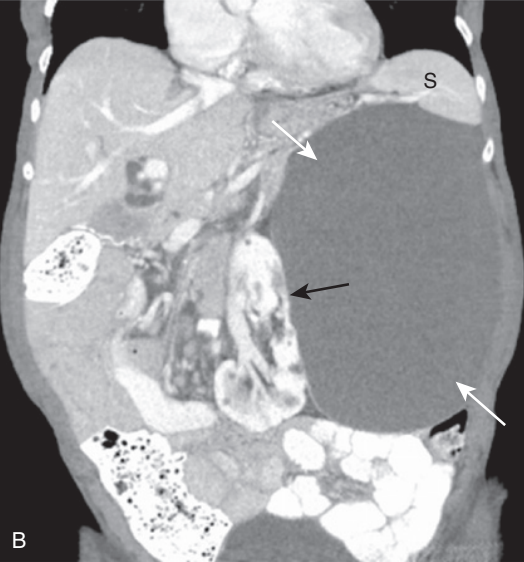
diagnosis
enlarged kidneys
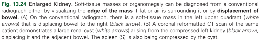
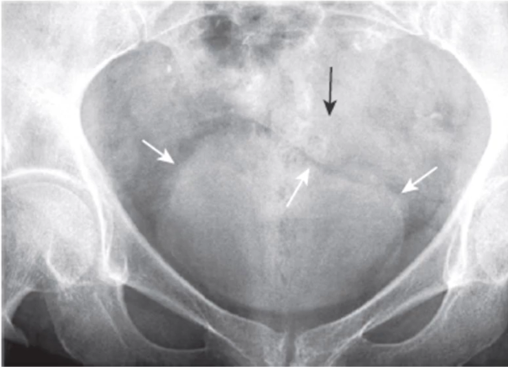
normal urinary bladder
diagnosis
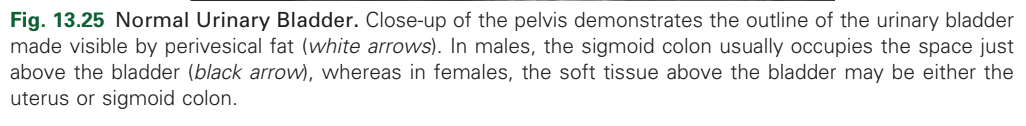
distended bladder
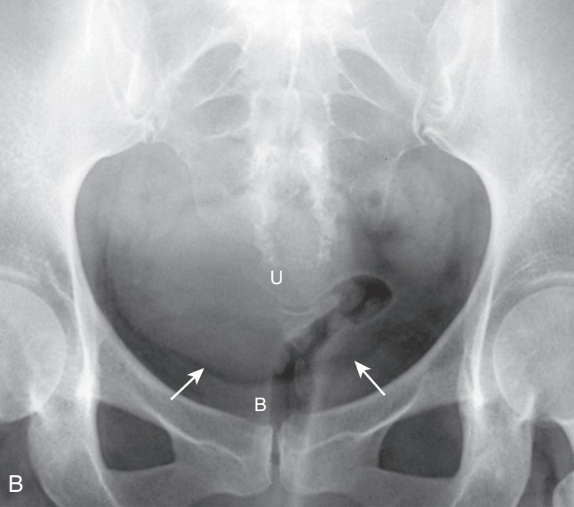
diagnosis
Enlarged Uterus.
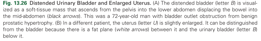
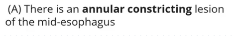
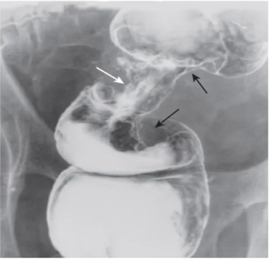
diagnosis
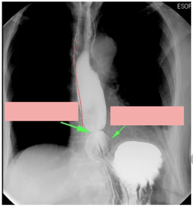
esophageal carcinoma - annular constriction
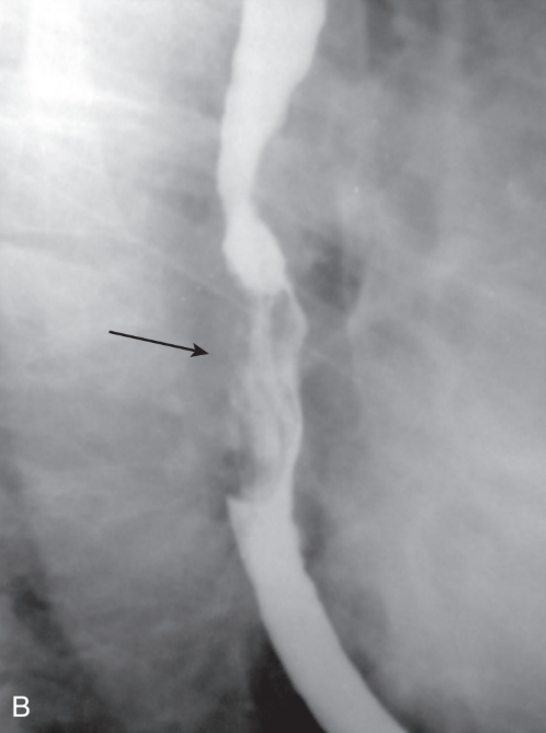
diagnosis
esophageal carcinoma - polyploid mass
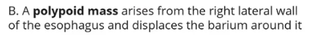
diagnosis
esophageal carcinoma - ulceration
diagnosis
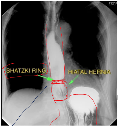
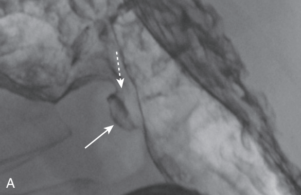
Benign Lesser Curvature Gastric Ulcer.
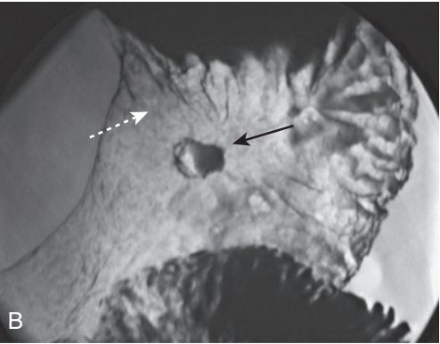
diagnosis
Benign Lesser Curvature Gastric Ulcer.
diagnosis
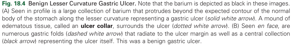
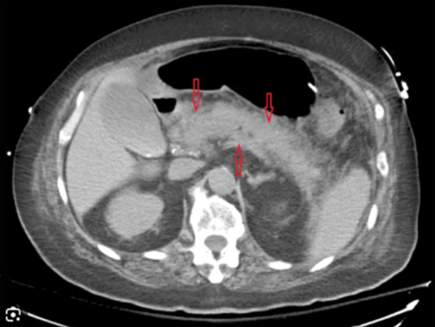
thickening and enhancement of the bowel wall
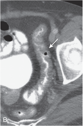
diagnosis
Submucosal infiltration
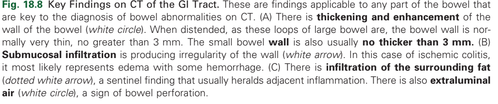
diagnosis
infiltration of the surrounding fat
extraluminal air (white circle),
diagnosis
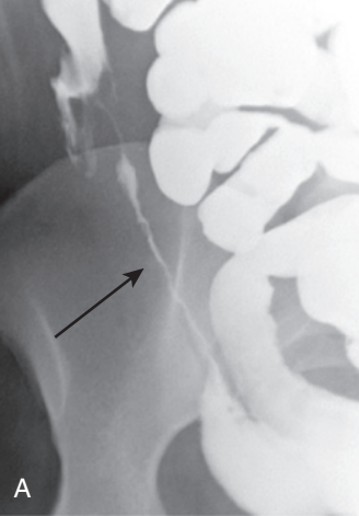
Crohn Disease

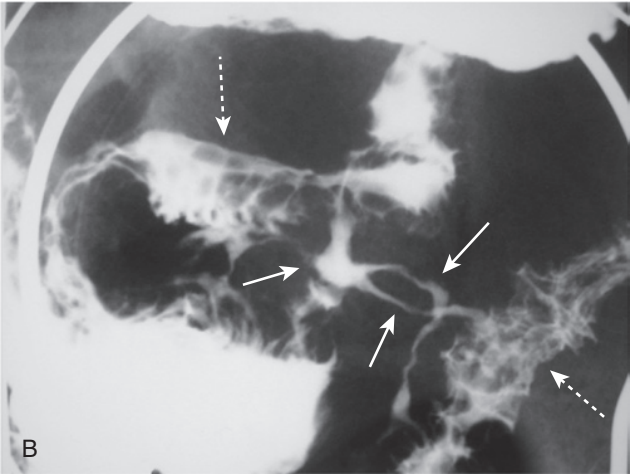
diagnosis
Crohn Disease
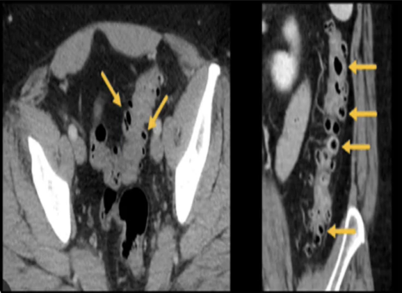
Diagnosis
diverticulosis
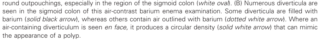
diagnosis
diverticulitis
diagnosis
colonic polyp
diagnosis
colonic carcinoma
diagnosis
colitis
diagnosis
colitis
diagnosis
colitis
diagnosis
appendicitis
diagnosis
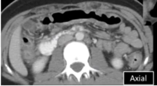
chronic pancreatitis
diagnosis
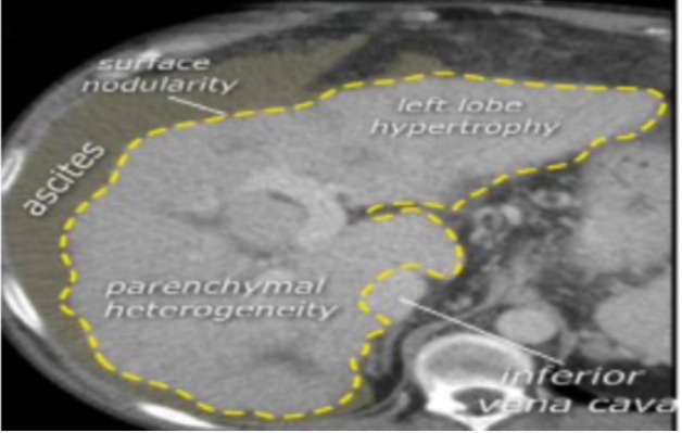
cirrhotic liver
diagnosis
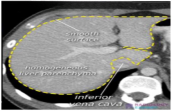
normal liver
diagnosis
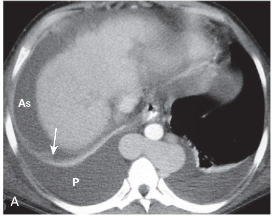
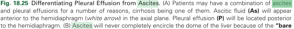
ascites and pleural effusion
diagnosis
Ascites

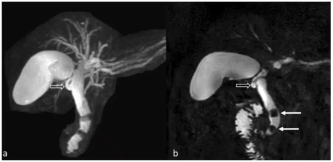
type of imaging used
Magnetic Resonance Cholangiopancreatography (MRCP)
Urinary bladder
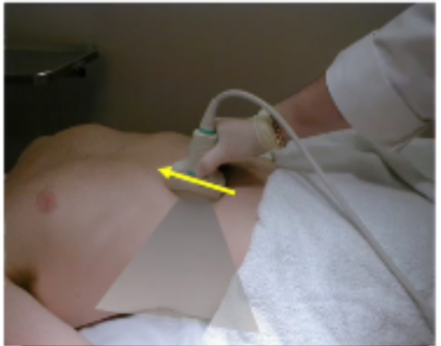
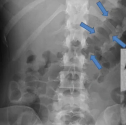
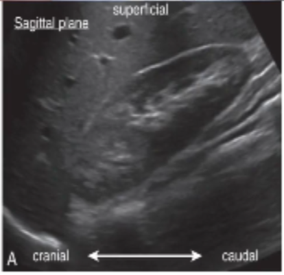
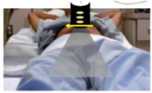
what imaging plane is used
sagittal/longitudinal plane
what imaging plane is used
sagittal/longitudinal plane
What imaging plane was used?
Transverse/Axial plane
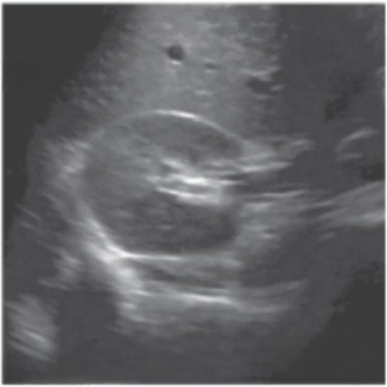
What imaging plane was used
Transverse/Axial plane
What imaging plane was used
Transverse/Axial plane
What imaging plane was used
sagittal/longitudinal plane
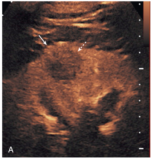
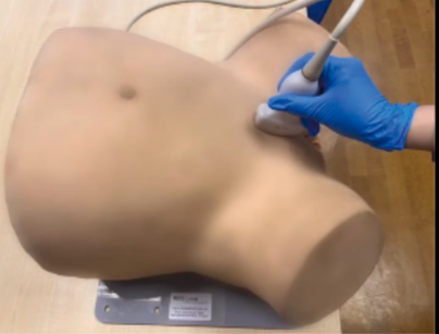
Specific imaging modality used
Contrast-Enhanced Ultrasound in color
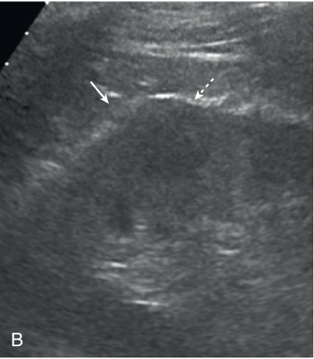
Specific imaging modality used
Contrast-Enhanced Ultrasound in grayscale
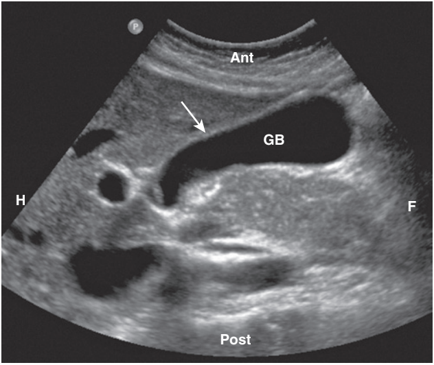
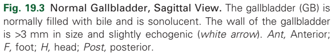
Describe radiograph
Normal Gallbladder
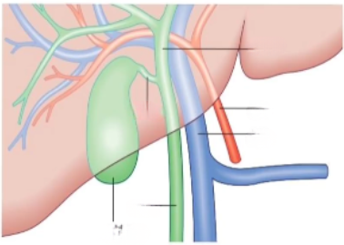
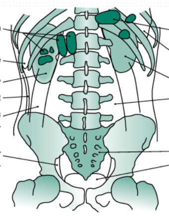
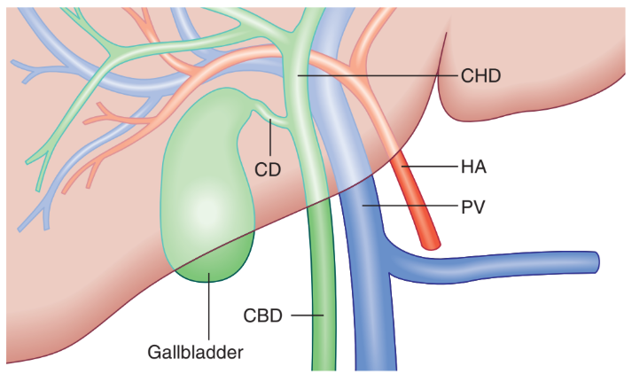
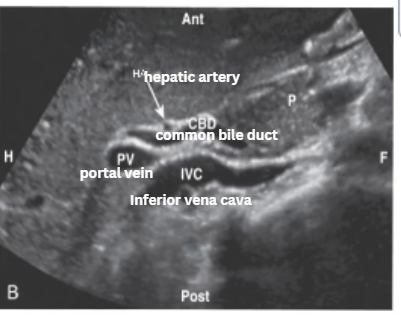
Label
Normal Common Bile Duct, Portal Vein, and Hepatic Artery: Illustration and Sagittal view.
(A) The illustration demonstrates the normal relationships of some of the structures in the porta hepatis.
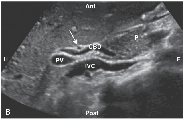
Label
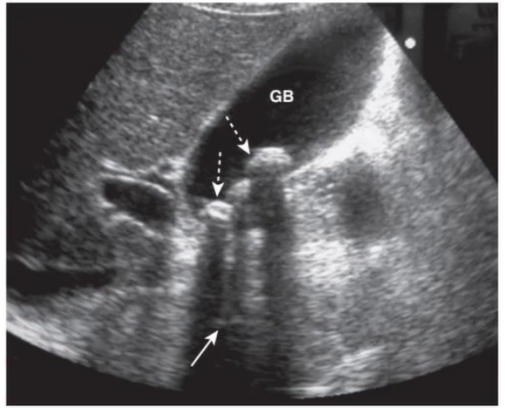
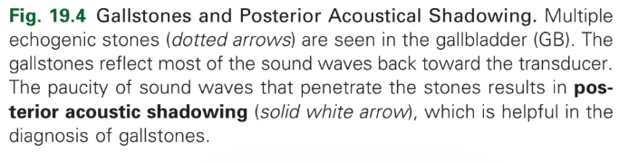
Diagnosis
Gallstones
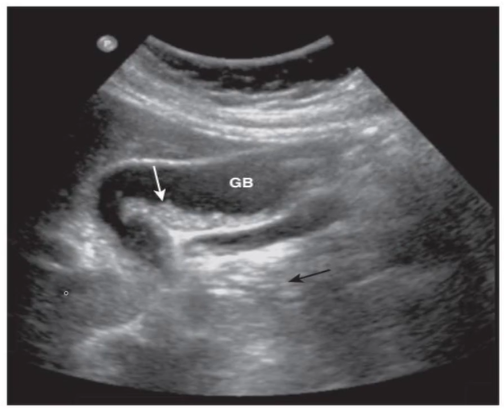
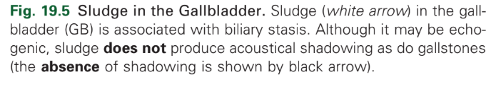
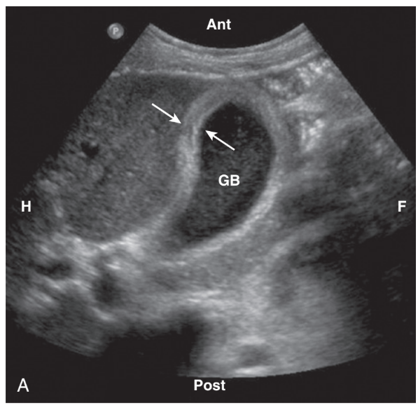
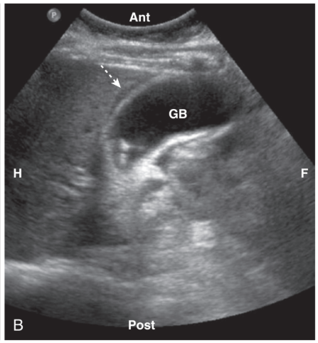
Diagnosis
Biliary sludge
diagnosis
Acute cholecystitis

diagnosis
Acute cholecystitis

diagnosis
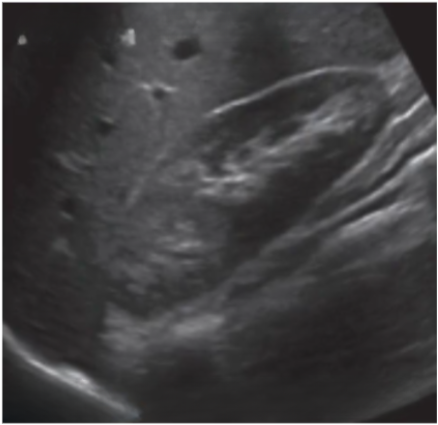
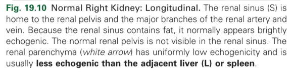
Normal right kidney - longitudinal
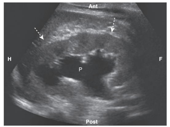
Diagnosis
Hydronephrosis
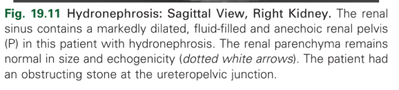
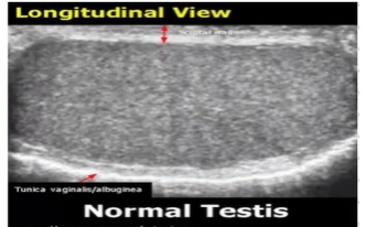
Normal Testis
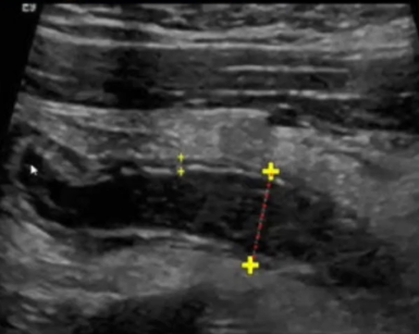
Diagnosis
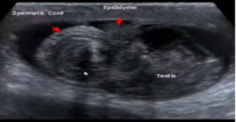
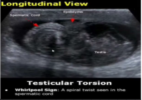
Testicular Torsion

Diagnosis
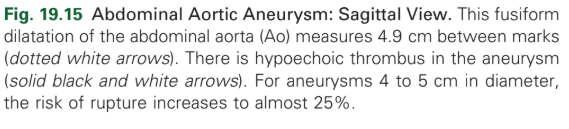
Abdominal Aortic Aneurysm
Diagnosis
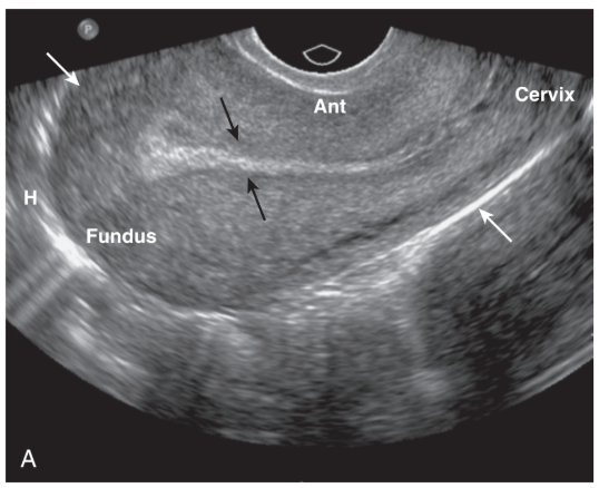
Normal Uterus

diagnosis
normal uterus

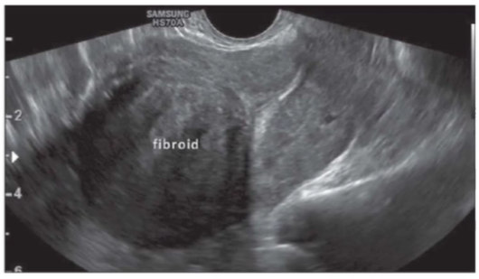
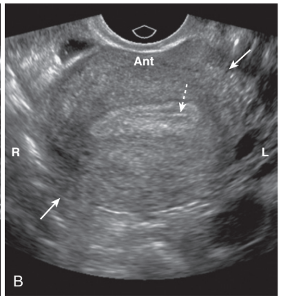
diagnosis
Leiomyomas
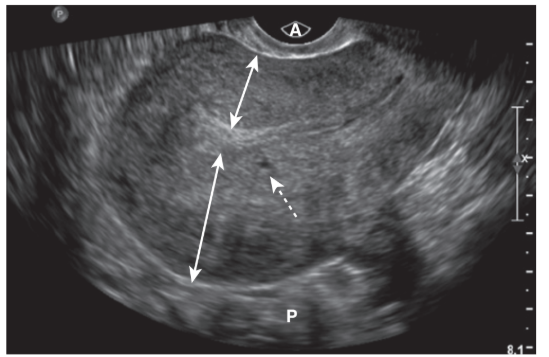
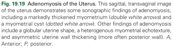
diagnosis
Adenomyosis
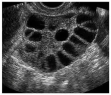
diagnosis
Polycystic ovarian syndrome (PCOS)
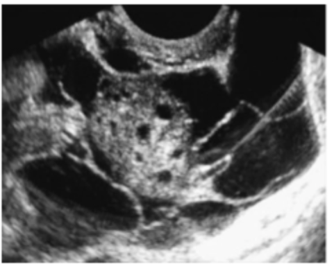
diagnosis
pelvic inflammatory disease
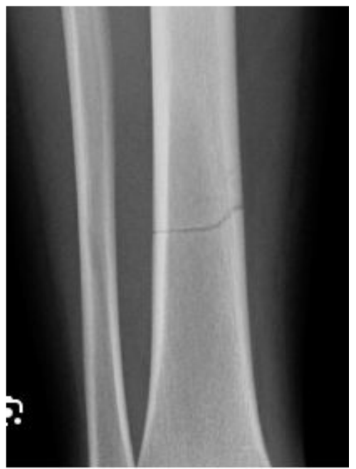
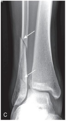
Describe
Fracture line
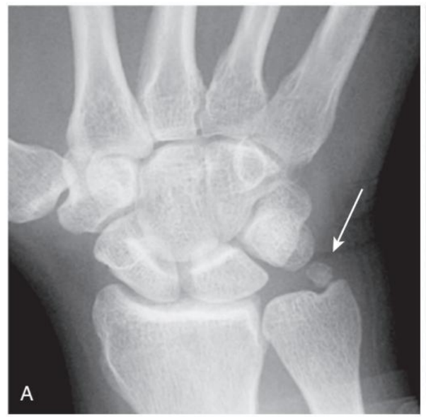
diagnosis
Old, unhealed fracture fragments
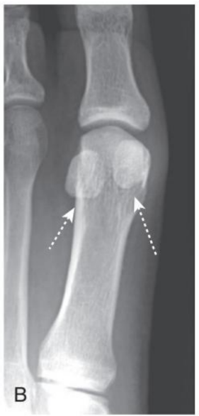
Describe
sesamoid bones that form in a tendon
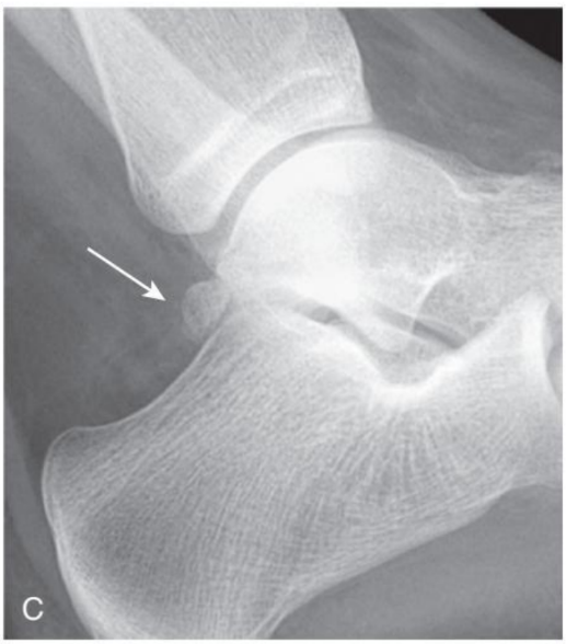
Describe
accessory ossicles that can mimic acute fractures

Normal
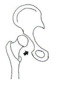
Subluxation
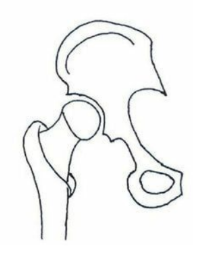
Dislocation
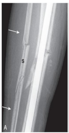
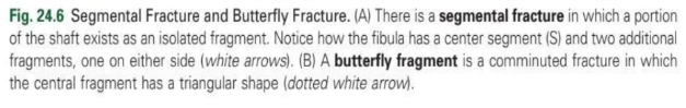
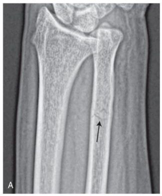
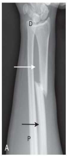
type of fracture
segmental fracture - comminuted fracture
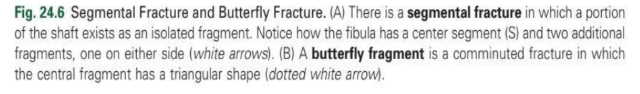
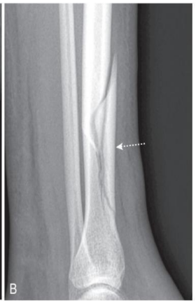
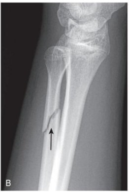
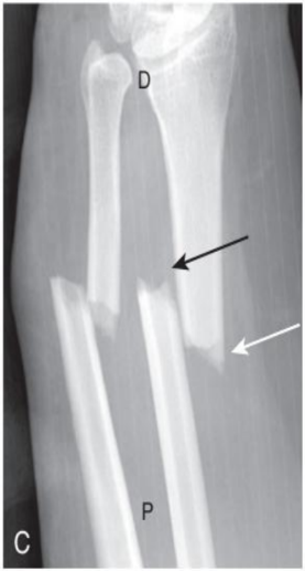
type of fracture
butterfly fracture - comminuted fracture
direction of fracture line
transverse fracture

direction of fracture line
oblique fracture

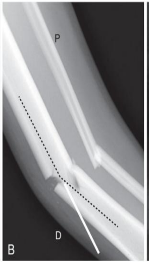
direction of fracture line
spiral fracture

fracture orientation
displacement

fracture orientation
Angulation

fracture orientation
shortening

fracture orientation
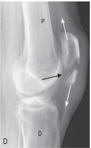
distraction
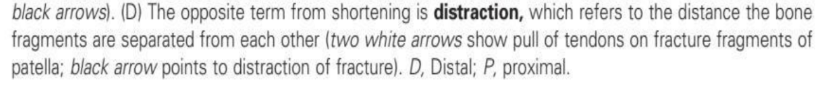
fracture orientation
rotation
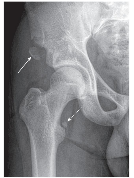
diagnosis
Avulsion fracture
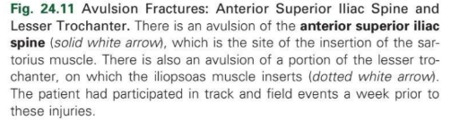
diagnosis
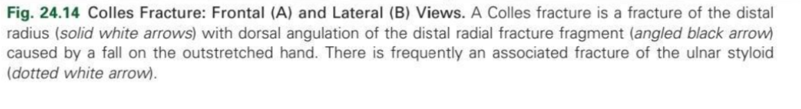
Colles fracture
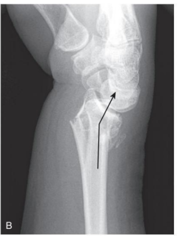
diagnosis
Colles fracture
diagnosis
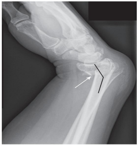
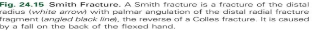
Smith fracture
diagnosis
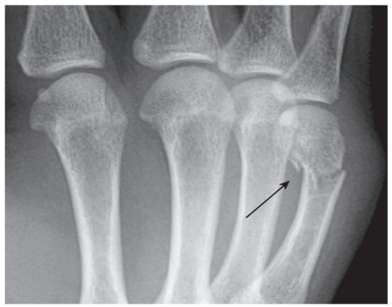
Boxer’s fracture
diagnosis
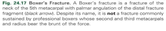
Jones fracture