8.4 Voluntary movements all processes involved
1/31
There's no tags or description
Looks like no tags are added yet.
Name | Mastery | Learn | Test | Matching | Spaced | Call with Kai |
|---|
No analytics yet
Send a link to your students to track their progress
32 Terms
Voluntary movements all processes involved
Upper motor neurons (UMNs)
Basal ganglia & cerebellum
Local circuit neurons (interneurons)
Lower motor neurons (LMNs)
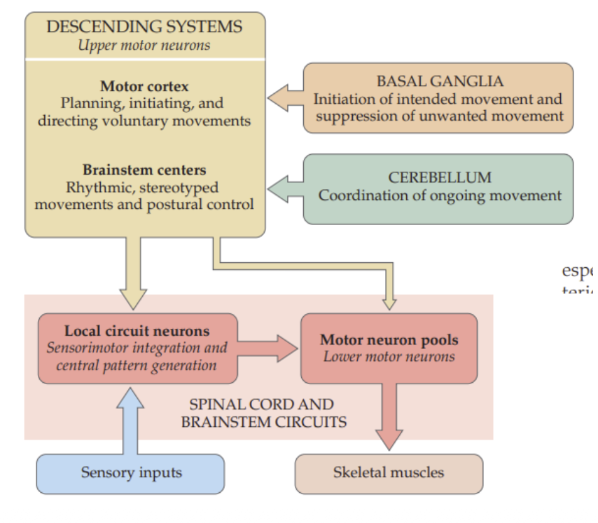
Upper motor neurons (UMNs):
Modulate the activity of lower motor neurons
They “order” the movements
Soma in motor cortex (or in some brainstem centers).
Axons synapse directly with lower motor neurons & with the local circuit interneurons.
Basal ganglia & cerebellum
help regulate UMNs, ensuring that movements are performed correctly (initiation, force, coordination).
Local circuit neurons (interneurons
They receive input from UMNs & sensory neurons.
They provide higher integration & can participate in reflex arcs.
Lower motor neurons (LMNs)
Soma in spinal cord or brainstem
Axons synapse with the skeletal muscle to activate contraction.
VOLUNTARY movement control involves
Decision-making & planning
Initiation of the movement
Execution of the movement
Motor unit
Lower motor neuron + skeletal muscle fibers innervated by the same motor neuron
muscles for fine motor actions containing
few fibers for precise control
muscles for gross motor actions
thousands of fibers allowing fro powerful simultaneous contraction.
Neuromuscular Junction
is the synaptic connection between the lower motor neuron & a muscle cell → resulting in a muscle contraction
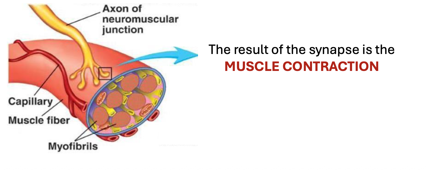
Neuromuscular junction neurotransmitters
ACETYLCHOLINE
Neuromuscular junction Receptor
Nicotinic Ach receptor
Neuromuscular junction Cessation of response
Acetylcholinesterase
Neuromuscular junction process →
The acetylcholine released from axon endings of the LMN diffuses across the synaptic cleft & binds to the ACh receptors located on the plasma membrane of the muscle cell, thereby stimulating the muscle cell & inducing a muscular action potential.
The neuromuscular synapse triggers the
excitation-contraction coupling
Excitation-Contraction coupling process
Motor neuron action potential
Action potential on skeletal muscle cell
Contraction (response)
Threshold stimulus for excitation-concentration
Minimum level of stimulation for a fiber to contract.
Subthreshold stimuli have no effect on the fiber contraction.
After the contraction, the muscle fiber immediately returns to the relaxed state.
Tetanization
The action potential in skeletal muscle does NOT have a refractory period
If a series of stimuli arrive in rapid succession, the muscle does not have time to fully relax before the next phase of contraction begins
Tetanus
When a motor unit is stimulated at such a high frequency that the individual muscle twitches fuse together → sustained contraction.
Mechanisms to increase the intensity of muscle contraction
Multi-fiber summation
Frequency summation (rate coding)
Frequency summation (rate coding)
Increasing the frequency of the action potential, until tetanization
Multi-fiber summation
Increasing the number of motor units that contract simultaneously.
Contractile Unit:
SARCOMERE (skeletal muscle)
SARCOMERE
Thin filaments (actin)
Thick filaments (miosin)
Regulatory proteins
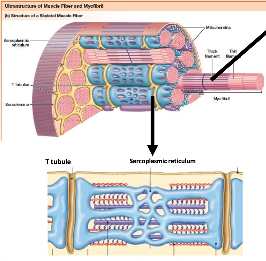
SARCOPLASMIC RETICULUM
The specialized endoplasmic reticulum of muscle cells, wrapping around the filaments.
Concentrates and stores Calcium (Ca2+), whose release will play a key role in contraction.
T-TUBULE
Invaginations in the plasma membrane of muscle cells, they help propagation of the action potential (depolarization) into the interior of the muscle fiber
MUSCLE CONTRACTION MECHANISM
Action potential arrives at the motor neuron.
The motor neuron releases ACh.
ACh binds to the nicotinic receptor in the muscle fiber.
Entry of Na+ through Ach nicotinic receptor channels initiates a muscle action potential.
Propagation of action potential into the muscle fiber by the T-tubules
Depolarization opens channels in the sarcoplasmic reticulum & Ca2+ is released into cytoplasm.
Ca2+ ions cause the thick & thin filaments to slide against each other, which constitutes the process of contraction
Ca2+ ions are pumped back into the sarcoplasmic reticulum by means of a calcium pump, & the filaments go back to the relaxed position.
MUSCLE TONE
muscles in tension even when we are at rest
Maintaining posture
Flaccid muscles (hypotonia)
Spastic muscles (hypertonia)
Flaccid muscles (hypotonia)
Lower tone than normal
Spastic muscles (hypertonia)
Higher tone than normal
Muscle tone is controlled by
negative feedback mechanisms in the spinal cord (motor medullary reflex)