Topic 4B: Vascular Mechanics
1/45
Earn XP
Description and Tags
We'll look at the mechanical properties and function of vasculature
Name | Mastery | Learn | Test | Matching | Spaced | Call with Kai |
|---|
No analytics yet
Send a link to your students to track their progress
46 Terms
What does the vasculature consist of a complex system of
arteries, arterioles, capillaries, venules and veins
What does the arterial system serves as a
conduit through which blood is transported from the heart to the capillary networks throughout the body
What is the arterial system split into
The arterial system is divide into two subsystems pulmonic and systemic arterial structure
What does the biomechanical behaviour depend on
strongly on specific location within the vasculature
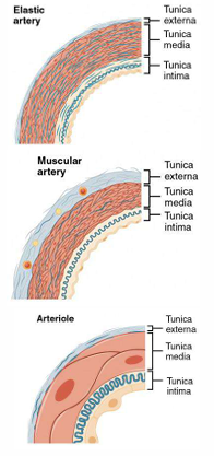
What in different arteries/veins share some common substructures called…
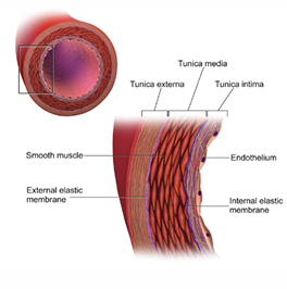
Tunicae
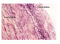
Tunica intima/interna
Tunica media
Tunica adventitia/externa
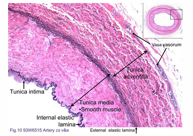
What is shown in pink
This slide is stained with Verhoeff’s stain to visualise the elastic fibers, and with eosin to show the cellular structures
What is the intima
Typically consisting of a monolayer of endothelial cells and an underlying thin basal lamina
What shape are endothelial cells
flat and elongated in the direction of blood flow
What is the lamina
fenestrated sheet of elastin that allows the transport of H2O nutrients and electrolytes across the wall as well as direct transmural cell to cell communication
What is the media
The media contains smooth muscle cells that are embedded in an extracellular piexus of elastin and collagen (primarily types I, III and V) as well as an aqeuous ground substance PG matrix
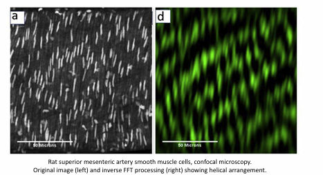
What are vascular smooth muscle cells (SMCs)
spindle-shaped cells that are typically 100um long and about 5um in diameter except near the nucleus where they are slightly thicker
Why does the orientation of vascular smooth muscles cells matter
Although the orientation and distribution of the medial constituents vary, vascular smooth muscle tends to be oriented helically, albeit nearly circumferentially in many vessels.
This preferential orientation is due to the primary role of SMC to contract and thus modify the distensibility of the large arteries or regulate the luminal diameter in medium and small arteries
What are elastic arteries made up of
the medial smooth muscle is organize into 5 to 15um thick concentric layers that are separated by thin fenestrated sheets of elastin
the outermost sheet of elastin is called the external elastic lamina: it separates the media and adventitia but is often considered to belong to the former
There may be as many as 40 to 70 concentric layers of smooth muscle in a thick elastic artery such as the human aorta.
What is the adventitia
The outermost layer of the wall, consists primarily of a dense network of type I collagen fibres with admixed elastin, nerves, fibroblasts and the vasa vasorum. The adventitial collagen fibres tend to have an axial orientation in most arteries.
Although the adventitia comprises only 10% and 50% of the arterial wall in most arteries, respectively, it is thought to limit acute overdistension in all vessels
What is the vasa vasorum
an intramural network of arterioles, capillaries and venules that serves the outer portion of the wall in arteries that are too thick for sufficient transport of O2, CO2, nutrients and metabolites from the intimal surface.
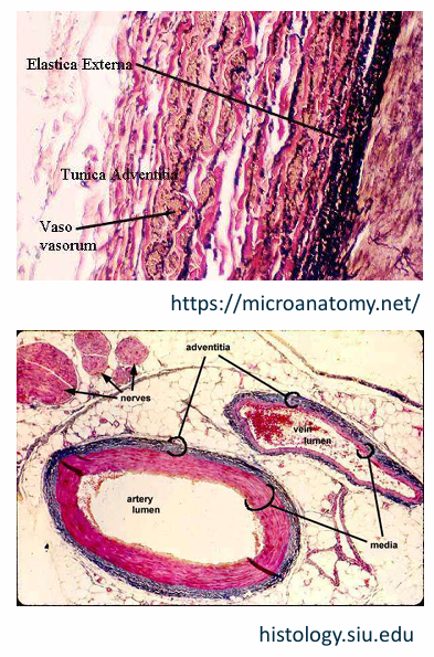
What order does the layers in the artery vascular wall
adventitia, media, intima and lumen
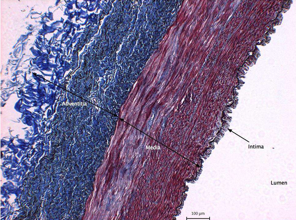
What does the blue, red and white represent in this image
The red is mostly the media with smooth muscle staining red. The blue is the thick collagen deposits in the adventitia with spare amounts in the media. The white is the lumen
What are the two general types of arteries
elastic and muscular
types of elastic arteries
aorta, main pulmonary artery, common carotids and common iliacs
types of muscular arteries
coronaries, cerebrals, femorals and renals
What size do elastic arteries tend to be
elastic arteries tend to be larger diameter vessels located closer to the heart
What size do muscular arteries tend to be
muscular arteries are smaller diameter vessels located closer to the arterioles
Elastic Arteries
are large arteries carrying a high volume of blood away from the heart. With its high content of elastin and few SMCs, the walls of elastic arteries are extremely resilient and can withstand the pulsatile flow and pressure fluctuations exerted during the cardiac cycle.
Elastic arteries distribute blood to muscular arteries, which supply the body’s skeletal muscles and internal organs. Smooth muscle contraction and relaxation allows the muscular arteries to control regional blood pressure and blood flow by actively changing the lumen size.
Elastic arteries are large enough to sustain systemic hemodynamic forces with lumen diameters up to 2.5cm
Muscular arteries range in size from 4mm to 0.5mm in internal diameter while arterioles are 30um or less
What causes of elastic arteries allows them to expand
the abundance in elastic fibers
What would happen if artery walls were rigid and unable to expand and recoil
Their resistance to blood flow would greatly increase and blood pressure would rise to even higher levels. The elastic recoil of vascular wall helps to maintain the pressure gradient that drives the blood through the arterial system
What are the specific orientations of certain structural constituents within the arterial wall
SMCs are oriented circumferentially/helically
Collagen in the adventitia is oriented longitudinally
Elastin is organised into thin concentric sheets
anisotropic
a physical property which has a different value when measured in different directions
Residual Stress
The stress that exists in a body in the absence of externally applied loads
In the artery when responding the radial cut what does the opening imply
It implies that the inner wall of the intact unloaded ring is in compression and the outer wall in tension.
The increased waviness in the internal elastic lamina in the unloaded ring is consistent with the existence of compressive residual stresses in the inner wall in this configuration
What is responsible for much of the residual strain in the normal arterial wall
elastin due to the selective digestion of different components of the arterial wall
What is the state of residual stress dependent on
The thickness and the composition of the artery. In fact, as arteries are remodelled in response to mechanical stress changes
what is the mark of the amount of residual stress
how much the blood vessel will open when cut. Since the blood vessel is under stress, when we cut the vessel, the stress holding the vessel together is removed and the blood vessel springs open
What is the effect of the remodelling (ligature was performed right below the diaphragm, to cause sudden increase in systolic pressure)
high effect on the aortic arch and descending aorta, where pressure was 25% above normal
lower effect below the diaphragm due to autoregulation
method for determining residual strains in an unloaded artery
optically measure the opening angle (a) of a cross-section taken from the artery
As the ring is cute, it opens the sector, on which the opening angle is defined as the angle between two radii, each drawn from the midpoint of the sector to the outer ends of the sector. Assuming no deformation in the axial direction, the circumferential stretch ratio can be calculated for a point in the vessel wall at a distance R from the centre
What is the equation of the circumferential stretch ratio
𝜃=pi r / O R where r and R are the radii of points in the wall in the unloaded and zero-stress states respectively and r and R the internal radii
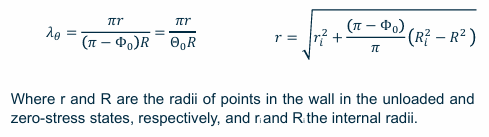
Whats in compression the inner or outer wall
the inner wall is in compression and the outer wall is in tension in the intact unloaded configuration
however, by accounting of the presence of intramural pressure, these values change and the actual value of circumferential stretch are lower than the nominal ones and more even
What does a larger opening angle correlate to
a more uniformly distributed circumferential stress
What happens to deformation while applying stresses
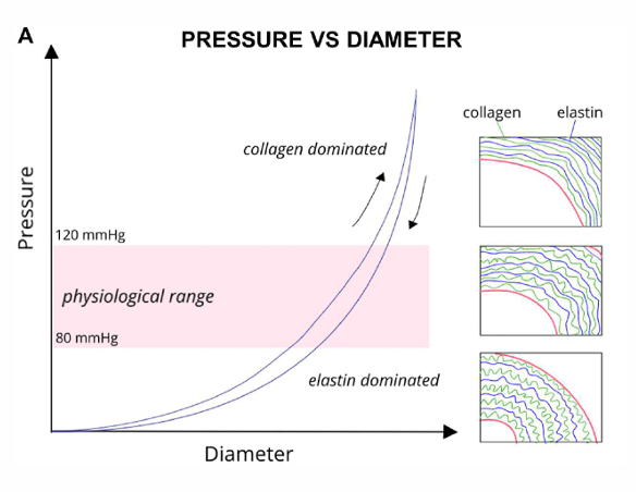
while at low applied stresses arteries are very easily deformed the arterial response becomes much stiffer at higher applied stresses.
As many other biological tissues, arteries exhibit an important variability in their mechanical response, both across species, organs, location or inter-individual
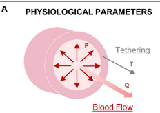
What are the physiological parameters in the artery
Physiological parameters (pressure, tethering) involved in the forces and stresses acting on the vascular wall.
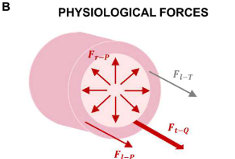
What are the physiological forces in the artery
Physiological forces acting on blood vessels resulted from the blood pressure and flow and surrounding tissues: radial force resulted from the blood pressure, longitudinal force resulted from the blood pressure, tangential force resulted from the blood flow, longitudinal force resulted from the tethering
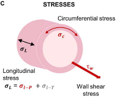
What are the stresses in the artery
Stresses generated in the vascular wall from the physiological forces: circumferential stress, longitudinal stress (due to pressure and tethering) and shear stress
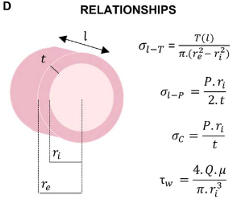
What are the relationships in the artery
Stresses relationships assuming incompressibility and uniform strain across the vascular wall
Uniaxial tensile testing
the artery is cut in either the circumferential or longitudinal direction and specimens are cut out of it and mounted on a standard tensile testing machine. this is to test the arterial mechanical properties
what are the important parameters for uniaxial tensile testing
preconditioning (5 cycles)
pre-load, to set a zero strain configuration
displacement rate
output (force, displacement/stretch/tissue strain if camera is available
end of test: peak strain, failure
What happens macroscopically for uniaxial testing
non linear response (toe, heel, linear)
longitudinal direction is way stiffer than the circumferential direction
important variability in the mechanical response, both across species, organs, location, or inter-individual
What happens microscopically for uniaxial testing
Collagen bundles in the tunica adventitia transition from a crimped to a straightened state under loading (fibre recruitment) allowing them to bear load alongside softer tissue components
Adventitial collagen bundles realign with load direction, facilitated by the elastin network. In the media, elastin lamellae, collagen fibers, and smooth muscle cells also reorient under load, aligning with the load direction
Collagen engagement begins earlier