Chapters 1,2,4,5 - Carter book
1/29
There's no tags or description
Looks like no tags are added yet.
Name | Mastery | Learn | Test | Matching | Spaced | Call with Kai |
|---|
No analytics yet
Send a link to your students to track their progress
30 Terms
Latent image
Captured the image but it has not be developed or digitized.
-Always sent to PACS
-formed by CR or PSP systems, when xrays interact with the PSP, they are captured and released in the reader
PSP
Photostimulable phosphor Plate
In CR you have a cassette → inside it is the PSP plate
Xrays hit the phosphor on the plate and store the image as trapped electrons
Then the plate goes into the CR reader, laser scans it, releases stored energy as blue light
Light gets turned into digital image
-Phospher is stimulated by light (ony in CR systems): uses storage phosphor plates to produce projection images
-Always sent to PACS
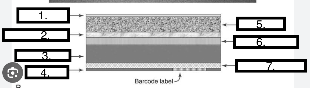
IP layers and purpose
IP - Imaging plate
Protective → protects phospher
Phosphor/active → PSP barium fluorohalide
Reflective → Sends light forward
Conductive→Absorbs/reduces static electricity
Color/light shielding→ Absorbs stimulating light
Support→Protects back of cassette
Backing→Protects back of cassette
Barcode→ Match image with patient
LABEL
Protective layer
Light reflective layer
Support layer
Backing layer
Phosphor layer
Conductive layer
Light shielding layer
Squence CR
IR placed in reader
Laser scans PSP, releasing the xrays captures by light
Photomultiplier amplies the light
Light is converted to digital signl via ADC
IP is erased by flooding it with light
ADC
Analog (light or electric) to digital converted (numbers)
TRANSLATOR
Photoconductors
Absorb xrays and convert to an electrical charge
-Direct system
Scintillators
Phosphors that produce light when absorbing xrays
-Indirect system
Direct Capture
Immediately converted into an electrical signal
-Photoconductor or radition conversion material = Amorphous selenium (aSE)
-Xrays converted to electrons and stored in TFT detectors
-TFT photosensitive array made up of DEL (detector elements or pixels), each pixel(DEL) contains a photodiode that absorbs electrons + generates an electric charge
-The FET (field effect transitor) sends the charge to image processor
Indirect capture
Two step process
-Uses a scintillator Gd202S or CSI that converts xrays to light
-Sent to a (Photodetector) photosensitive array made up of small pixels (Amphous silicon) to convert light to electrons
-TFT photosensitive array made up of DEL (detector elements or pixels), the DEL absorbs electrons + generates an electric charge
-The FET (field effect transitor) sends the charge to image processor
BLUE= same as direct capture
Gd202S (Turbid)
-Unstructed
-Allows light to escape
-Very rugged so good for portables
-Scintillator
CSI
-More popular
-Structured, thin needles
-Greater detection
-Once very delicate but improvements have been made
-Scintillator
Analog capture
Exposure latitude is based on characteristic response of film which is NONLINEAR
Digital capture on a graph?
Linear capture (straight line)
DQE
Detective Quantity Effeciency - MAs
How efficiently a system converts the X-ray input signal into a useful output image
DQE is a measurement of the percentage of X-rays that is absorbed when they hit the detector.
The takeaway - is the phosphors used in any system (film/PSP systems/Cassettless DR systems) and the way they interact and release x-ray energy impacts DQE
Direct - Highest
Indirect
CR - Lowest
Pixel
the smallest element in a digital image
Pixel bit depth
-the number of bits within a pixel
-Each pixel contains pieces of information
Matrix
square arrangement of numbers in columns or rows, numbers correspond to discrete pixel values
Field of view (FOV)
the amount of body part included on the image
-Larger FOV = more area is imaged
-Change FOV = no change is matrix size
Relationship = Spatial resolution and pixel size
Increase spatial resolution = Decreased pixel size
Relationship = matrix and pixel size
increased matrix size= decreased pixel = increase spatial resolution
Relationship = matrix and number of pixels
increased matrix size = increase number of pixels
Relationship = matrix and spatial resolution
Increase matrix size = Increased spatial resolution
Kstd - air kerma or gray
Standard radiation exposure
measurement of the beam afer it is filtered
Kind radiation
What strikes the IR or the detector
-Measurement of the radiation that was the incident exposure on the IR for the exposure
KTGT
Target exposure Index
target values established by the equipment system vendor- what is supposed to hit the detector
Deviation Index
kind/KTGT
To determine if an image is overexposed or underexposed
want it to be 0
Spatial resolution is affected by what?
SID
OID
Tube angle
Focal spot size
DICOM
Digital imaging and communications in medicine : PACS can accept any image that is DICOM
Standard to make sure: images can open at any hospital, patient info stays attached, PACS systems know how to store images, and different machines all communicate the same way - Using the same format for all
CR - Konica system
Indirect system
Range 100-300