Female Reproductive System
1/43
There's no tags or description
Looks like no tags are added yet.
Name | Mastery | Learn | Test | Matching | Spaced | Call with Kai |
|---|
No analytics yet
Send a link to your students to track their progress
44 Terms
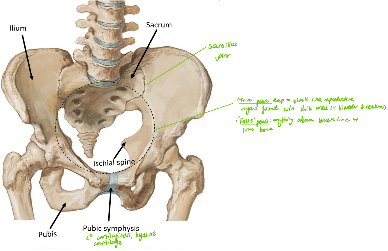
Label the following diagram
What is the role of the pelvic inlet
Separates the true pelvis from the false pelvis
True pelvis: structures below the pelvic inlet, including the reproductive organs within this area, bladder & r3ctum
False pelvis: anything above the pelvic inlet to the iliac bone
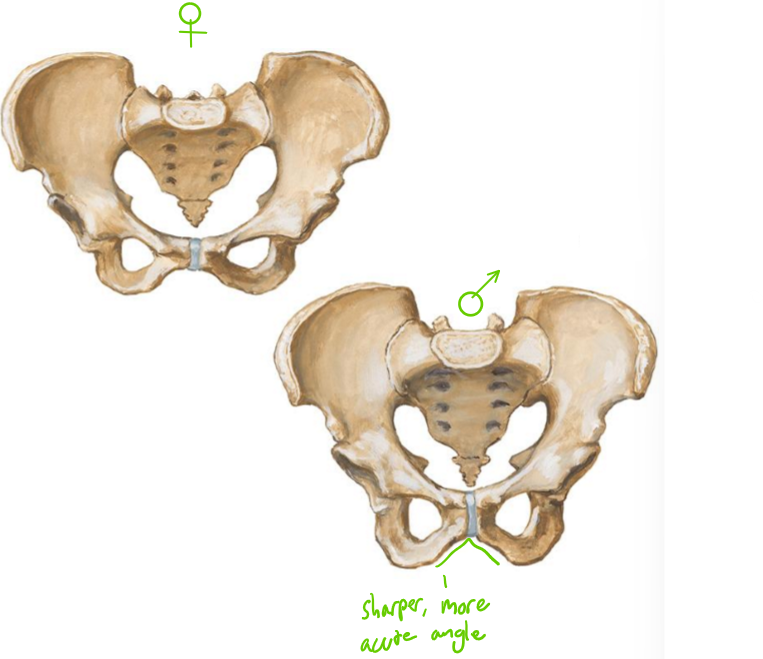
Name the 4 main pelvic shape variations
Gynaecoid: Wider, more round
most common type in females
Android: narrower, more tapered pelvis
Anthropoid: oval-shaped pelvis
Platypelloid: wider side-to-side, flatter front-to-back
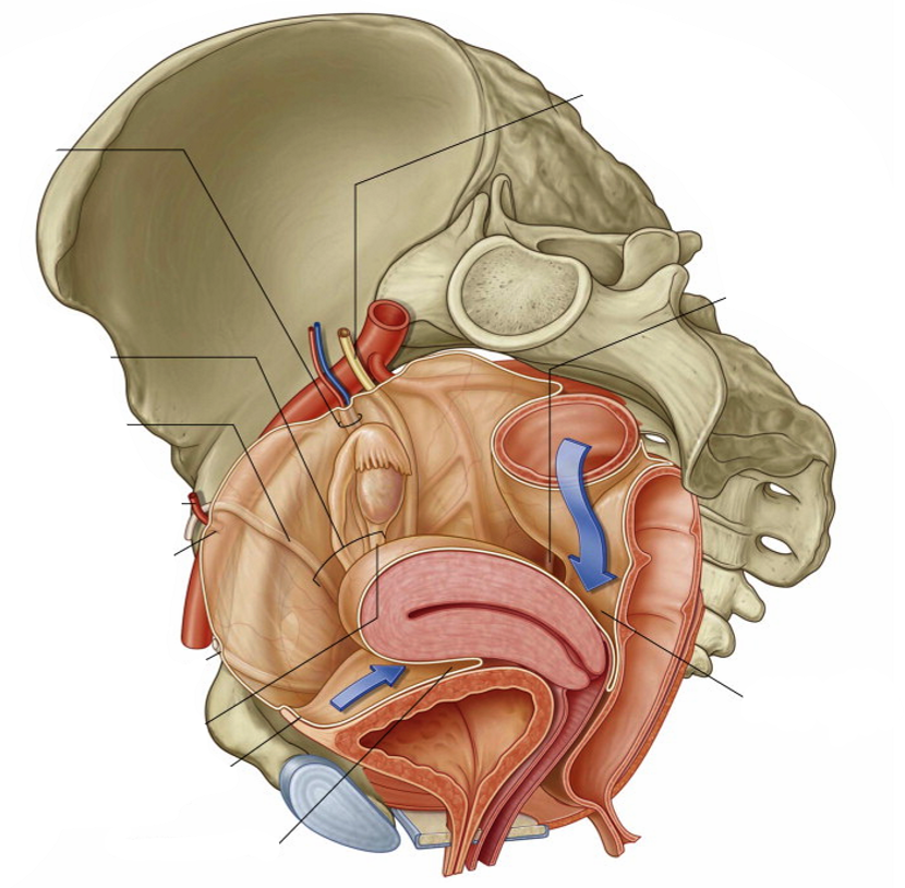
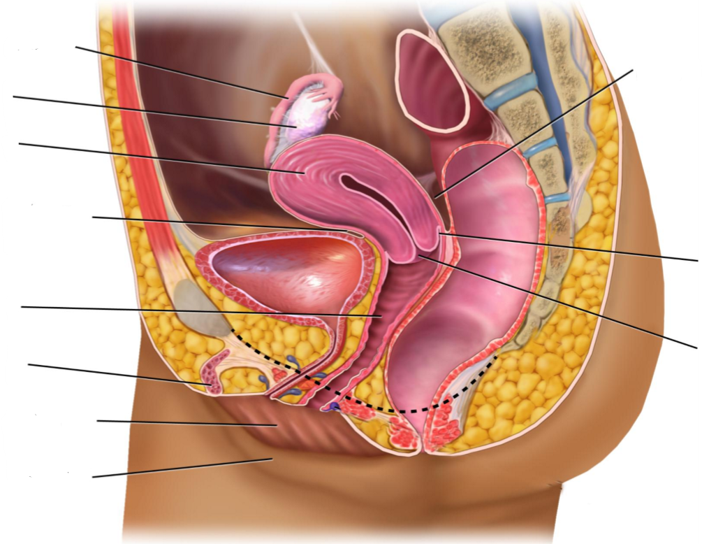
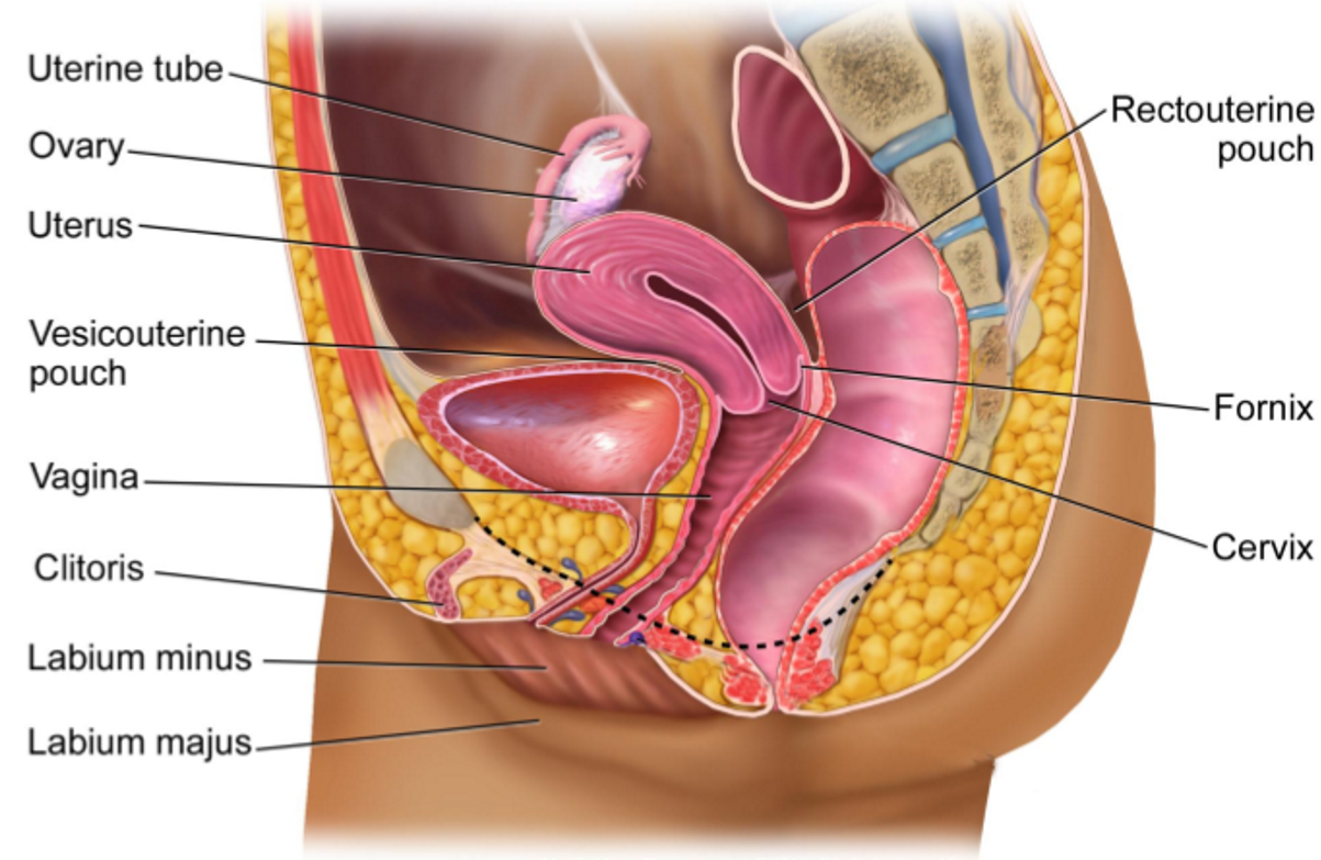
Label the following diagram
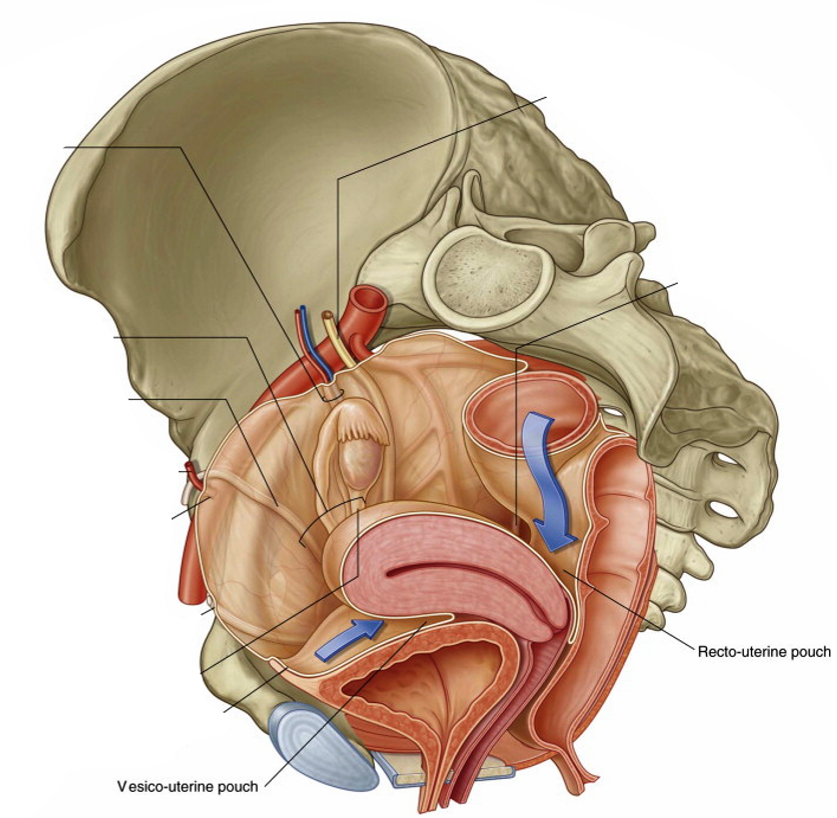
What are the 2 peritoneal pouches & why are they clinically relevant?
Peritoneal pouches: where the peritoneum reflects over pelvic organs, forming pouches
Vesicouterine pouch: the pouch between the bladder & uterus
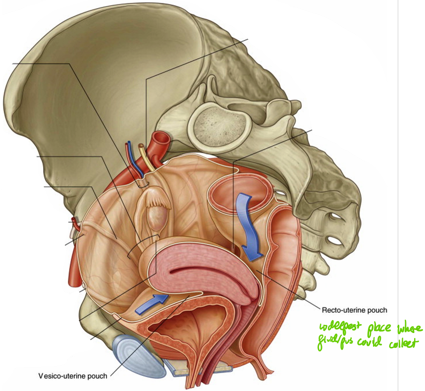
Rectouterine pouch (pouch of Douglas); the pouch between the uterus & r3ctum
Clinically relevant due to fluid accumulation sites (more common in rectouterine poch - deeper)
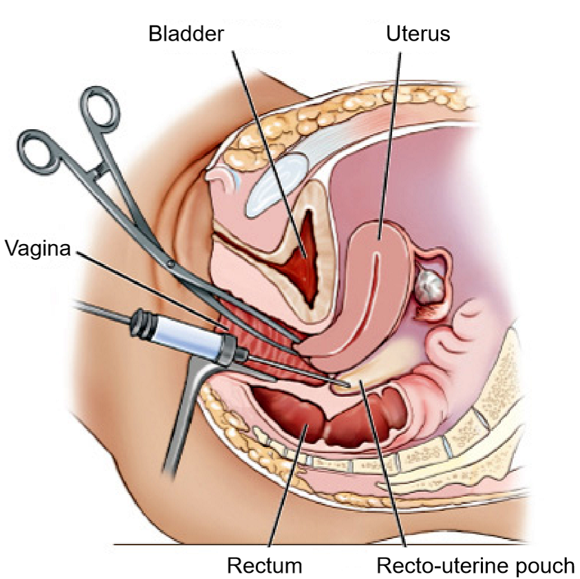
How can fluid be drained from the rectouterine pouch?
By puncturing the back of the vag!na with a probe to drain fluid
What are the ovaries?
Paired almond-shaped gonads located in the lateral pelvis, which are the site of oocyte production & hormone (oestrogen & progesterone) secretion
Positioned near lateral pelvic wall
Connected to uterus via ligaments (mesentery) → mobility
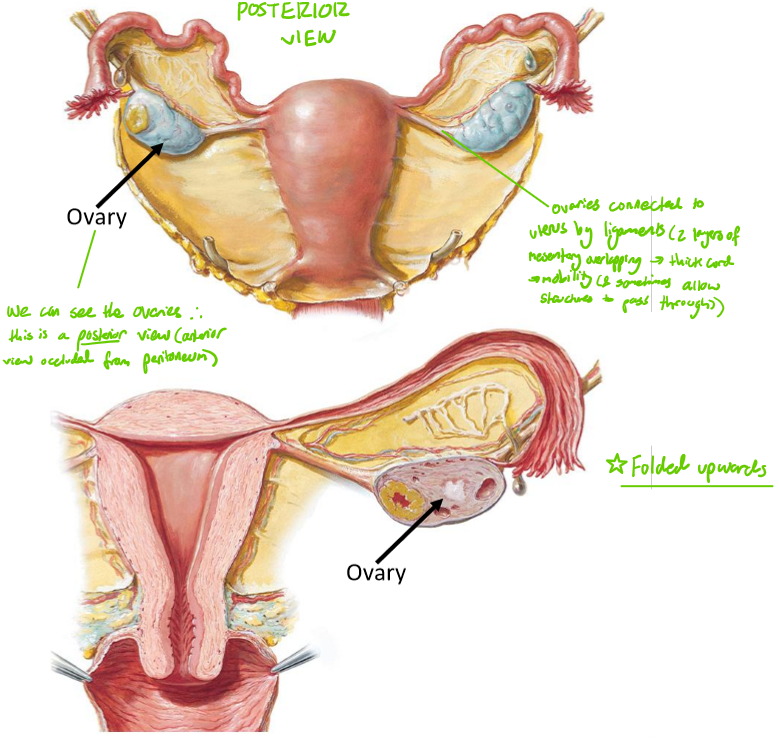
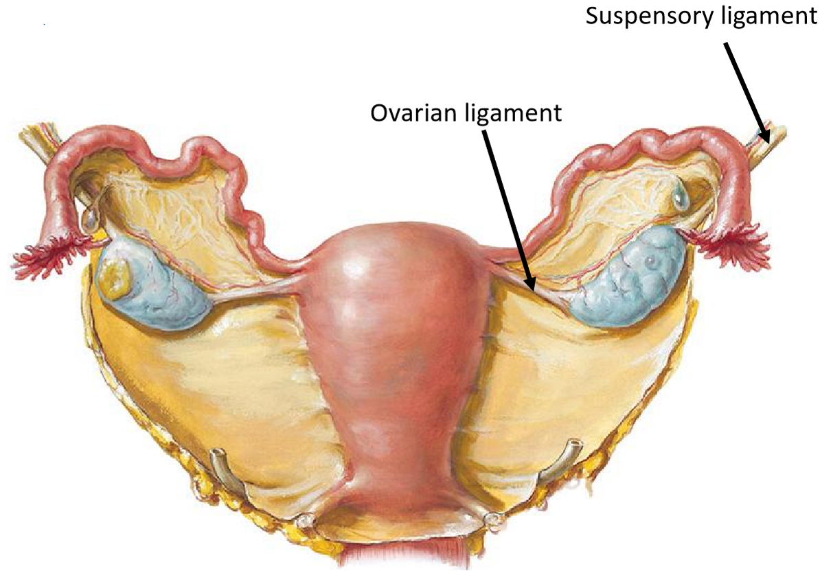
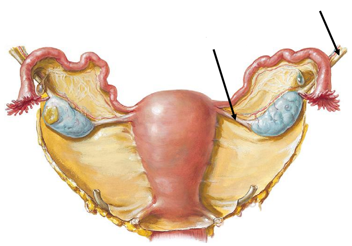
Describe the 2 attachments of the ovaries
Ovarian ligament: connects ovary to uterus
Suspensory ligament: connects ovary to lateral pelvic wall
Contains ovarian vessels = major blood supply
*Structures help maintain ovarian position
Label the following diagram

For the uterine tubes, state:
What they are
What they are also known as
Where they are located
What they open into
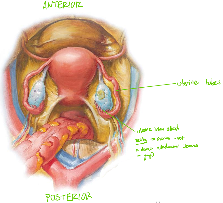
Uterine ligaments = paired muscular tubes extending from uterus → ovary
AKA Fallopian tubes (though this term is not used anymore)
Located in superior aspect of broad ligament
Open into peritoneal cavity near ovary
Not actually connected to ovary; ‘waft’ the egg from the ovary into the tube
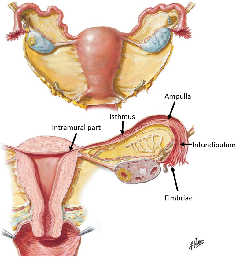
Name & describe the 5 regions of the uterine tube
Fimbriae: finger-like projections near the ovary
Infundibulum: funnel-shaped lateral end
Ampulla: widest & longest segment (site of fertilisation)
Isthmus: narrow medial segment
Intramural part: passes through uterine wall

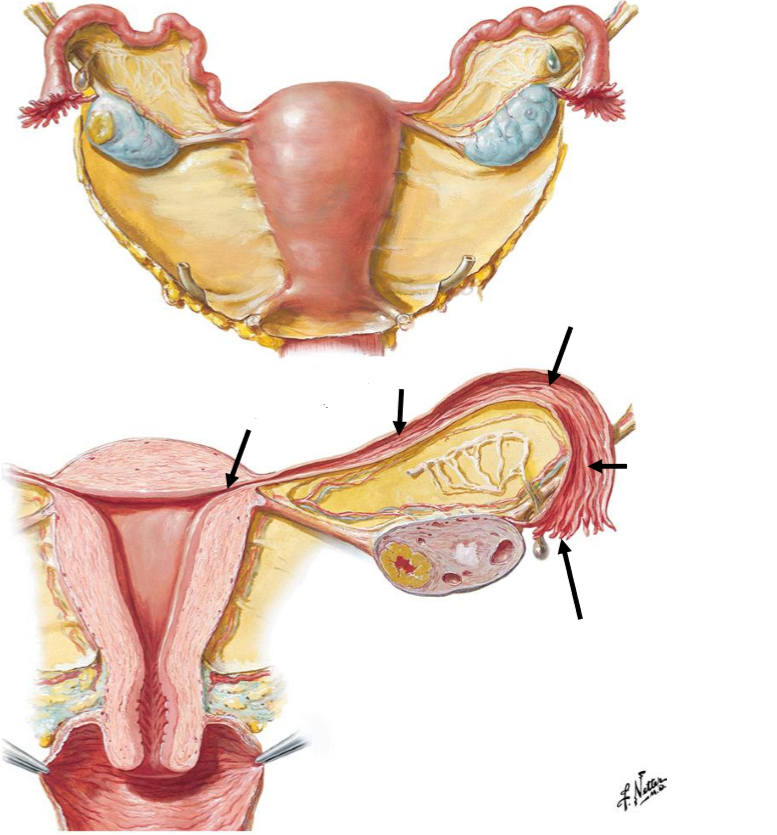
Label the following diagram of the regions of the uterine tube
Where can ectopic pregnancies occur?
Anywhere outside the uterus;
Abdominal cavity b/w ovary & uterine tube
Anywhere along uterine tube
Fimbriae
Infundibulum
Ampulla
Isthmus
Intramural part
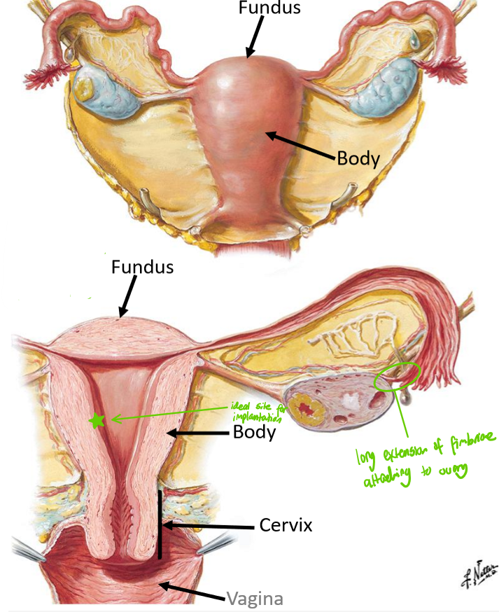
For the uterus, state:
What it is & where it is located
What it is divided into
Key role
Hollow muscular organ in midline pelvis
Divided into:
Fundus: superior region
Can be felt after third month; measure height of foetus
Body: main portion
Cervix: inferior portion
Directly connected to uterus
Premature births if placenta (highly vascular) grows over base cervix (if implantation occurs to low)
Key role: site of implantation

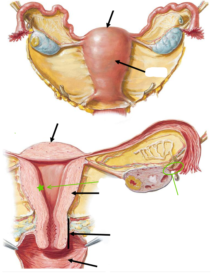
Label the following diagram of the key regions of the uterus
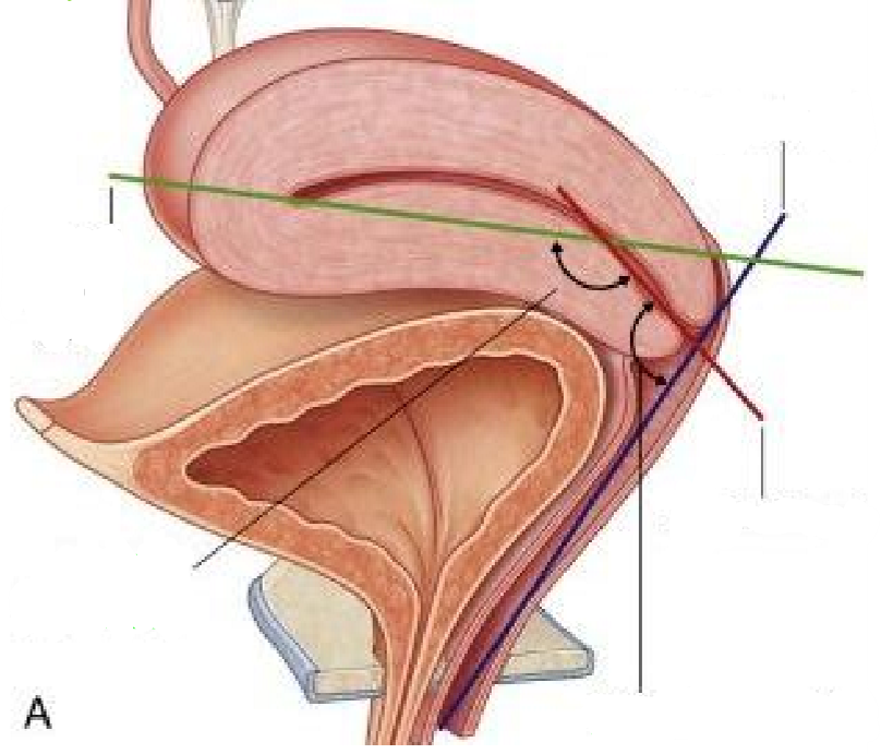
Label the following diagram of the axes & angles of the uterus
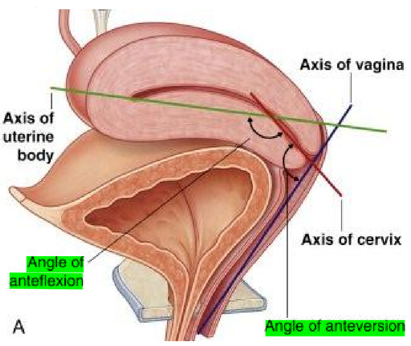
Describe the typical position of the uterus, in terms of:
Relation to vag!na
Relation to bladder
What it is position in between
What the cervix opens into
Normally anterverted (tilted forward relative to vag!na)
Normally anteflexed (flexed forward over bladder)
Positioned b/w bladder (anterior) & r3ctum (posterior)
Cervix opens into anterior wall of vag!na

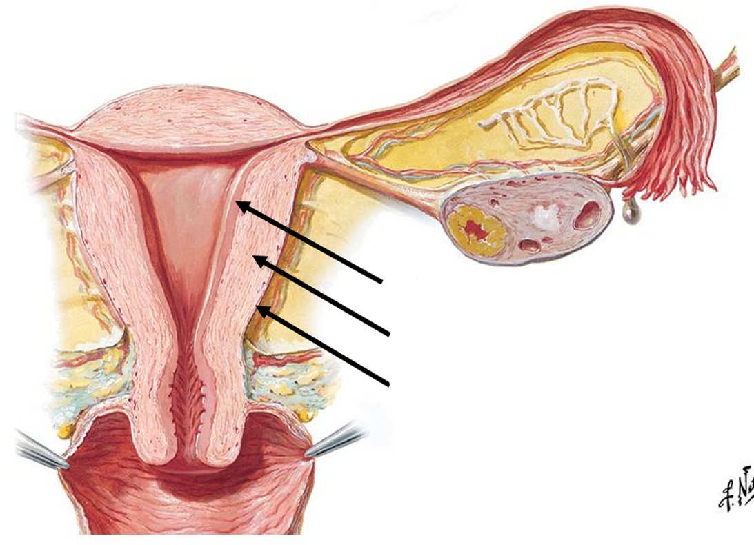
Label the following diagram of the uterine wall layers
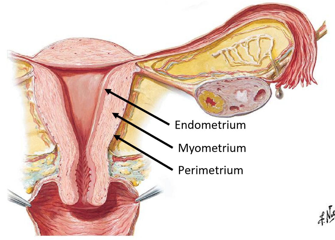
Name & describe the uterine wall layers
Endometrium: inner mucosal layer
Site of implantation
Undergoes cyclical changes (menstruation)
Myometrium: thick, smooth muscle layer
Dominant layer
Leads to uterine contractions
Perimetrium: outer serosal layer (i.e. visceral peritoneum)

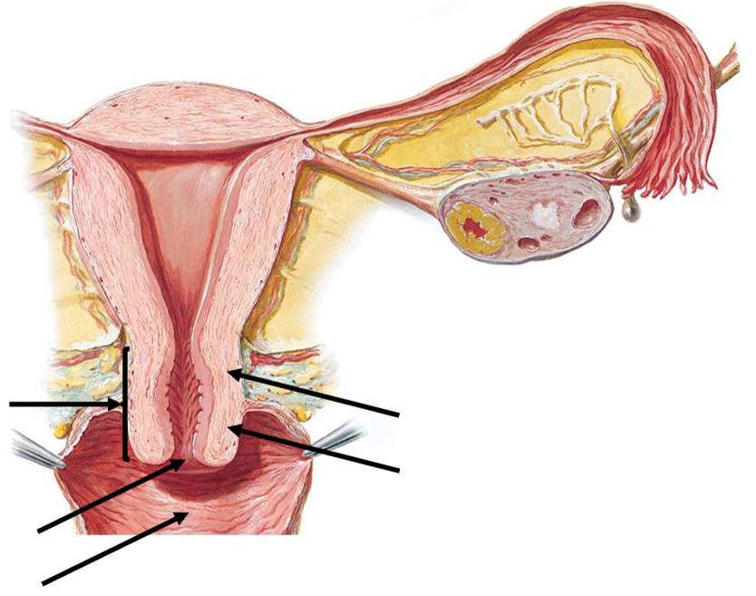
Label the following diagram of the cervix & surrounding structures

For the cervix, state:
What it is
What it contains
What it is divided into
What is opens into
Inferior, narrow portion of uterus
Contains cervical canal
Divided into supravaginal & vaginal parts
Opens into vag!na via external os

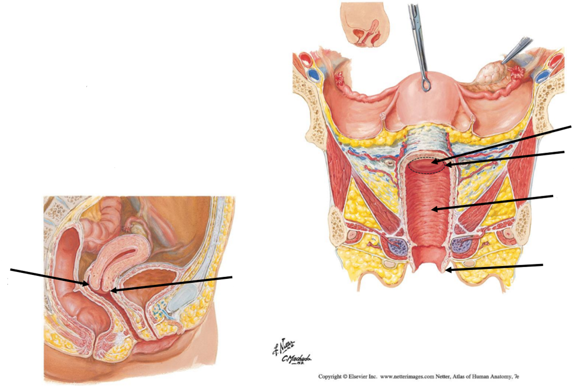
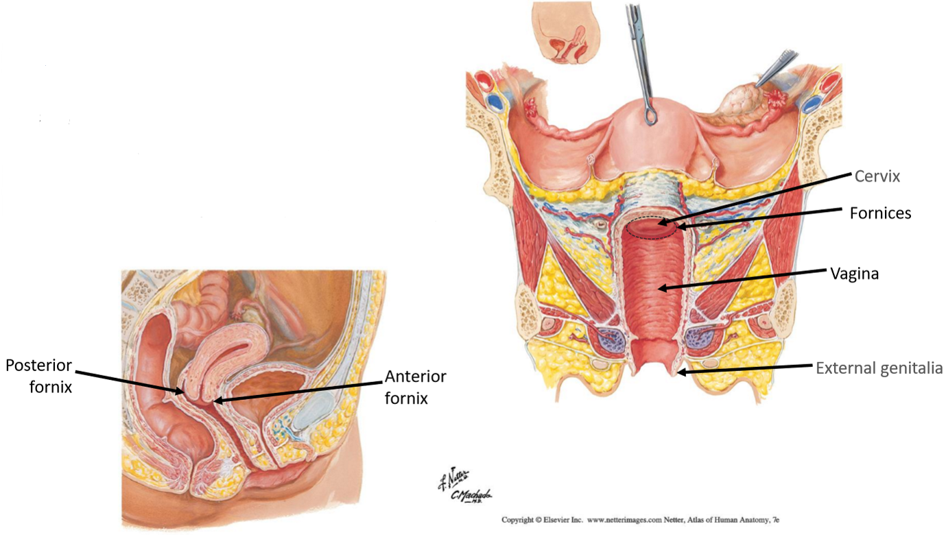
Label the following diagram of the vag!na
For the vag!na, state:
Its structure
Where it extends
Where it is positioned
What it surrounds & what this forms
Fibromuscular tubular structures
Extends from cervix → external genitalia
Posterior to bladder & urethra
Surrounds cervix → vaginal fornices

What are the vaginal fornices?
Depressions around the vaginal part of the cervix

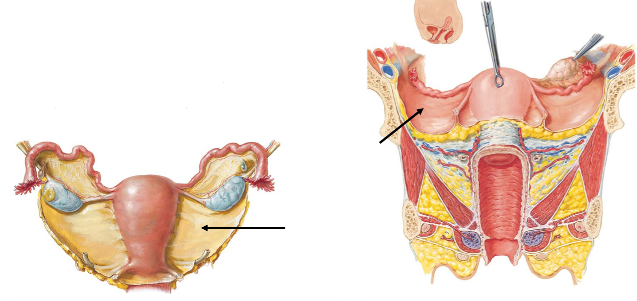
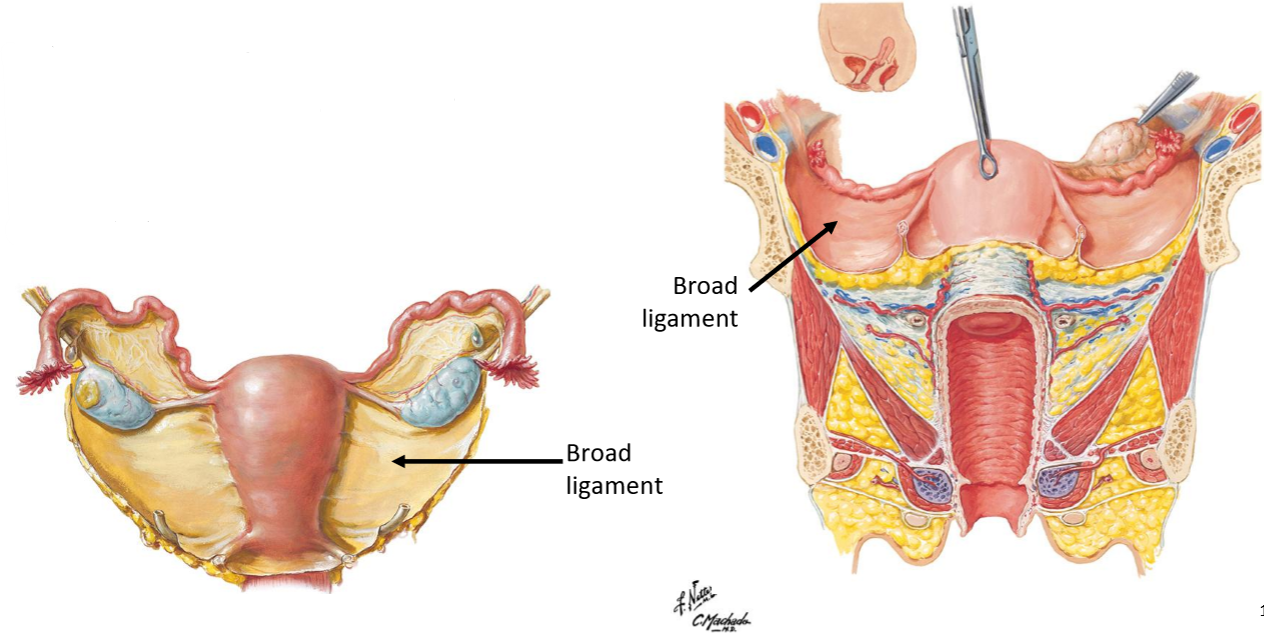
Label the following diagram

For the broad ligament, state:
What it is
Where it extends
Its role
What it contains
Double-layer of peritoneum
Extends from lateral uterus to pelvic walls
Provides supportive mesentery-like structure → keeps uterus in the right location while allowing for some movement during pregnancy
Contains vessels & uterine tube
Aside from the broad ligament, what are the other 3 main ligaments in the female reproductive anatomy & what are their roles?
Round ligament OF THE UTERUS: maintains uterus anteversion
Closed tube, doesn’t carry anything (= to spermatic cord in males)
Goes through inguinal canal towards external genitalia
Runs within the broad ligament
Ovarian ligament: connects ovary to uterus
Suspensory ligament: carries ovarian vessels
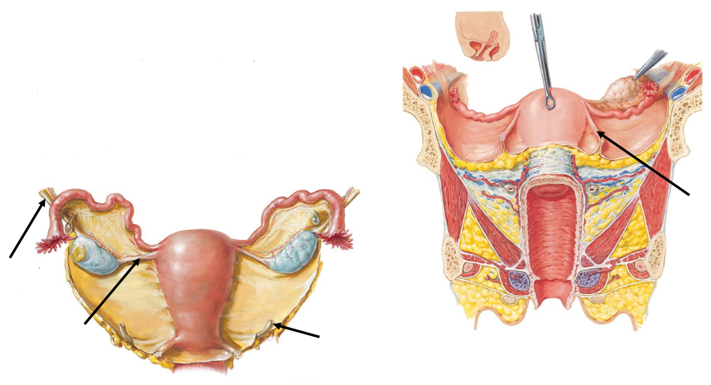
Label the following diagram
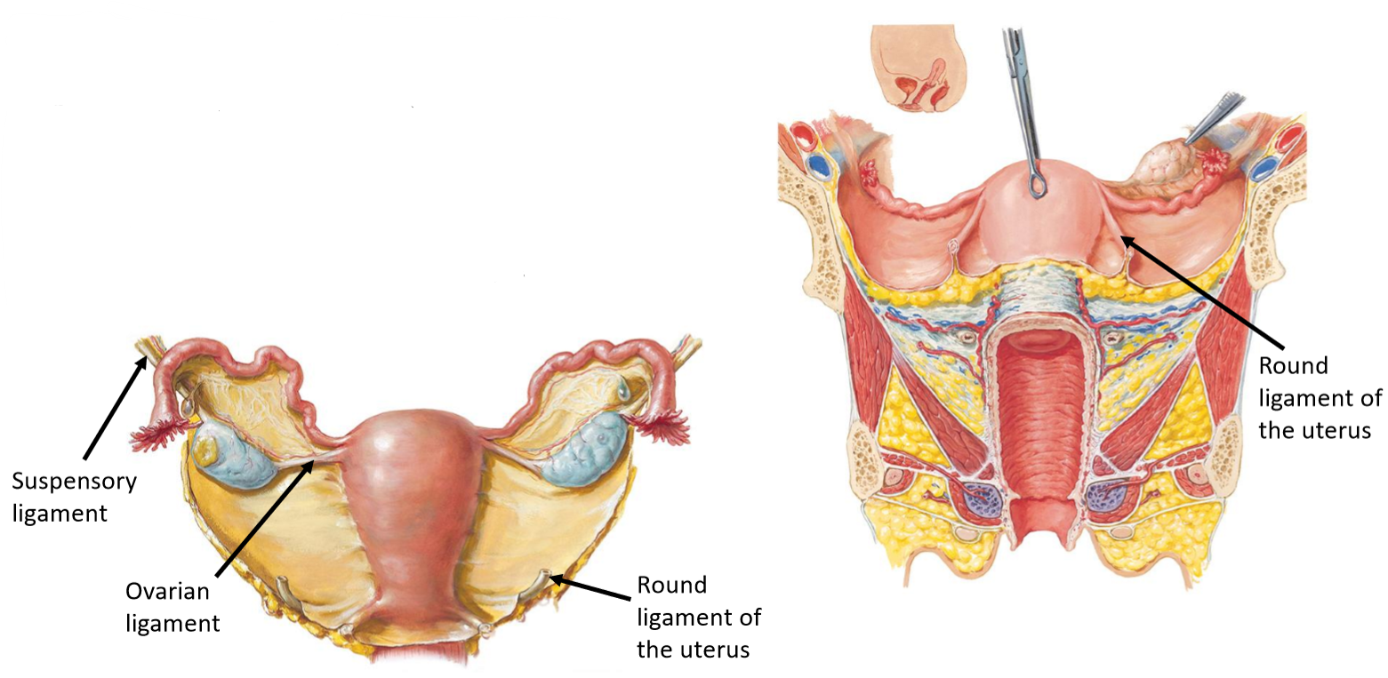
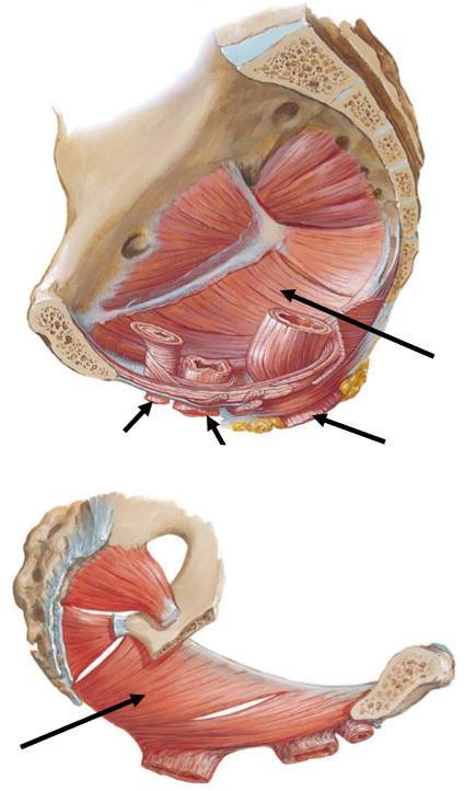
Label the following diagram

For the pelvic floor, state:
What it forms
What it is composed of
What it does
Key muscle group
Forms inferior boundary of pelvic cavity
Composed of muscles & connective tissue (seals off bottom of pelvis)
Supports pelvic organs & keeps abdominal organs in place
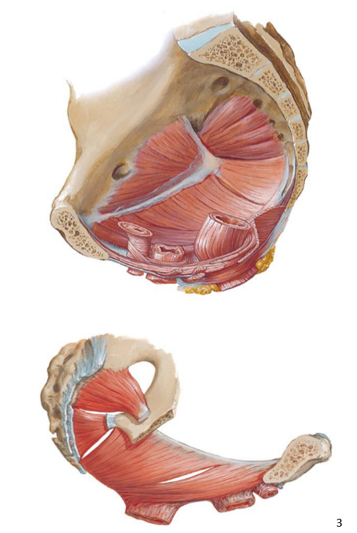
Key muscle group = levator ani (3 subgroups of muscles)
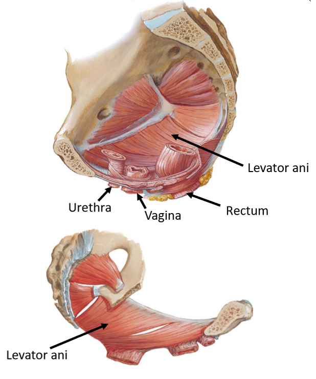
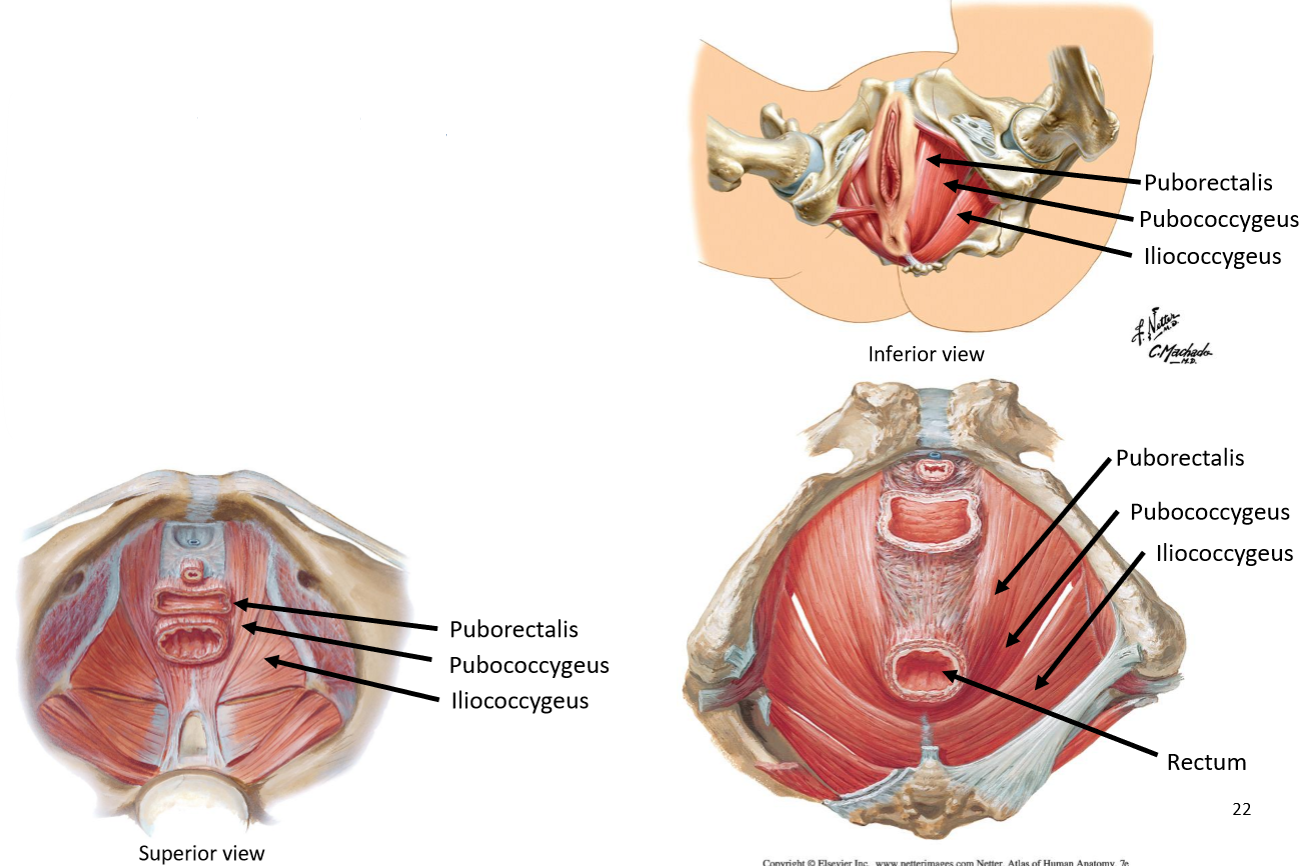
Name the 3 levator ani muscles & where they come from
Puborectalis: pubic bone
Concentric contraction → functional sphincter → close rectum
Pubococcygeus: moves towards bone @ back (doesn’t form a sling)
Iliococcygeus: raphe of iliac area → coccyx bone
*All attach to pubic bone

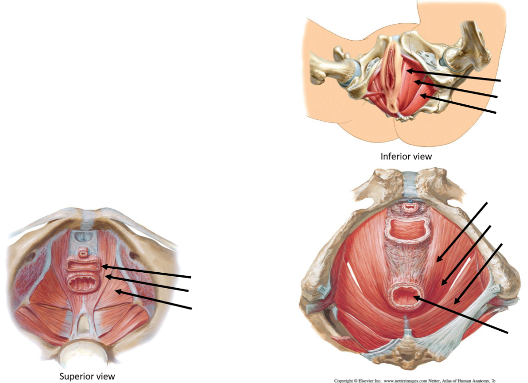
Label the following diagram of the levator ani muscles
Describe the key function of the pelvic floor
Supports pelvic organs
Maintains continence (urine & faeces)
Puborectalis acting as functional ‘sling’ around r3ctum → maintain anorectal angle & contribute to continence
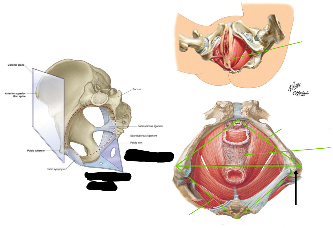
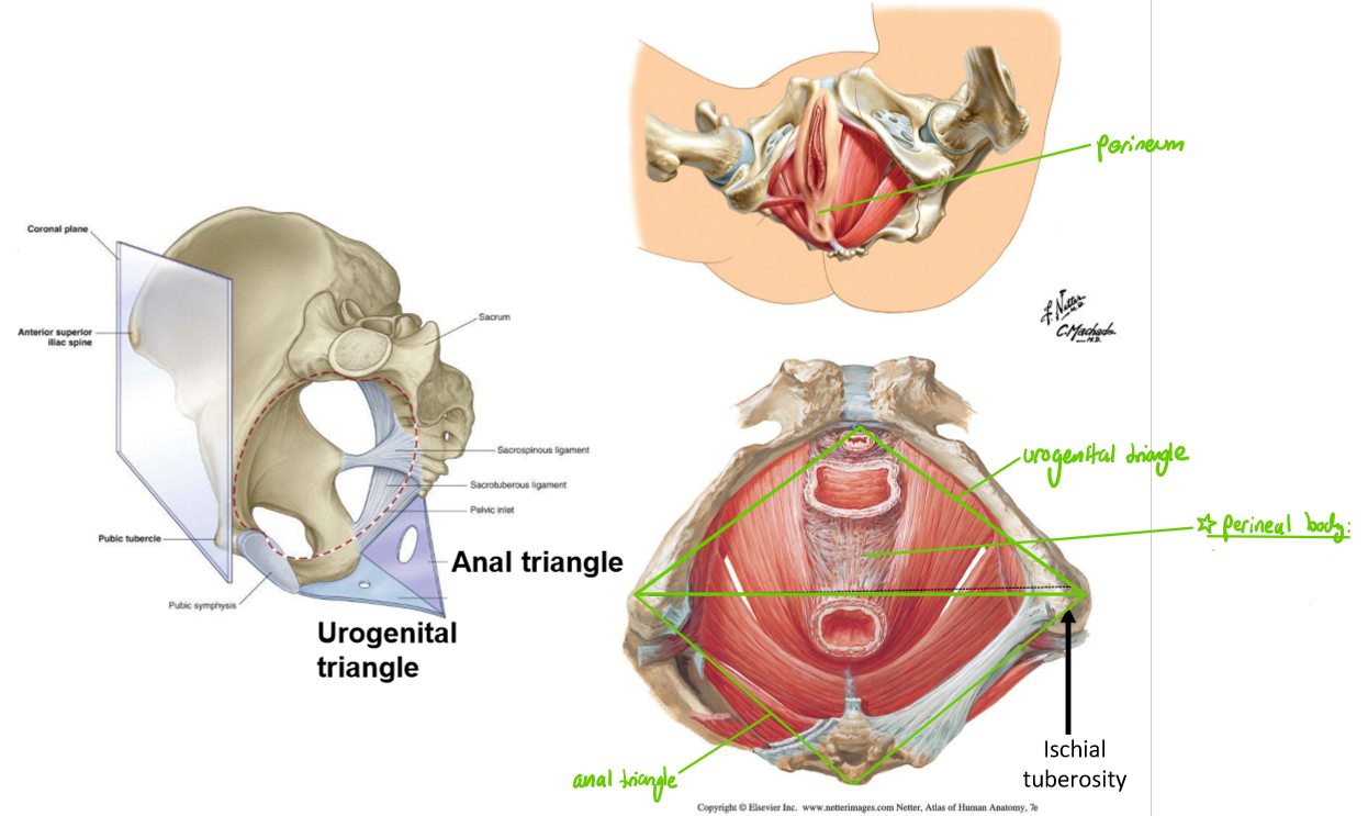
What is the perineum & what does it do?
Diamond-shaped region inferior to the pelvic floor
Supports continuity b/w pelvic cavity & the exterior
Important route for vessels
State what the ischial tuberosities of the pelvis separates the perineum into, & what each of these features contain
Divided into:
Urogenital triangle: contains external openings of urethra & vag!na
An@l triangle: contains external openings of @nus

What is the perineal body, & what is a major clinical implication of this area during birth?
The major anchorage point for levator ani
Birth → becomes stretched → tears → loss of support → involuntary defection & urination
Might have to be manually cut during pregnancy so that the baby’s head has enough space to pass, & then be easily stitched back

Label the following diagram
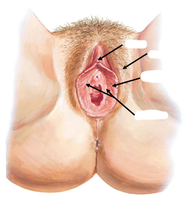
Label the following diagram of the vulv@
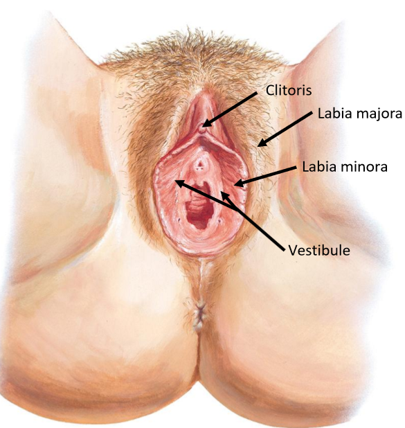
Name & describe the 4 key regions of the vulv@ (external genitalia)
Lab!a majora: outer skin folds
Where the round ligament of the uterus inserts within
Protects deeper genitalia
Lab!a minora: inner mucosal folds
Glands → lubricant
Do not grow hair
Cl!toris: anterior erectile structure
Similar to male pen!s
Vestibule: area b/w lab!a minora, containing urethra + vag!nal opening

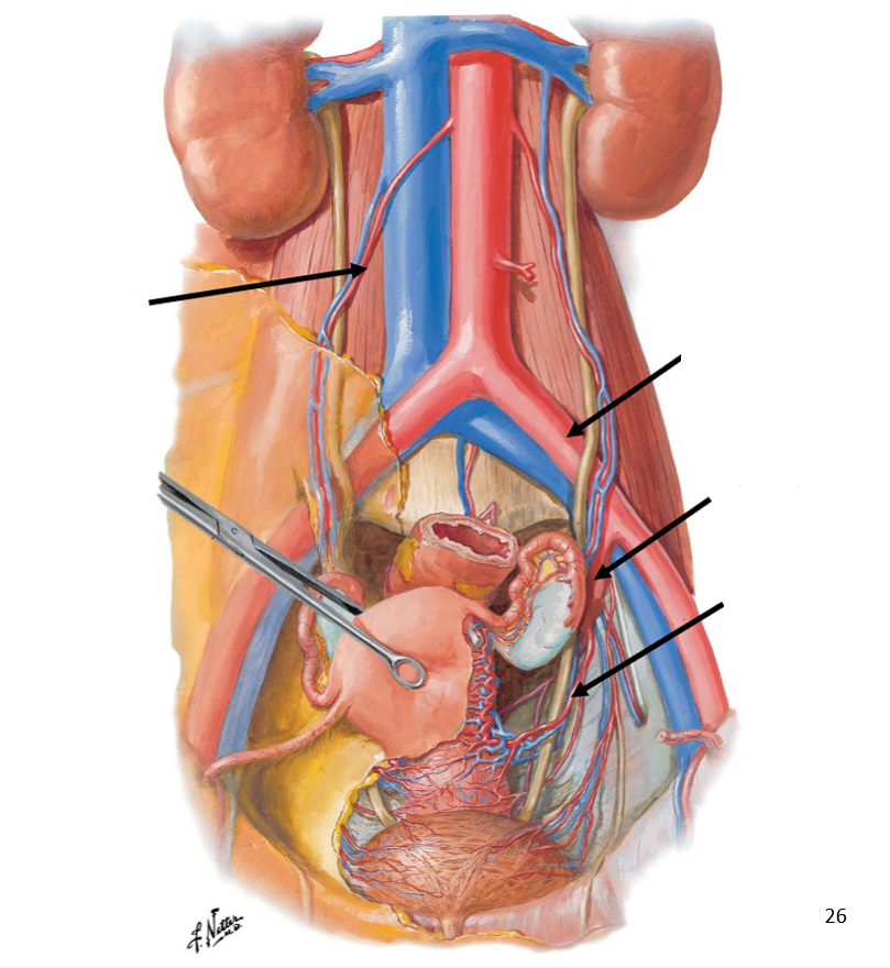
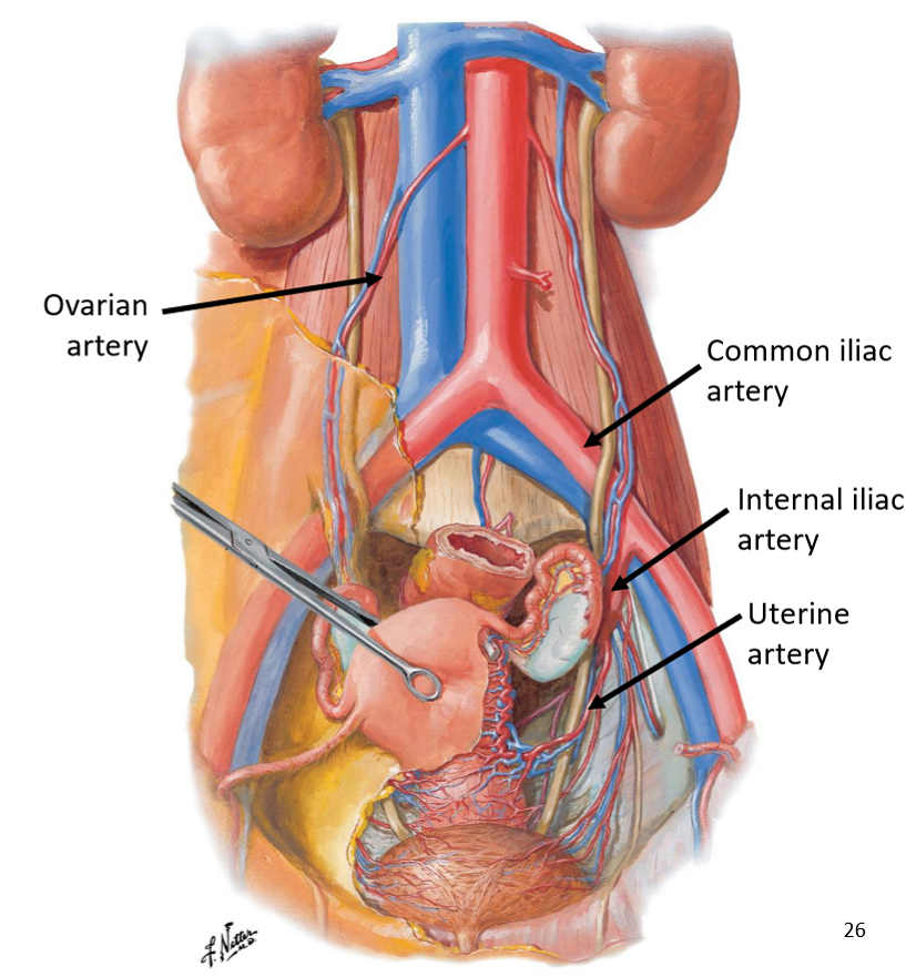
Label the following diagram of the vascular supply of the female reproductive system

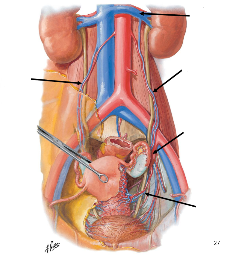
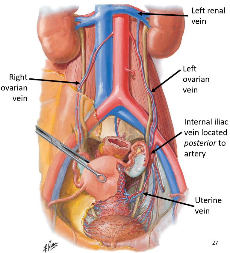
Label the following diagram of the venous drainage of the female reproductive system
For the vascular supply of the female reproductive system, state:
the 2 main arteries and where they arise from
Where vessels run
Where the arteries supply
Main arteries:
Uterine artery arising from internal iliac artery
Gives rise to vag!nal artery
Ovarian artery arising from gonadal paired vessels of abdominal aorta
Runs over common iliac vessels & pelvic brim
Vessels run within supporting ligaments
Supply corresponds to the organ location
For the venous drainage of the female reproductive system, state:
Where veins follow
Where the right ovarian vein drains to
Where the left ovarian vein drains to
What the uterine drainage follows
Veins follow arterial pathways
Right ovarian vein → inferior vena cava
Left ovarian vein → left renal vein
Uterine drainage follows internal iliac system

State the anatomical pathway of the female reproductive tract
Ovary → uterine tube → uterus → cervix → vag!na