Medical physics
 Call Kai
 Learn
Learn Practice Test
 Spaced Repetition
 Match
Match Flashcards
 Knowt Play
1/133

Earn XP
Description and Tags
134 Terms
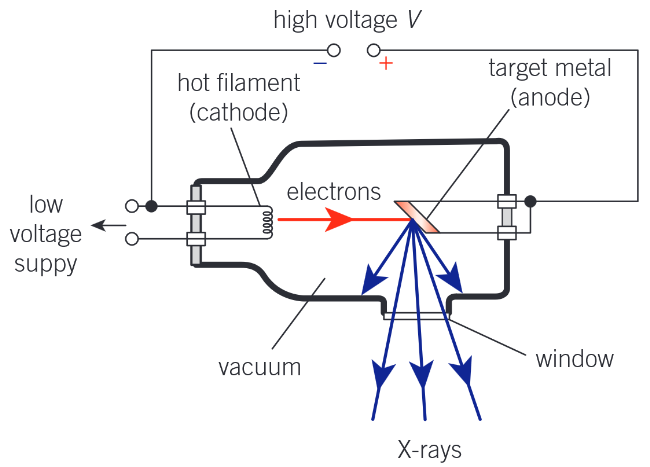
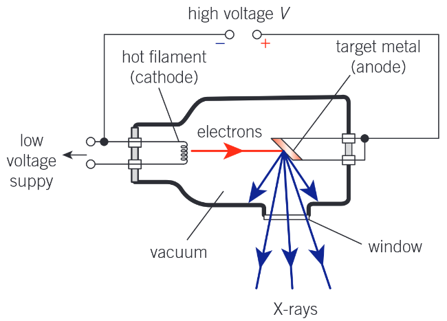
Diagram showing how X-rays are produced
Key elements:
High voltage power supply
Hot filament (cathode)
target metal (anode)
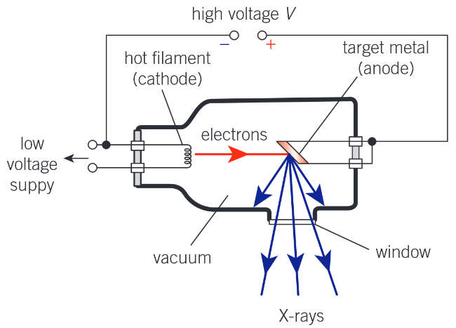
What is the purpose of the high voltage power supply when producing X-rays?
To create a large p.d. between the cathode and anode to accelerate electrons towards the anode.
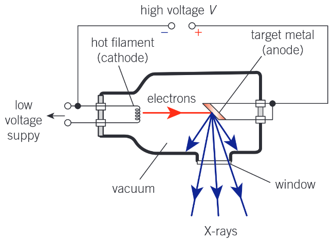
What is the purpose of the hot filament / heater when producing X-rays?
To produce electrons by thermionic emission
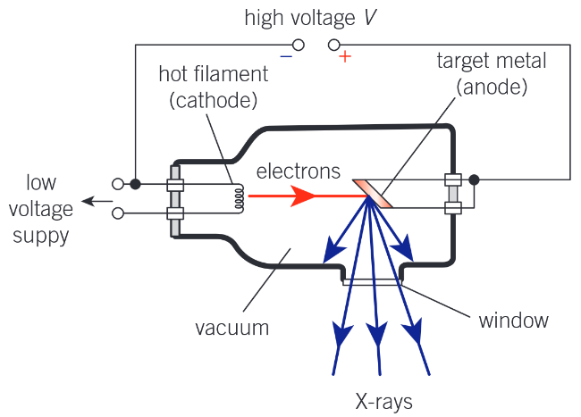
What is the purpose of the target metal when producing X-rays?
To decelerate the electrons and cause them to produce X-ray photons
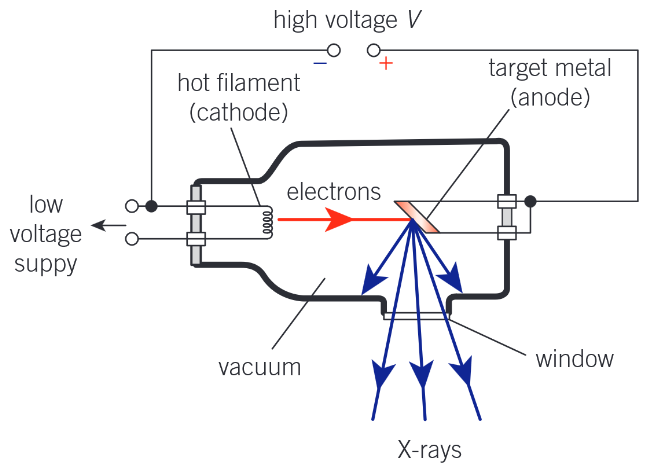
What is the purpose of the led shield around an X-ray tube?
To shield the radiographer from X-rays by aborbing X-rays not passing through the window
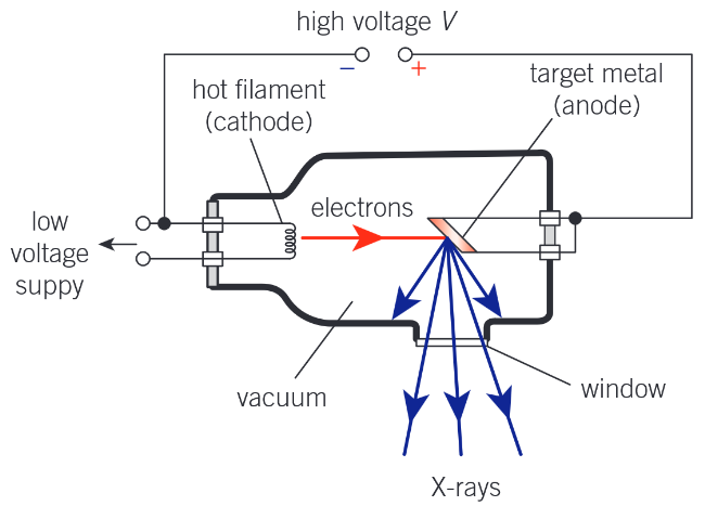
How is a collimated beam of X-rays produced?
The cathode is a heater which produces electrons by thermionic emission
These electrons are accelerated towards the anode by a large p.d. from the high voltage power supply
The anode is the target metal which decelerates the electrons, causing them to produce X-ray photons
The lead shielding aborbs all X-rays other than those passing through the window, producing a collimated beam of electrons
What range of potential difference is used to generate X-rays for imaging?
30 to 120 kV
Derive the equation for shortest wavelength produced by a X-ray machine with accelerating p.d. V
Work done on electrons = QV = eV
One electron releases one X-ray photon ⇒ maximum energy of X-ray photon = maximum KE of electron from principle of conservation of energy
Energy of X-ray photon = hc / λ ⇒ eV = hc / λ ⇒ λ = hc / eV
What is the reletioship between accelerating p.d. in a X-ray tube and the minimum wavelength produced?
λmin ∝ 1/V i.e. they are inversely proportional
How does increasing the current in an X-ray tube affect the X-rays produced?
It increases the intensity
No effect on wavelength
How does decreasing the current in an X-ray tube affect the X-rays produced?
It decreases the intensity
No effect on wavelength
How does increasing the voltage in an X-ray tube affect the X-rays produced?
No effect on intensity
Reduces wavelength
How does decreasing the voltage in an X-ray tube affect the X-rays produced?
No effect on intensity
Increases wavelength
Why is the X-ray tube evacuated?
So that electrons pass through the tube without interacting with gas atoms.
What are the two ways that electron energy is released on hitting the target anode?
X-rays (<1%)
Increasing the thermal energy of the anode
How is the anode cooled?
Often oil is circulated to cool the anode
Alternatively the anode is rotated to spread the heat over a large surface area
How are X-rays collimated?
The anode is shaped to emit X-rays in the desired direction
A lead shield absorbs any X-rays emitted in other directions
Sketch a typical X-ray spectrum of intensity vs wavelength for an X-ray tube running at a particular supply voltage
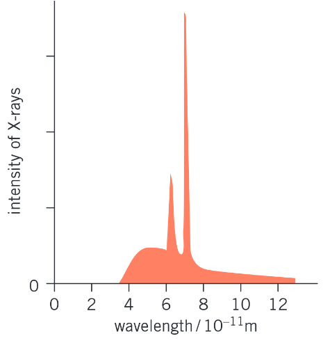
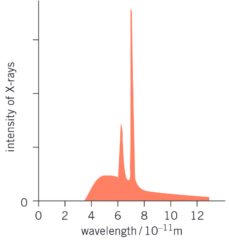
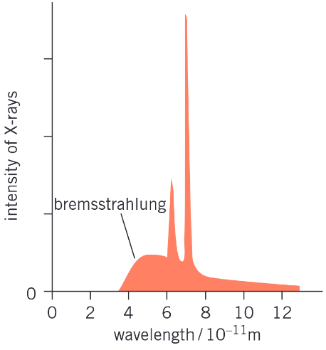
Identify the bremsstrahlung on this X-ray spectrum
The background hump - not the 2 spikes
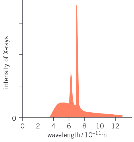
Identify the K lines on this X-ray spectrum
The 2 spikes and not the background hump
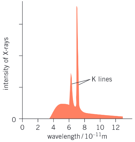
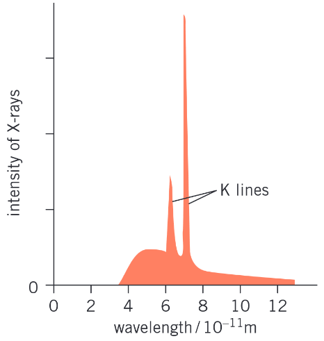
Describe how K-lines are produced
The electrons incident on the anode can remove electrons in the metal atoms close to the nuclei, creates gaps in the lower energy levels.
These gaps are quickly filled by electrons dropping from higher energy levels.
These transitions release photons of specific energies and therefore wavelengths, creating spikes in intensity at these wavelengths
What are the four X-ray attenuation mechanisms?
Simple scatter
Photoelectic effect
Compton scattering
Pair production
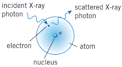
Simple scatter of X-rays (diagram and description)
The X-ray photon interacts with electron in the atom, but has less energy than the work function, so the X-ray photon simply bounces off without any change to its energy.
What is the range of energies over which simple scattering occurs?
X-ray photons with 1-20 keV
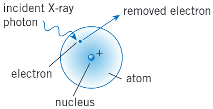
Photoelectric effect with X-rays (diagram and description)
The X-ray photon is absorbed by one of the electrons in the atom. The electron uses this energy to escape from the atom.
What is the range of energies over which the photoelectric effect occurs?
X-ray photons with 20-100 keV
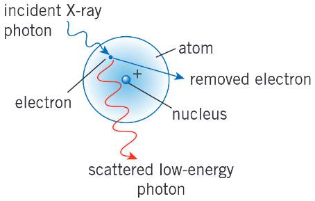
Compton scattering (diagram and description)
An electron is ejected from atom
The photon is scattered with lower energy.
What is the range of energies over which compton scattering occurs?
X-ray photons with 0.1-5 MeV
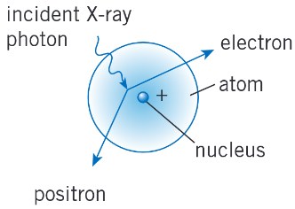
Pair production from X-rays (diagram and description)
An X-ray photon interacts with the nucleus of the atom. It disappears and the electromagnetic energy of the photon is used to create an electron and its antiparticle, a positron.
What is the range of energies over which pair production occurs?
X-ray photons with >= 1.02 MeV
Which attenuation mechanism is most likely in a hospital X-ray machine?
Photoelectric effect
Hospital X-ray machines typically used 30-100 kV producing X-ray photons with <100 keV
Hence the photoelectric effect is dominant
Which attenuation mechanism is least likely in a hospital X-ray machine?
Pair production as this requires a p.d. > 1 MV which is ten times higher than the typical p.d.s used in hospital X-ray machines.
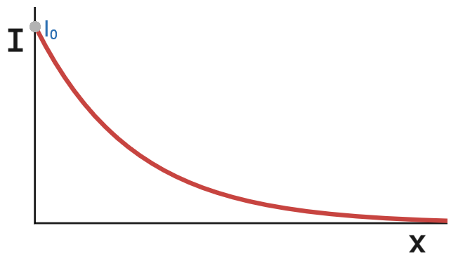
State the equation for transmitted intensity of X-rays in a given substance. Identify each quantity
I = I0 e-μx
I is the transmitted intensity
I0 is the innitial intensity before absorption
μ is the attenuation coefficient of the substance
x is the thickness of the substance
What is the attenuation coefficient also known as?
The absorption coefficient
The higher the attenuation coefficient, the ___ X-rays a material will aborb per unit length
more
Draw a sketch of transmitted intensity of X-rays I versus thickness of substance x
What is Z?
Atomic number - the number of protons in a nucleus
How is μ related to Z?
μ ∝ Z³
What is the average Z of soft tissue?
7
What is the average Z of bone?
14
What is the atomic number (Z) of iodine?
53
What is the Z of barium?
56
What is a contrast medium?
A subtance that improves the visibility of a material (like soft tissue) in an X-ray image
Why are iodine and barium used as contrast media?
Ziodine = 53 and Zbarium = 56
For X- rays, attenuation coefficient μ ∝ Z³
The average Z of soft tissues is 7, so both iodine and barium absorb many more X-rays than soft tissue, making them much more visible than soft tissue
Both are relatively harmless to humans
These two properties makes them good contrast media
What does CAT stand for?
Computerised axial tomography
Compare the radiation dose to patient of a X-ray and CAT scan
CAT scans take much longer so expose the patient to much more radiation than an X-ray (years of background rad. vs days)
Compare the cost of a X-ray and CAT scan
A CAT scan takes longer and involves taking many individual X-rays so is more expensive than a simple X-ray
Compare the time of scan of a X-ray and CAT scan
X-ray is much quicker than CAT scan (<1 min vs >10min)
Compare the quality of image produced by a X-ray and CAT scan
X-ray produces 2D image, CAT scan produces 3D image
CAT scans show more details of soft tissues than X-rays
What are the advantages of a CAT scan over a X-ray?
It produces a 3D image
Has better contrast so shows more details of soft tissue
What are the advantages of a X-ray over a CAT scan?
Cheaper
Quicker to carry out
Less exposure to patient
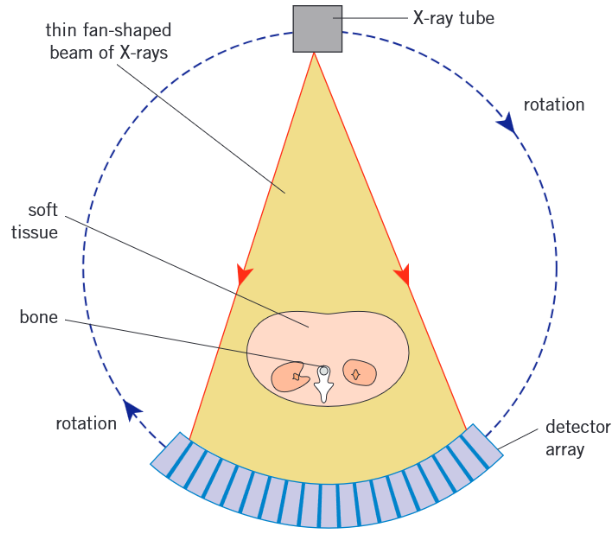
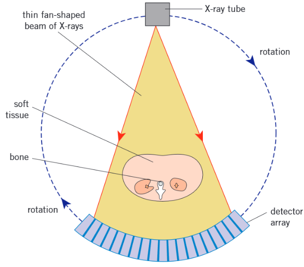
Diagram of how CAT scans take an image
Describe how CAT scans take a 3D image of the patient
The X-ray tube rotates around patient, firing thin fan-shaped beams through patient.
Each full rotation produces an image of a cross-section of the patient (a slice)
The patient slowly moves through ring as it rotates, so each rotation produces an image of a new slice
A computer combines the 2D slices to give a detailed 3D image of the patient in which soft tissues can be identified.
What are the two ways that radioisotopes are used in medical physics?
Diagnosis (finding what’s wrong with patient)
Therapy (treating patient)
Name the process where radiation is used to target tumours inside the body
Brachytherapy
Why are alpha and beta minus sources are less useful for imaging techniques?
They are almost completely absorbed by the patients body before escaping
They are too ionising so are too dangerous
Why is using radioisotopes with a short half-life is important for medical imaging?
So the source has high activity so that only a small amount needs to be used
Prevents patient from receiving high radiation dose that continues after procedure has ended
Who needs to be protected from radiation?
Radiographers as they will carry out many scans in a day so would have a very high, persistent (every day) exposure if not protected
What are the properties of Technetium-99m?
Its nucleus has more energy than a stable technetium nucleus so wants to undergo gamma decay
Emmits a gamma photon of energy 140 keV when it decays
Has a half life of ~6.0 hours
What are the uses of Technetium?
Can be used to image major organs
What does the m stand for in Techtenium m?
Metastable
What does metastable mean?
A nucleas that stays in a high energy state, with more energy than the stable nucleus, for longer than expected
What is the source of Technetium-99m / how is it produced?
The natural decay of Molybdenum-99
What is the decay equation of the Molybdenum-99 into Technetium-99m (including half-life)?

What is the symbol for Technetium-99m?
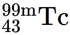
What is the decay equation of Technetium-99m (including half life)?

Describe the use of medical tracers to diagnose the condition of organs
The Tracer is injected into the body and circulates in the blood
It is absorbed by organ / shows blockage
Beta detector or gamma camera used to detect radiation from the body and form an image of the organ
Describe how a medical tracer reaches the cells in the brain
It is injected into the patient and travels through their bloodstream to the brain.
It is absorbed by the brain tissue so remains in the brain
What is a radiopharmaceutical?
A radioisotope chemically combined with elements that will target particular tissues in order to ensure that the radioisotope reaches the correct organ or tumour
Diagram of the key components of the gamma camera
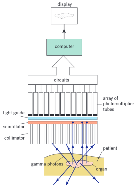
What is the collimator and its purpose (in a gamma camera)?
Honeycomb of long, thin lead tubes
Only gamma photons parallel to the axis of the tubes to pass through (absorbs any of axis photons)
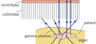
What is the scintillator and its purpose in a gamma camera?
A material that produces many visible light photons when hit by a gamma photon
Produces a flash of light when a gamma photon strikes it
Only 1 in 10 incident photons interact
What is the purpose of the light guide in a gamma camera?
To guide light from the scintillator into the photomultiplier tubes
What is the purpose of the photomultiplier tubes in a gamma camera?
To convert each photon of light entering the tube into an electrical pulse which is sent to the computer
What is the purpose of the computer in a gamma camera?
To process the electrical pulses coming from the photomulitplier tubes and produce an image from them
How is the image from a gamma camera different to a X-ray or CAT scan?
It shows the function and processes of the body instead of the body’s anatomy
What is the name given to a radioisotope chemically combined with another element?
Medical tracer or radiopharmaceutical
Gamma rays and visible light are referred to as ____ inside a gamma camera
photons
The electrodes in the photomultiplier tube are called …
Dynodes
Why is the decay of Tc-99m within a patient not a major concern during a Gamma scan?
Tc-99 is the product formed which is quite stable with a half life of ~210 000 years
What are the advantages of using technetium-99m for imaging organs?
Emits gamma photons
Has a small half-life
Can diagnose the function of organ
Can detect tumours
Gamma rays are the least ionising least ionising
What does PET stand for?
Positron emission tomography
What is the decay equation of Fluorine-18 (including half life)?

What is the half life of Fluorine-18?
~110 minutes
How is Fluorine-18 made?
Made with a particle accelerator: high-speed protons collide with oxygen-18 nuclei to produce fluorine-18 nuclei and neutrons.
What is the nuclear equation for the production of Flourine-18?

Describe FDG
Stands for fluorodeoxyglucose
A medical tracer used in PET scans
Molecule similar to glucose but tagged with a radioactive fluorine-18 atom in place of one oxygen atom.
Our body treats it like normal glucose
How is FDG used?
Injected into patient
Our body treats it like normal glucose, so it gathers in tissue with a high respiration rate
Gamma detectors are used to monitor the FDG’s activity
The absorption of FDG is like glucose, so it accumulates in organs where a lot of ____ occurs
Respiration
What are 2 examples of medical tracers used for PET scans?
FDG - glucose made with a flourine-18 in place of one oxygen atom
Carbon monoxide made with carbon-11
Difference between use of FDG and carbon monoxide in PET scans
FDG accumulates in tissue with a high respiration rate
Carbon monoxide binds to haemoglobin in red blood cells
What is the half life of carbon-11?
~20mins
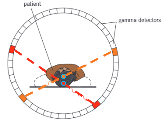
Diagram showing structure of a PET scanner
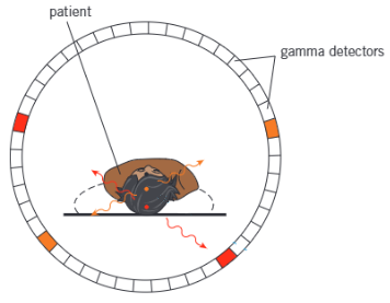
Describe the annihilation process utilised in PET scans
The medical tracer emits positrons, which annihilate with electrons producing two gamma photons traveling in opposite directions (so that momentum is conserved)
How does a PET scan work?
The medical tracer emits positrons
The positrons annihilate electrons within the patient, producing two gamma-photons travelling in opposite directions
The difference in the arrival times at the detectors is used to locate the point of annihilation which is close to the tracer nucleus
A computer uses these points of annihilation to form an image where different colours correspond to different concentrations of the tracer
What are the advantages of PET?
It is non-invasive
Can detect cancer
Allows the function of the brain/organs to be observed
Produces a 3D image
The effect of new medicines can be assessed
Can help plan heart surgery
Can diagnose Alzheimer’s
What are the disadvantages of PET?
Expensive as special facilities needed to produce medical tracers (these must be at the hospital or nearby as f-18 has a short half life)
What is one advantage of using non-invasive medical imaging techniques for diagnosis?
No surgery so no risk of infection
What is ultrasound?
Longitudinal waves with frequency greater than 20 kHz, beyond the range of human hearing