TCD (14)
1/32
There's no tags or description
Looks like no tags are added yet.
Name | Mastery | Learn | Test | Matching | Spaced | Call with Kai |
|---|
No analytics yet
Send a link to your students to track their progress
33 Terms
what is TCD?
transcranial doppler
non invasive, non ionizing, inexpensive, ultrasound method used to examone the blood circulation within brain
what can TCD detect for?
intracranial stenoses and occlusion
assess collateral circulation
eval intracranial arteriovenous malf
detect emboli
brain death
subclavian steal assessment
classify strokes
risk factors for TCD
hypertension
diabetes
cardiac disease
hereditary
smoking
oral contraceptives
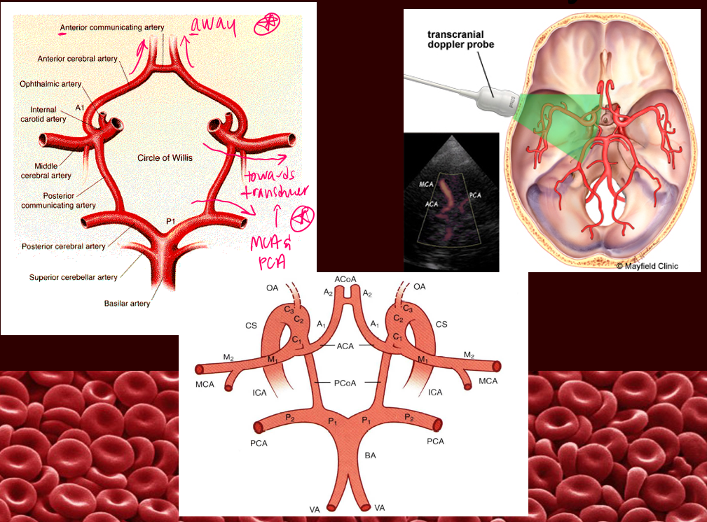
what transducer is used for TCD imaging?
2MHz cardiac probe
what are the three windows used in TCD?
transtemporal
transorbital
transforamenal (suboccipital)
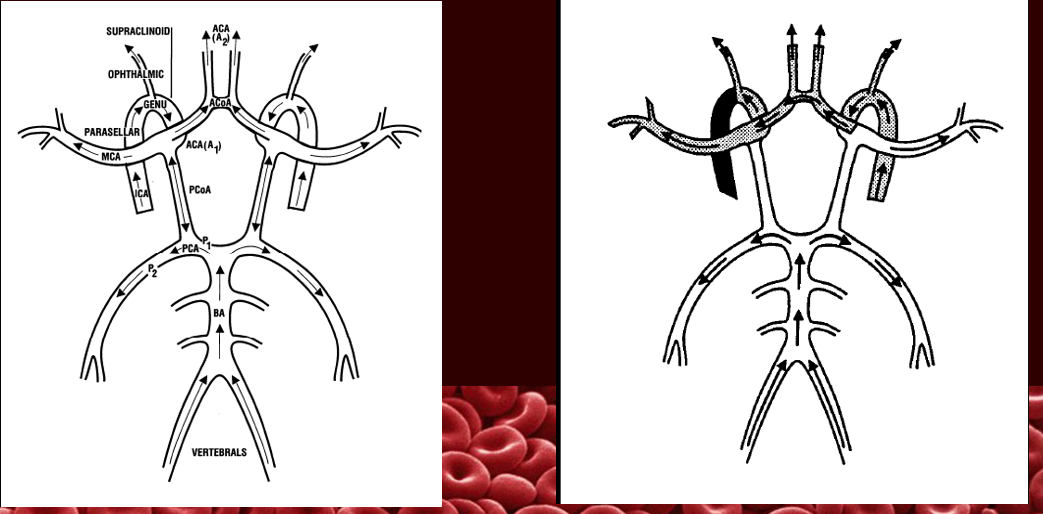
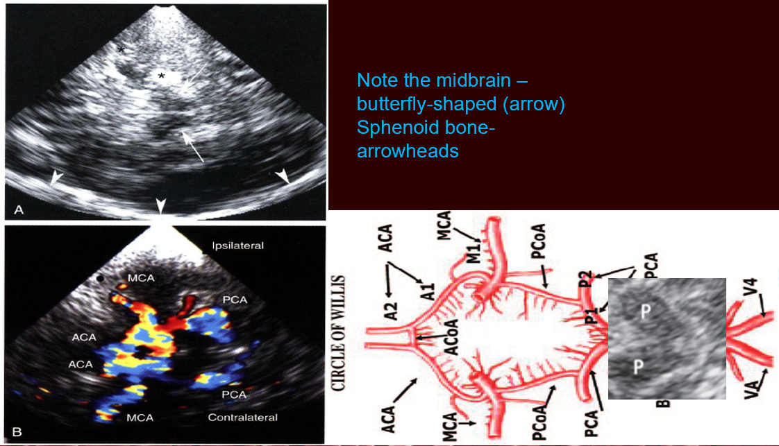
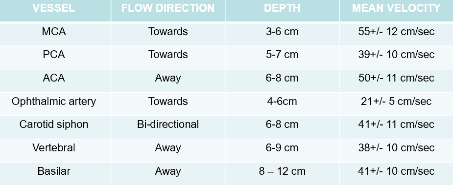
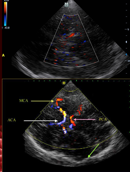
what vessels are scanned in transtemporal window?
MCA
ACA
ACoA
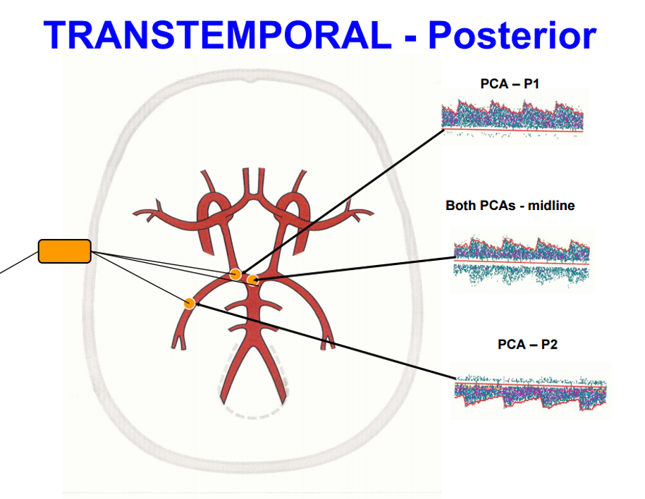
PCA
PCoA
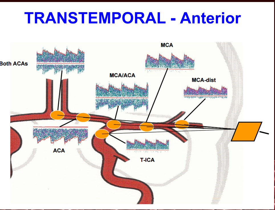
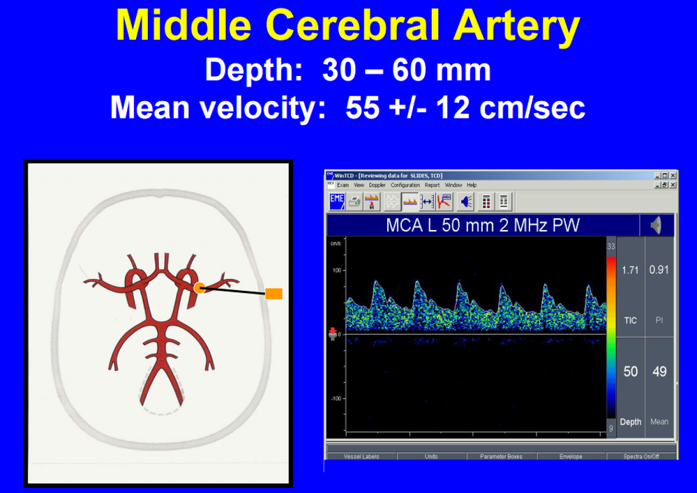
what is the depth and direction of the MCA?
3-6cm
towards transducer/above baseline → RED
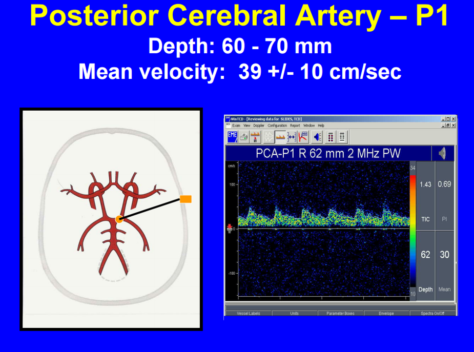
what is the depth and direction of the PCA?
5-7cm
towards transducer/above baseline → RED
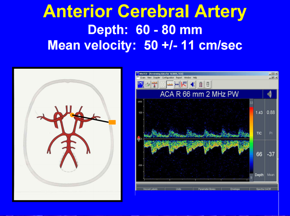
what is the depth and direction of the ACA?
6-8cm
away from transducer/below baseline → BLUE
what if there is a blue vessel seen below 3cm?
might be collaterals
what is the landmark in the transtemporal window?
cerebral peduncles
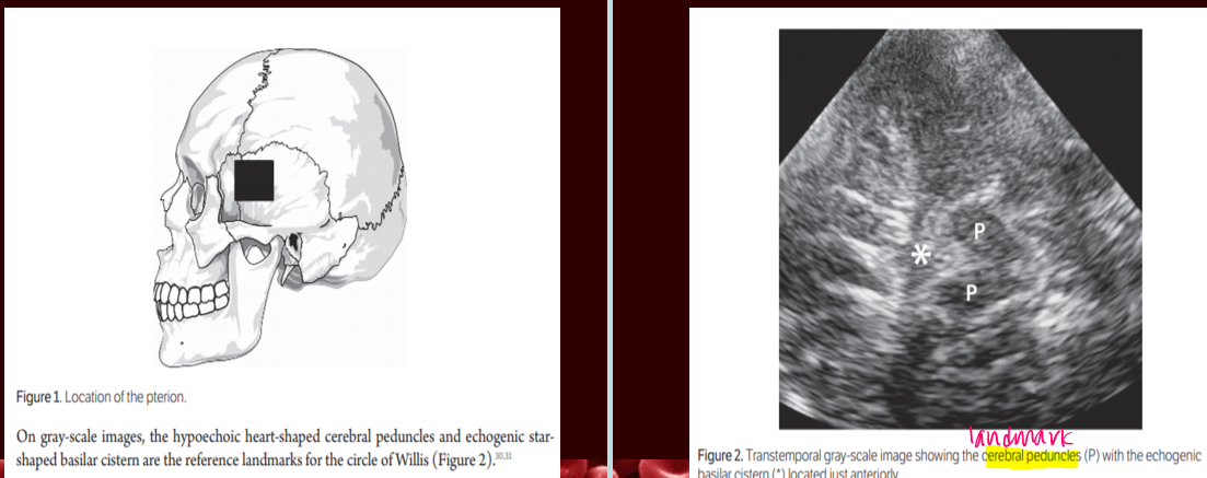
what do the arrow and arrowheads represent?
arrow : midbrain
arrowheads : sphenoid bone
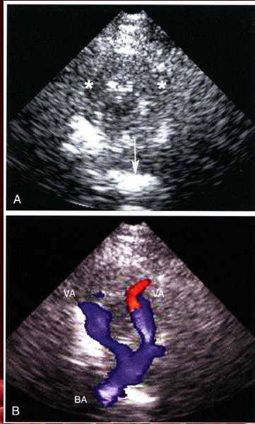
what vessels are scanned in the suboccipital view/transforamenal?
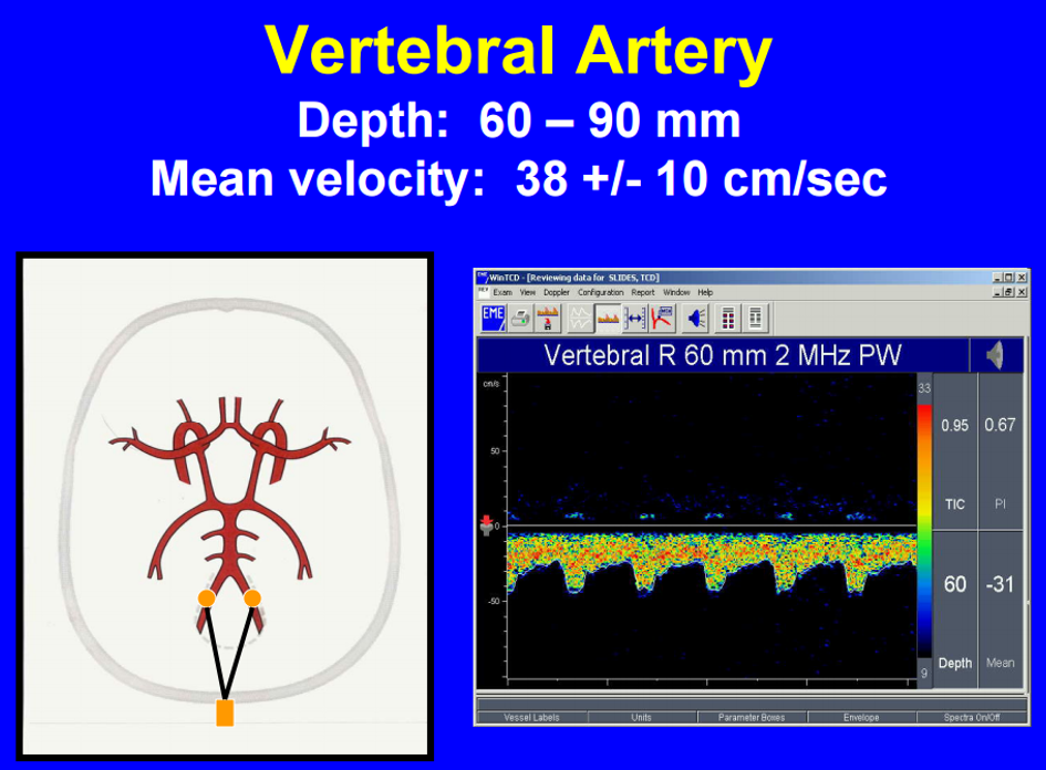
distal vertebral artery
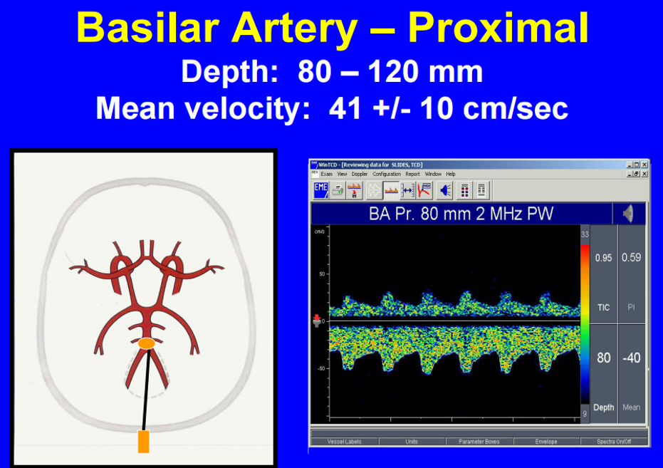
basilar artery
** in asterick is the foramen magnum
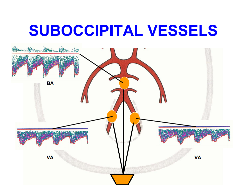
what is the direction and depth of vertebral artery in suboccipital view?
depth : 6-9cm
direction : away from transducer/below baseline → BLUE
what is the direction and depth of basilar artery in suboccipital view?
depth : 8-12cm
direction : away from transducer/below baseline → BLUE
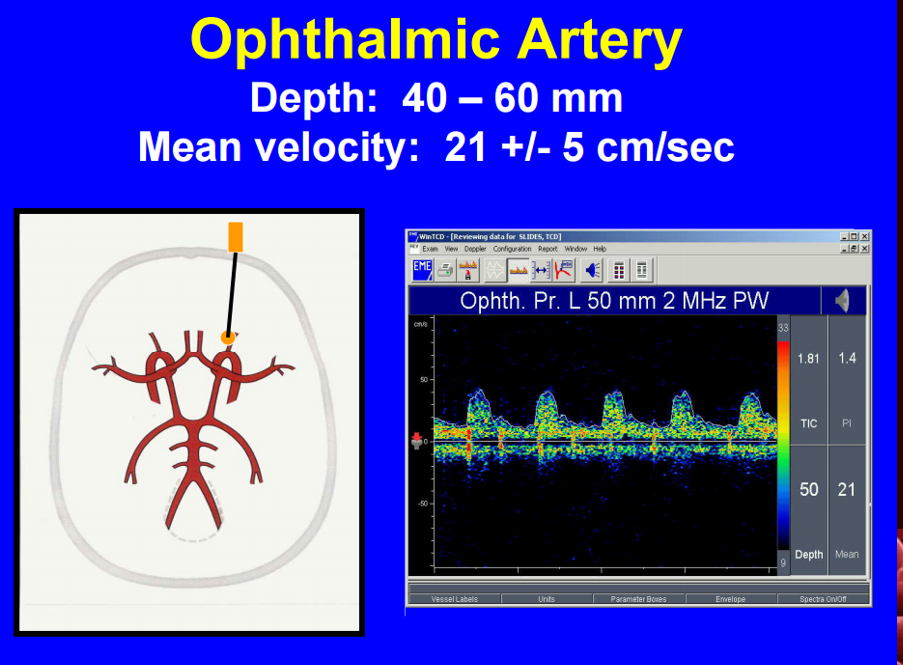
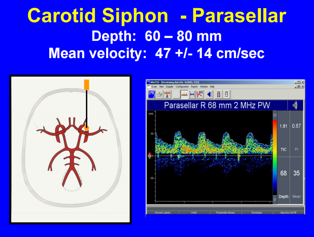
what vessels are scanned in transorbital view?
opthalmic artery
carotid siphon
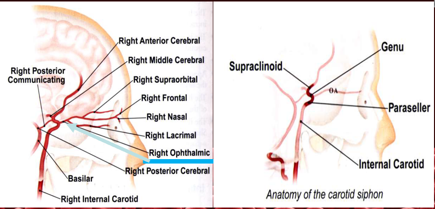
what is the direction and depth of opthalmic artery in transorbital view?
depth : 4-6cm
direction : towards transducer/ above baseline → RED
what is the direction and depth of carotid siphon in transorbital view?
depth : 6-8cm
direction : bidirectional
when doing a transorbital, mechanical index should not exceed
.23MI (AIUM)
compare normal velocity relationships between MCA, ACA, PCA, VA, BA, Carotid siphon, OA
MCA > ACA > PCA ~ Carotid siphon & VA & BA > OA
Qs what are common pitfalls in TCD scanning?
lack of knowledge
lack of sight
depth
spatial anatomy
phsiology
poor acoustic windows
operator dependent with large learning curve
how can we differentiate between PCA and MCA?
measure velocities
MCA > ACA > PCA
what are common pitfalls regarding collaterals?
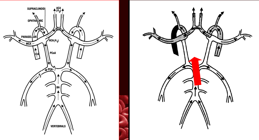
posterior to anterior collateral through posterior communicating artery
external carotid to internal carotid thorugh reversed opthalmic artery
crossover collateralization through ACA
why would TCD be used/applied?
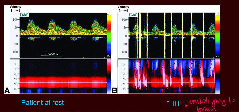
emboli monitoring
carotid endartectomy, stenting
cardiopulm bypass surgery
detection of cardiac shunts
confirmed when HITS (high intensity transient signal) detected in MCA
brain death
used to correlate with other diagnostic tests
brain death is clinical diagnosis, TCD is a confirmatory test
sickle cell disease
early detection of abnormal MCA velocities
what are the causes of intracranial stenosis?
atherosclerosis
thrombus
dissection
vasculitis
vasospasm
extrinsic vessel compression
how is the MCA affected when there is occlusion?
lack of signal
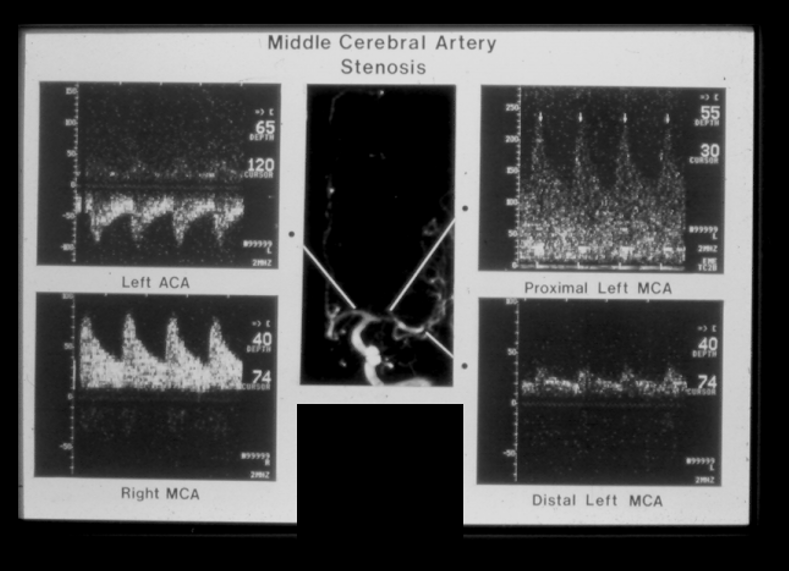
how is the ACA/PCA affected when there is occlusion?
velocities are unreliable
collaterals can cause reversed, increased or absent flow
what is a vasospam and what is it associated with?
pathologic response due to irritation of cerebral arteries by blood breakdown products
associated with subarachnoid hemorrhage
results in structural wall changes and lumenal narrowing
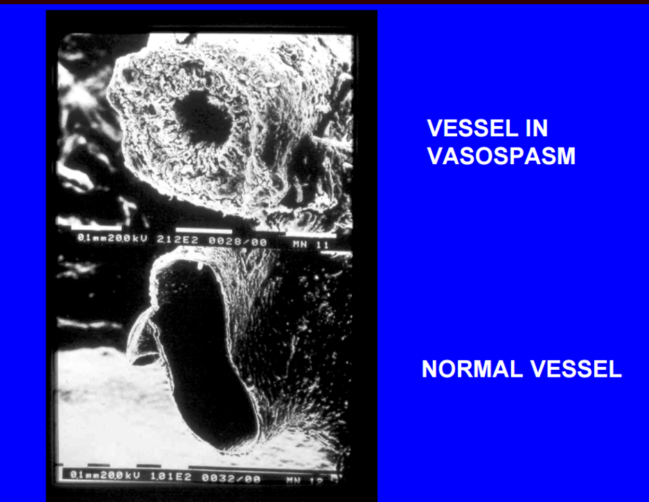
20% of population and women are more likely to have poor
temporal windows
embolic events are characterized by
an audible chirp and simultaneous visual HIT on screen
Qs explain crossover collateralization through the anterior communicating artery
when blood from unaffected side flows across ACA to supply compromised side