WSU BIO 315 Lab Exam 4 (respiratory system and mediastinum, with BQ)
1/130
There's no tags or description
Looks like no tags are added yet.
Name | Mastery | Learn | Test | Matching | Spaced | Call with Kai |
|---|
No analytics yet
Send a link to your students to track their progress
131 Terms
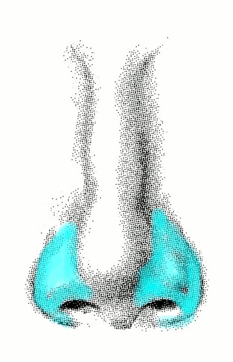
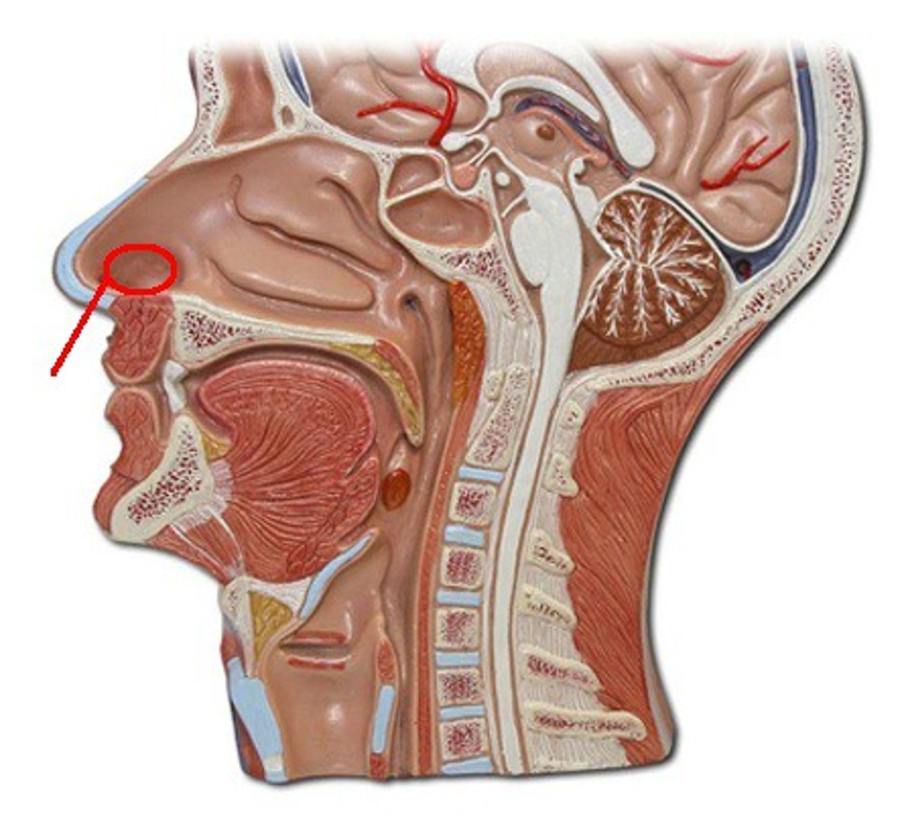
dorsum of nose
bridge of the nose
ridge
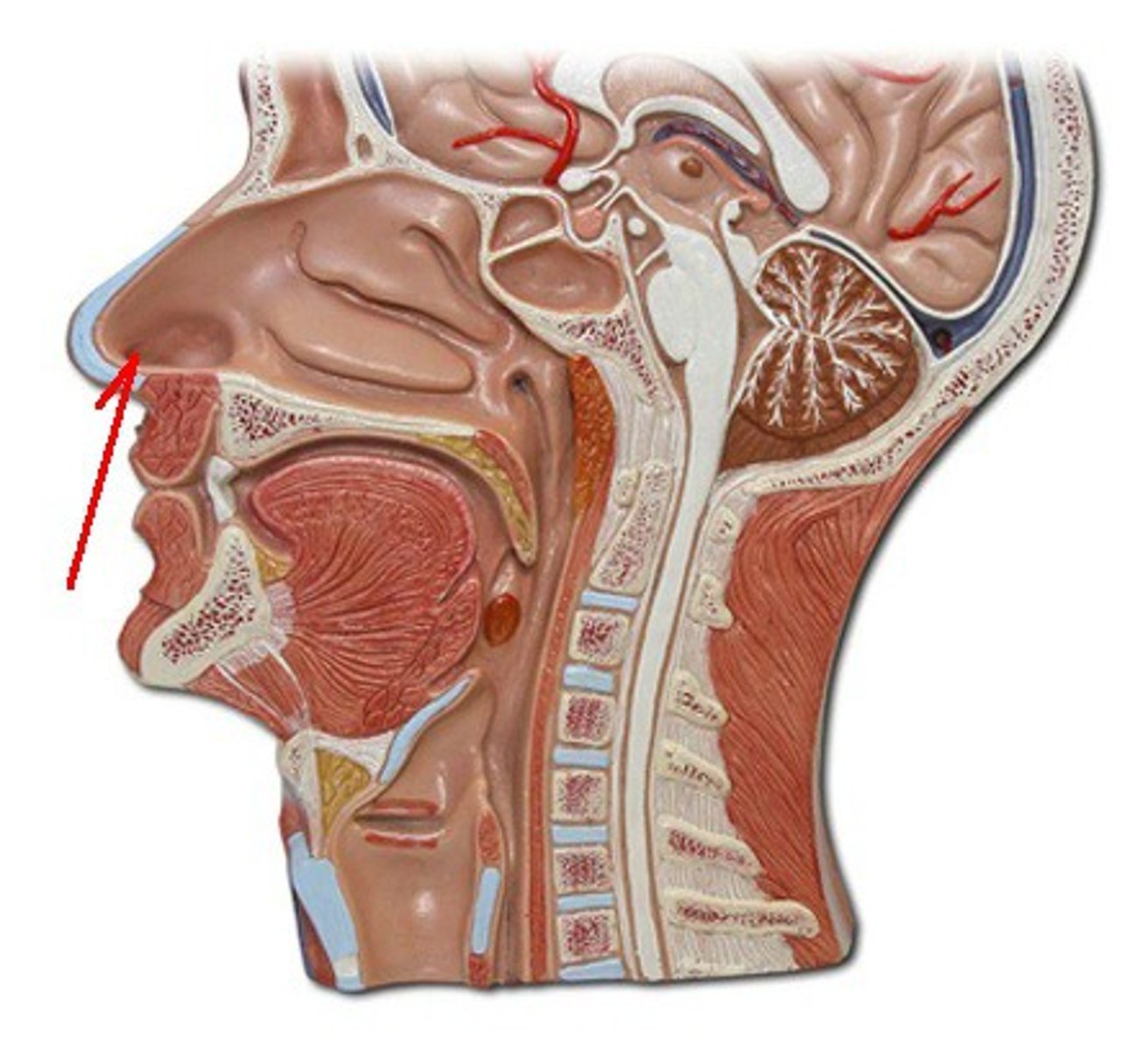
apex of nose
boop
feature
ala of nose
where u get a stud nose piercing
feature
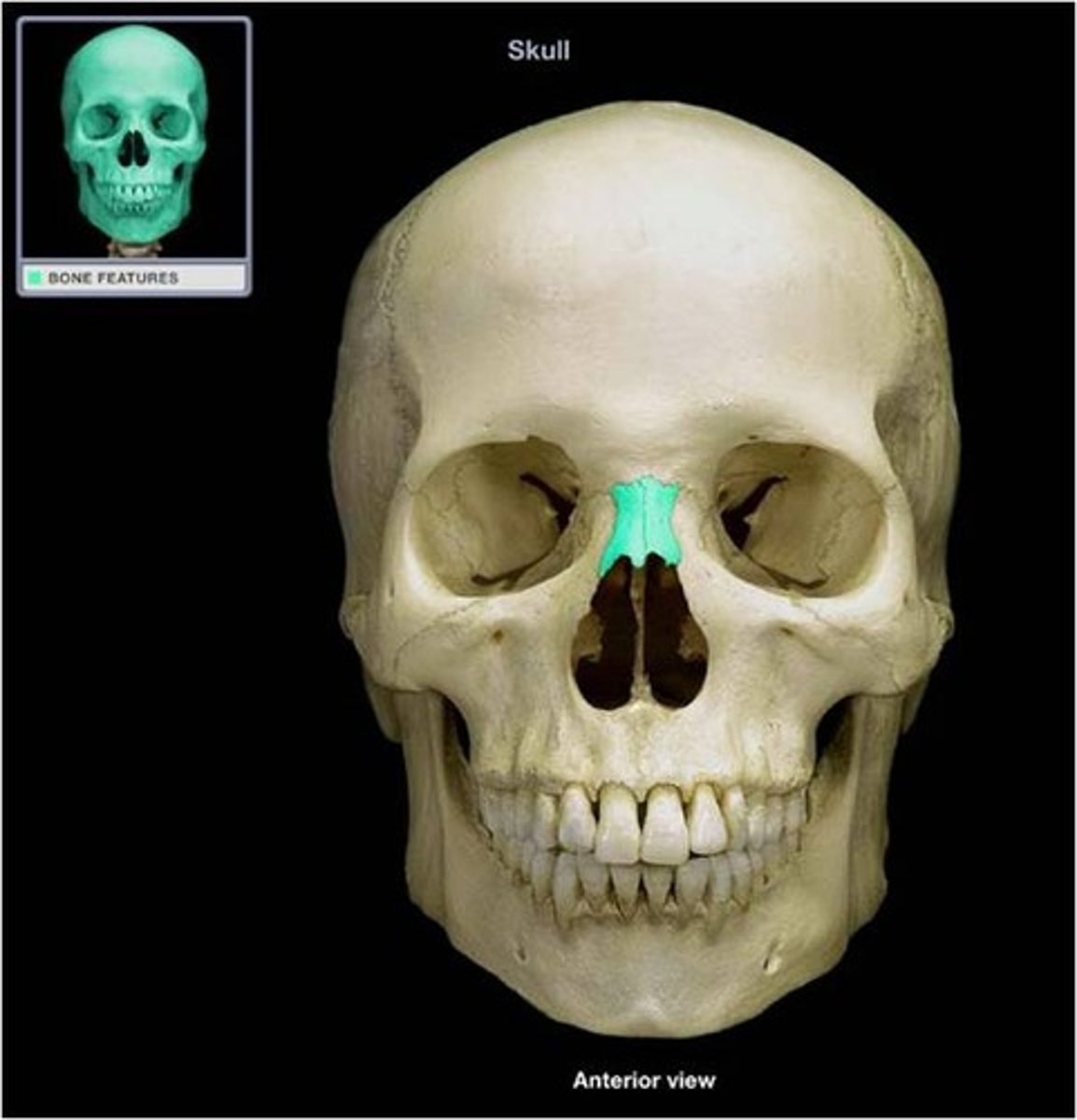
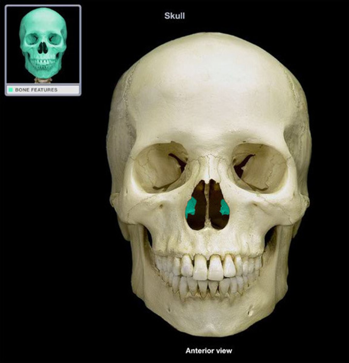
nasal bones
white front bones on rainbow skull
structure
major alar cartilages
the sides of the nose where glasses would sit
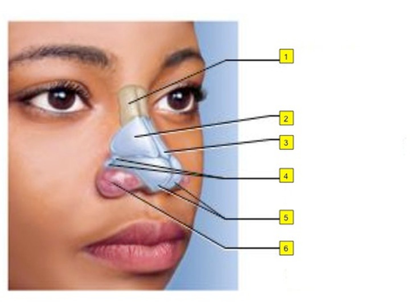
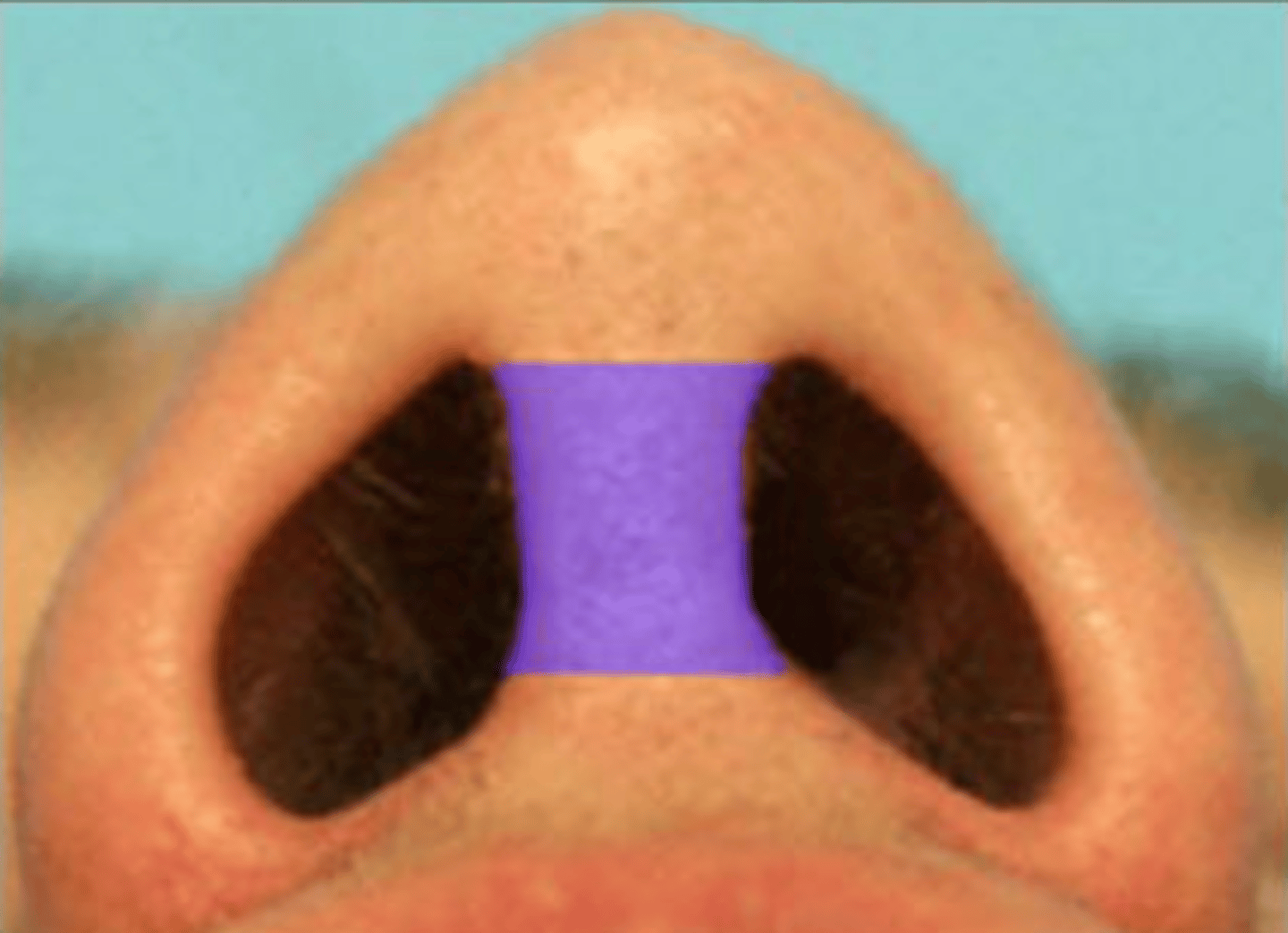
mobile nasal septum
right above philtrum where septum piercing would go
structure
bony part of the nasal septum
yellow piece in the middle of rainbow skull nose
structure

nares (nostrils)
TA will lay probe inside the nostril
2D opening
vestibule of the nose
booger space same as nares but 3D not 2D
3D SPACE
vibrissae of the nose
BQ ONLY: fancy name for “nose hairs”
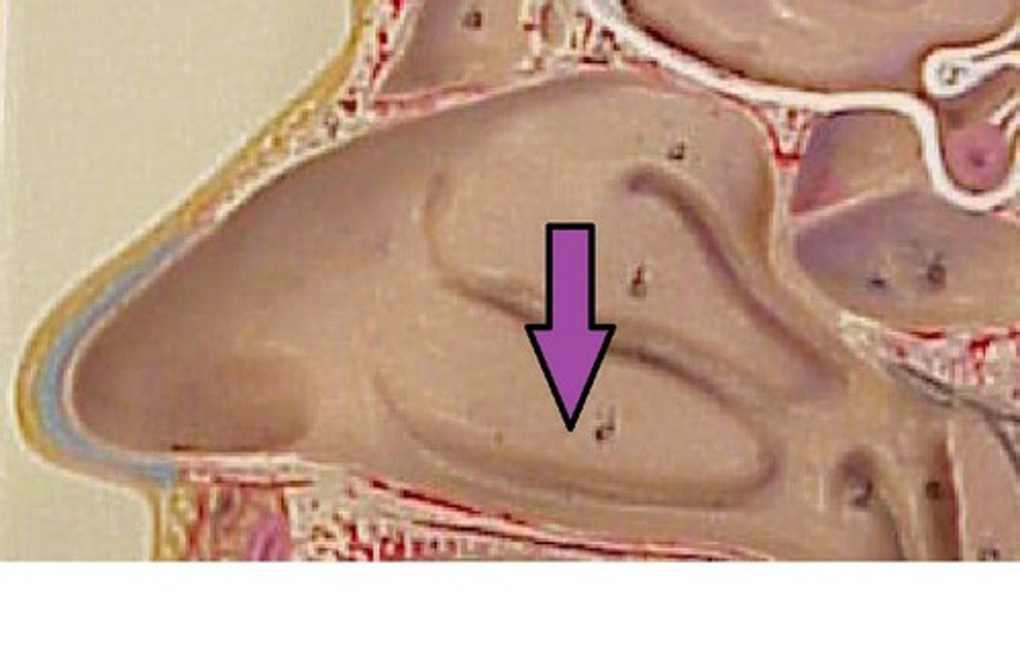
nasal conchae
the three ridges of the nose cross section model
collective structure
superior nasal conchae
#6 top ridge
strucutre

middle nasal conchae
#7 middle ridge, longest of the three
strucutre

inferior nasal conchae
#8 the thickest
structure
nasal meatuses
the three spaces on nose model
collective passageway
superior nasal meatus
#9 the smallest passageway
passageway

middle nasal meatus
#10 middle passageway
passageway
inferior nasal meatus
#11 bottom passageway
Opening of nasolacrimal duct
hole in the orange
3D space
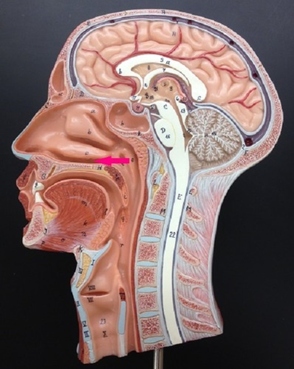
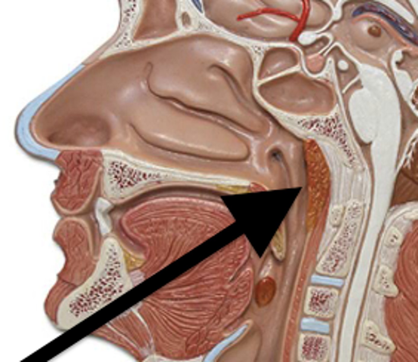
respiratory region
BQ ONLY: The epithelium here warms, humidifies, and filters the air / lined with respiratory epithelium
olfactory region
BQ ONLY: receptors here for smell, lined with olfactory epithelium
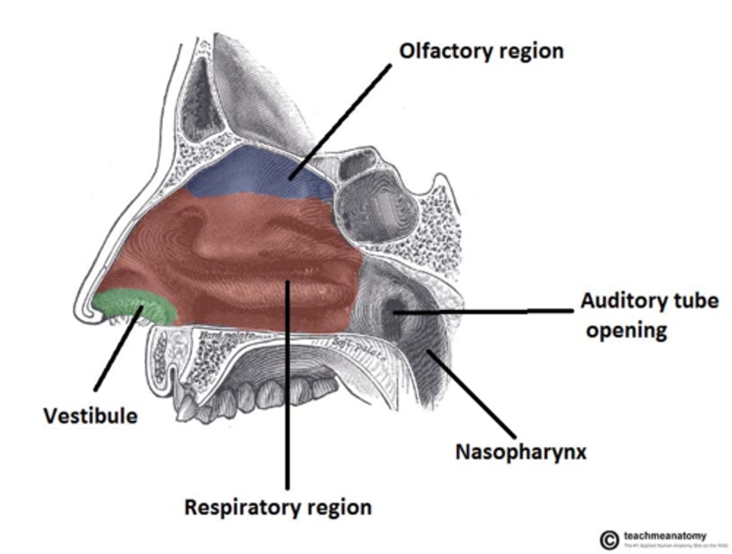
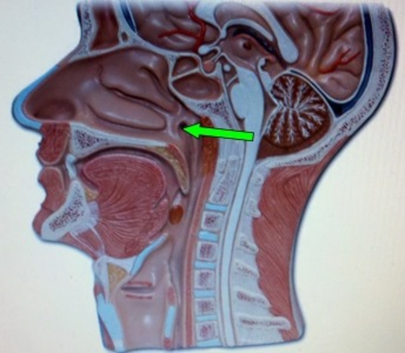
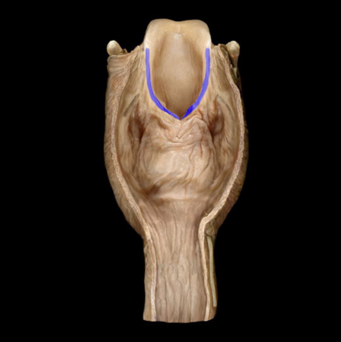
choanae
#17 white lined depression
depression
frontal sinuses
TA will tap the forehead
SPACE
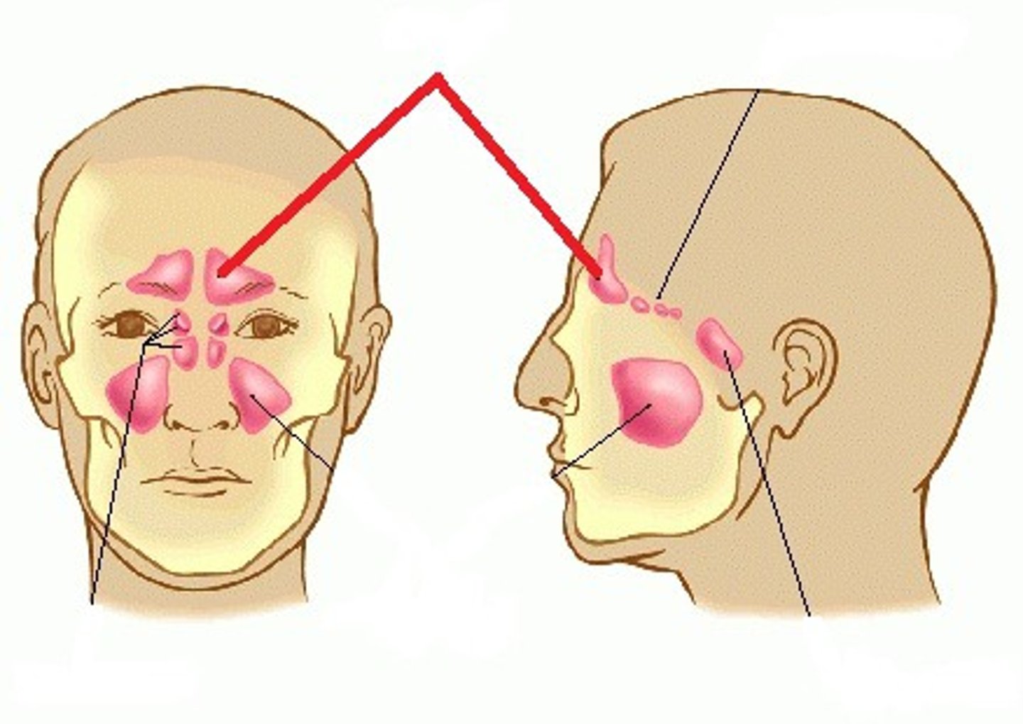
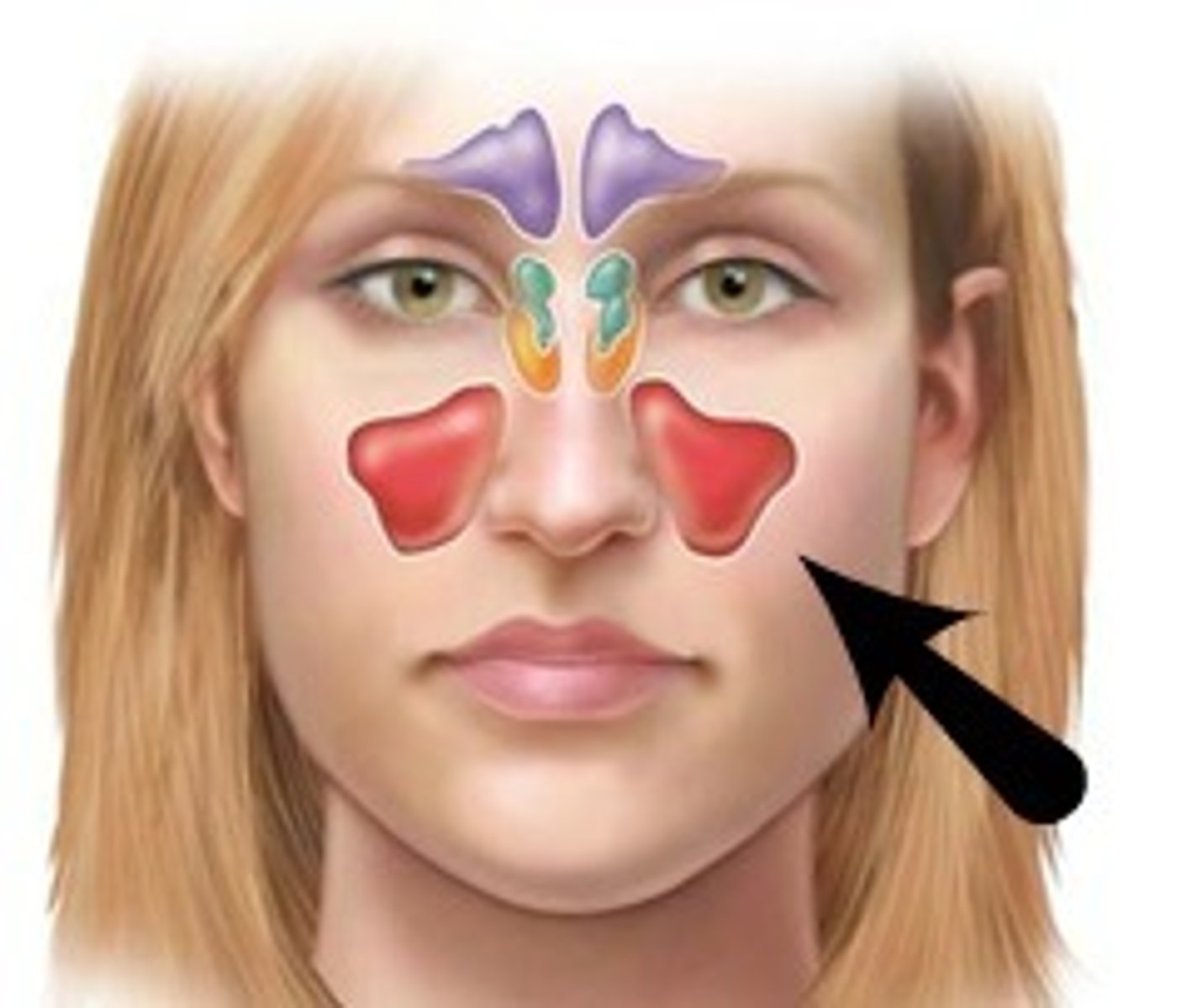
maxillary sinuses
BQ: the largest of the paranasal sinuses
located underneath eyes on the maxilla bone
SPACE
sphenoidal sinuses
TA will flip skull and tap red pokey bit
space
ethmoidal cells
TA will pinch between nose
space
ethmoidal cells
BQ: as many as 20 small sinuses.
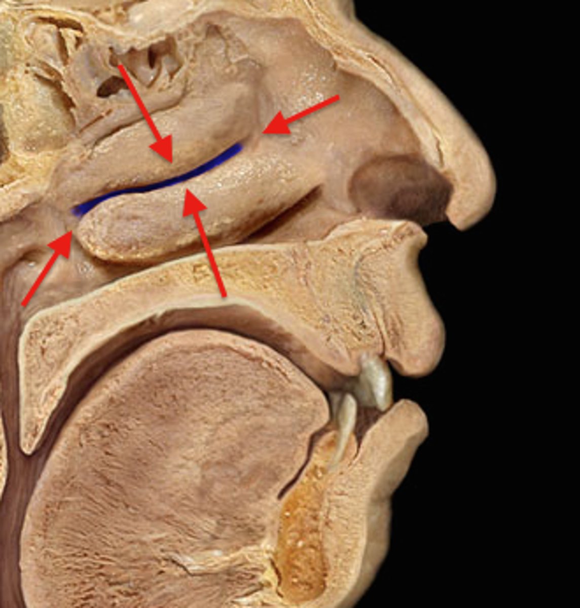
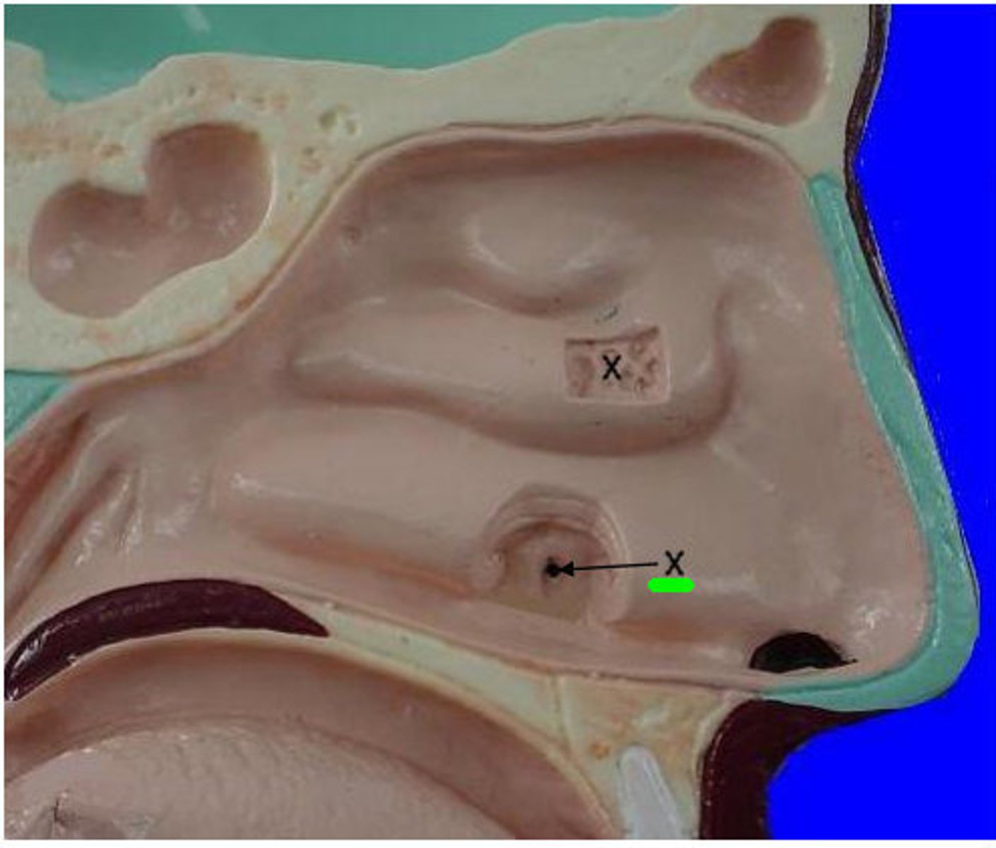
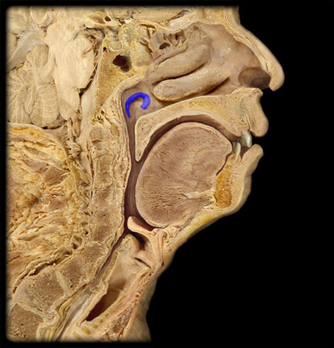
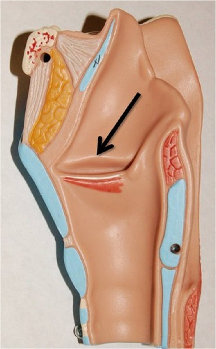
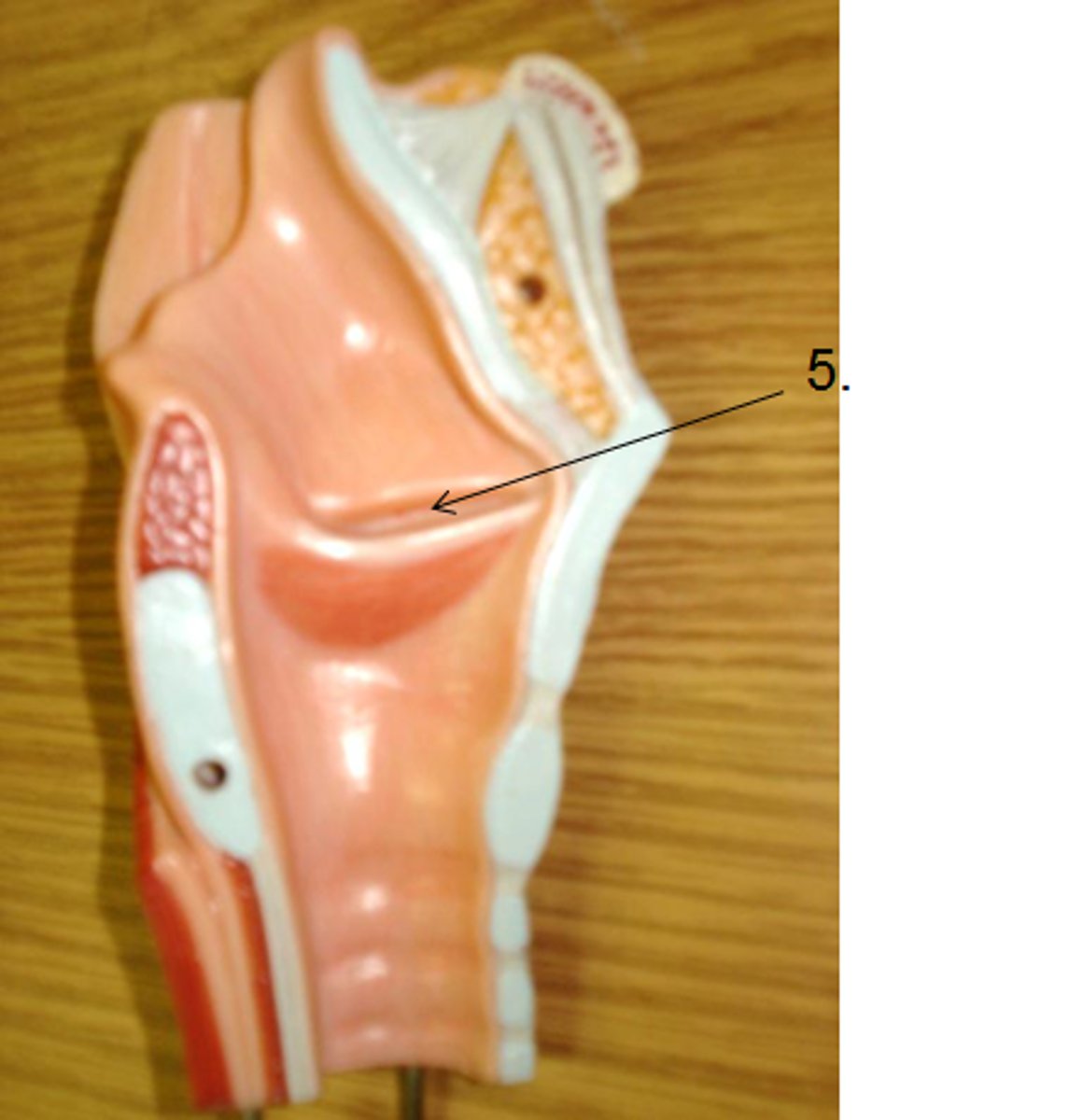
opening of pharyngotympanic tube (eustachian tube)
#12 the most posterior
2D space
torus tubarius
ridge surrounding the Eustachian tube
ridge
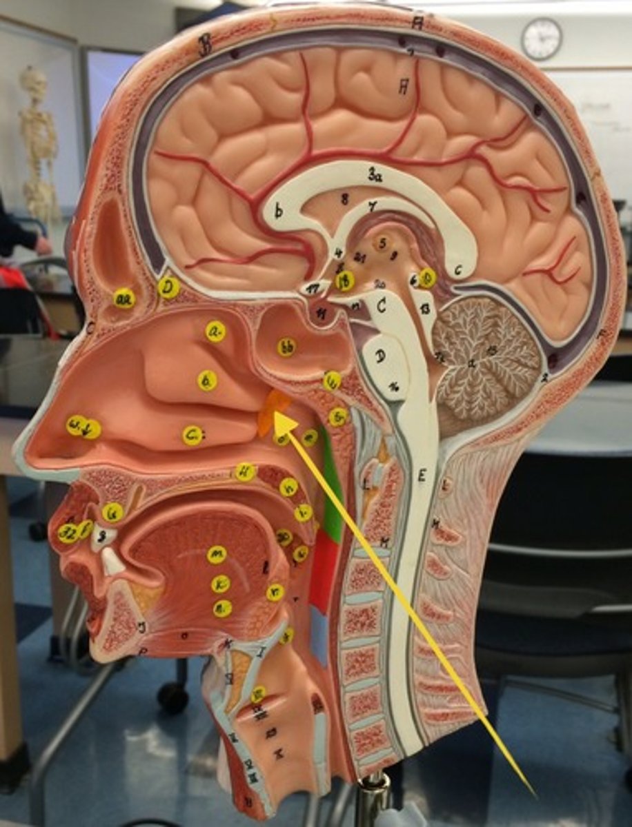
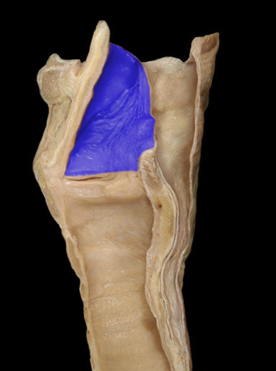
pharyngeal tonsils
roof on the model
structure
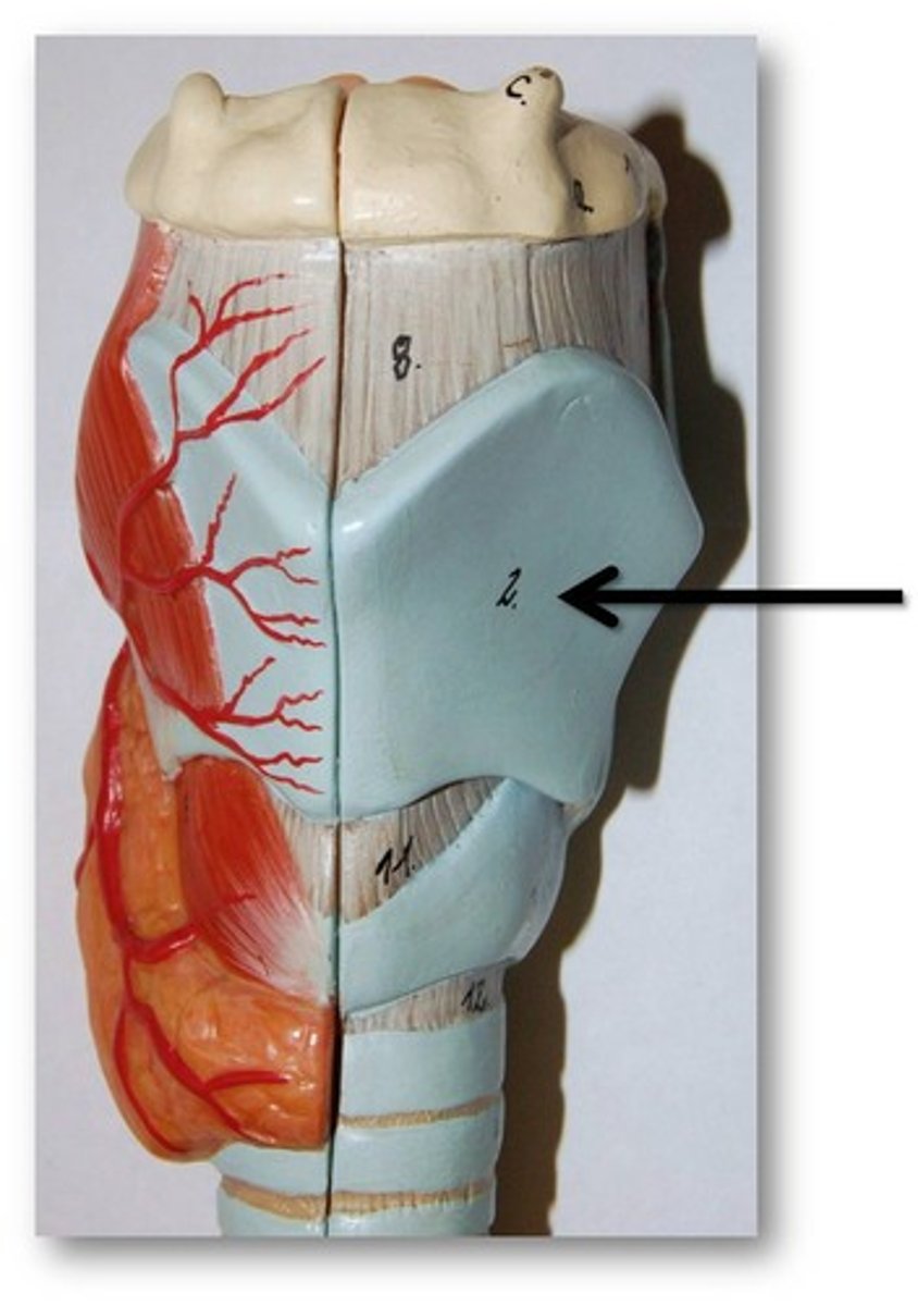
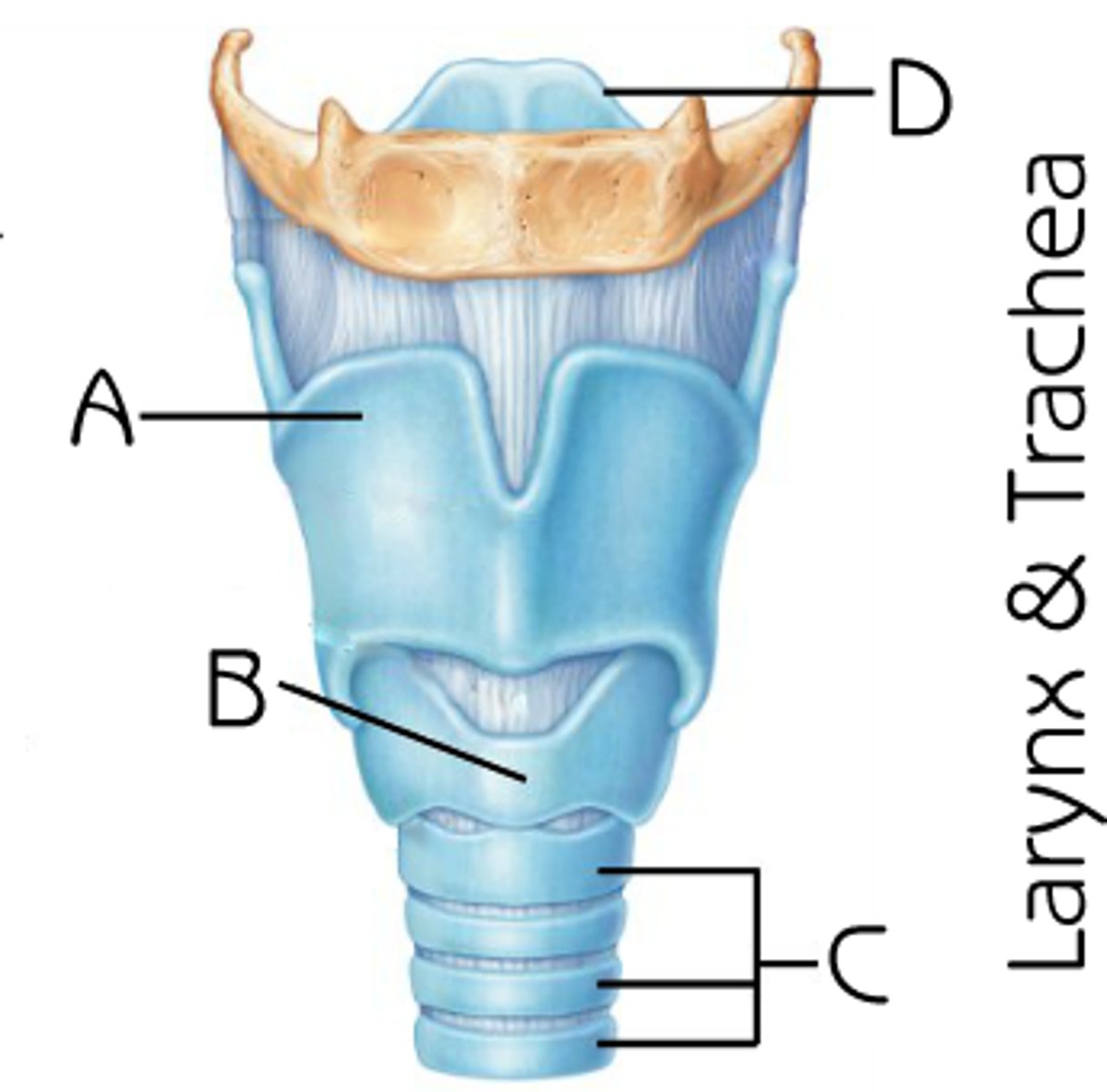
thyroid cartilage
On larynx model
in front of the thyroid gland, almost like a “shield”
structure
laryngeal prominence
point where thyroid shields meet
the point of the thyroid
structure
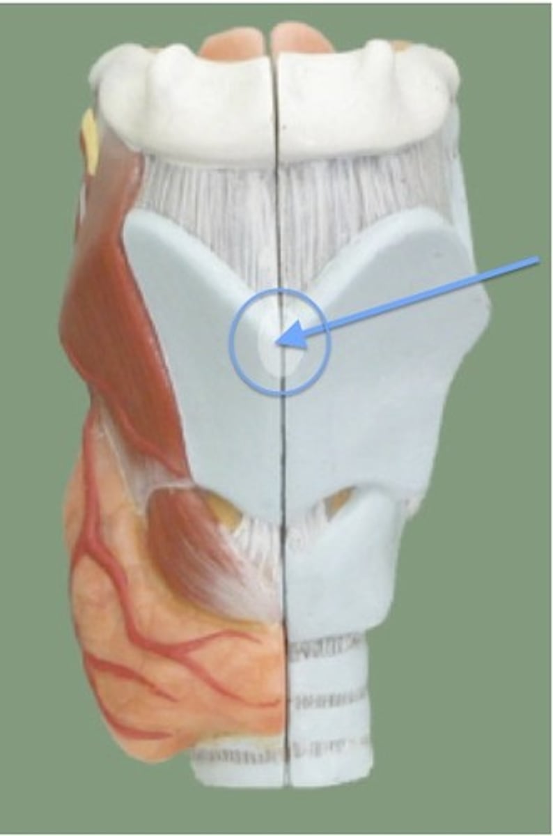
laryngeal prominence
BQ: adam's apple
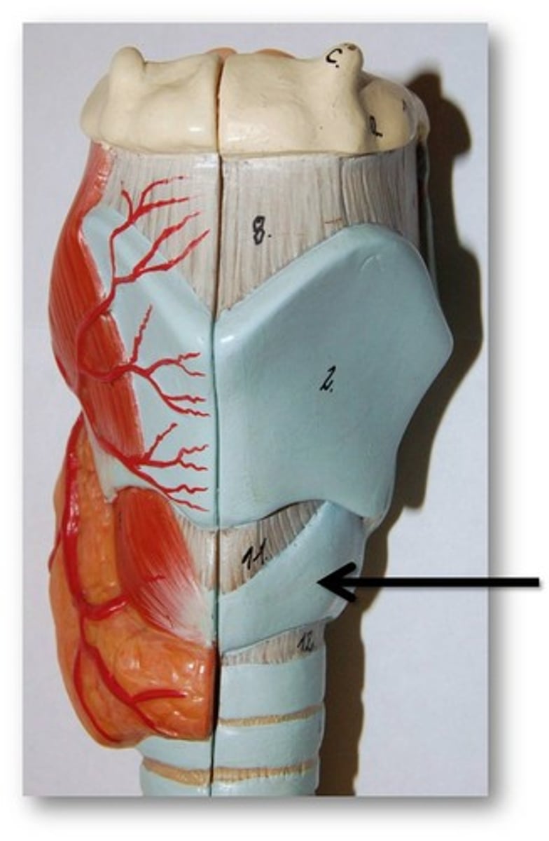
cricoid cartilage
hourglass shape on the posterior side of the model
structure
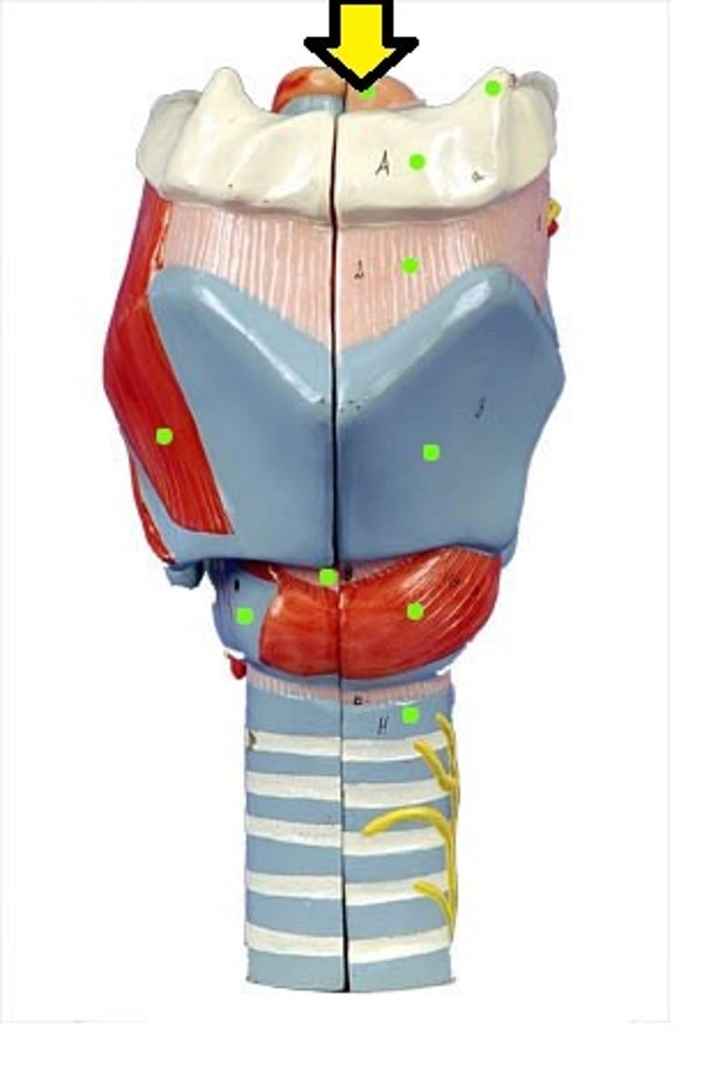
epiglottis
the tongue like structure
structure
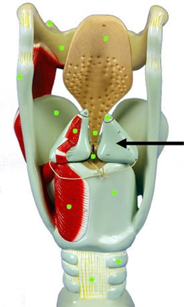
arytenoid cartilage
small blue fangs on larynx model
structure
muscular process of arytenoid cartilage
BQ ONLY: projet LATERAL
vocal process of arytenoid cartilage
BQ: project ANTERIOR for attachment of vocal folds
cricothyroid joint
small bony pointy nub on thyroid cartilage
articulation
thyrohyoid membrane
almost baleen looking striations
structure
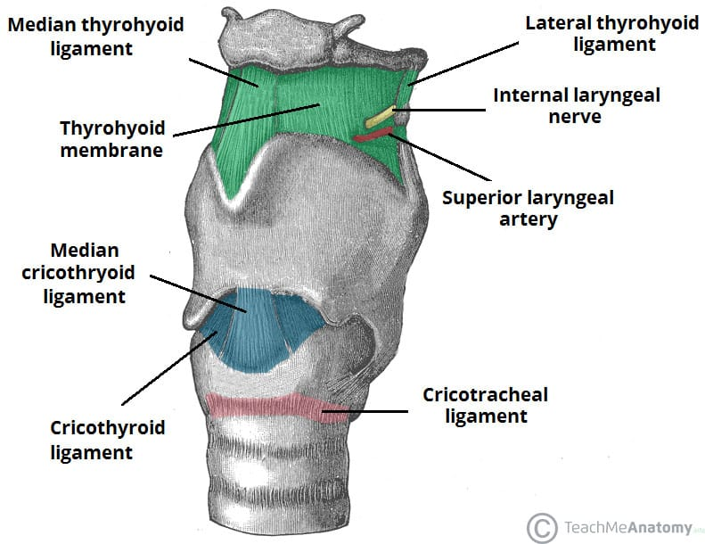
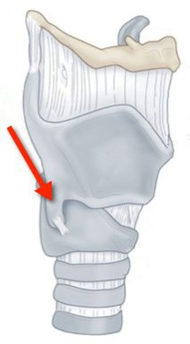
cricothyroid ligament
TA will take off the thyroid cartilage to show the “baby whale teeth”
strucutre
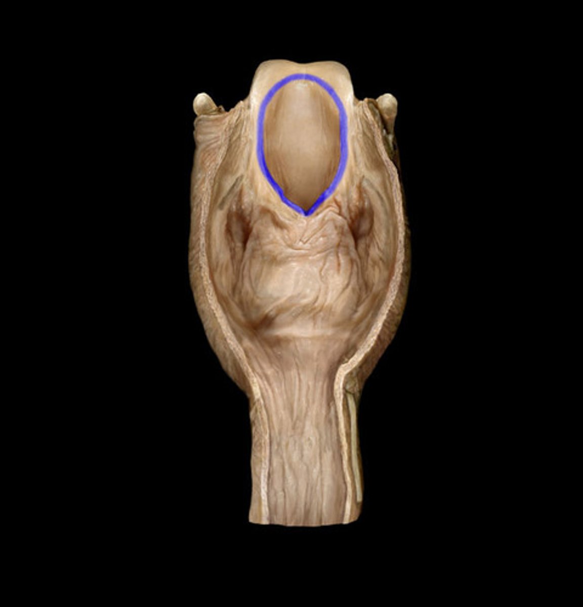
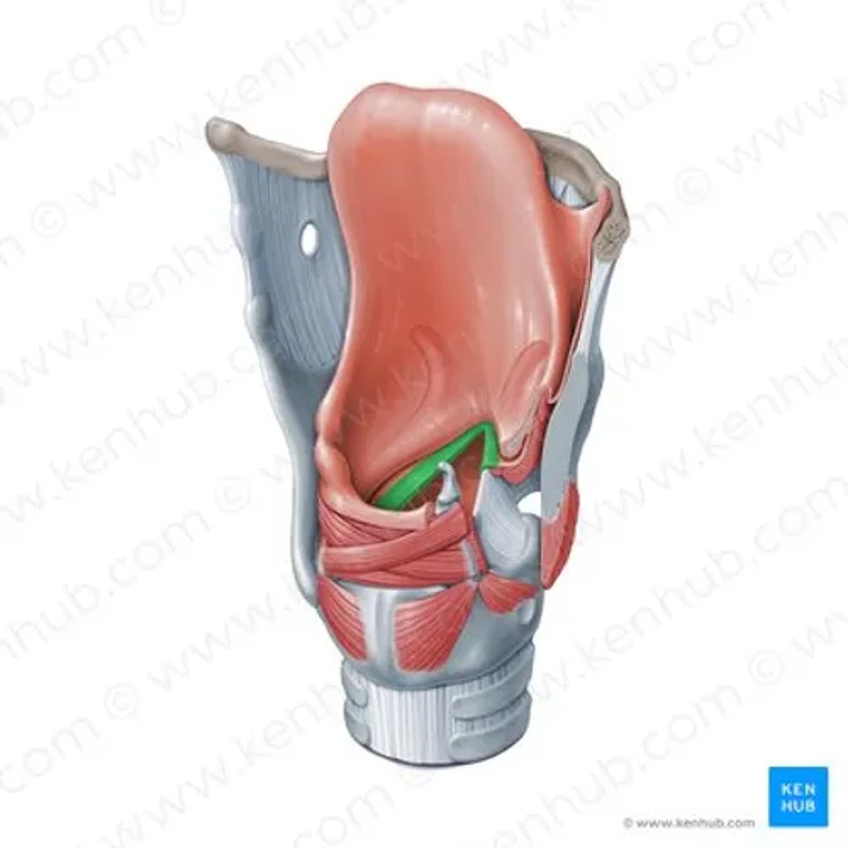
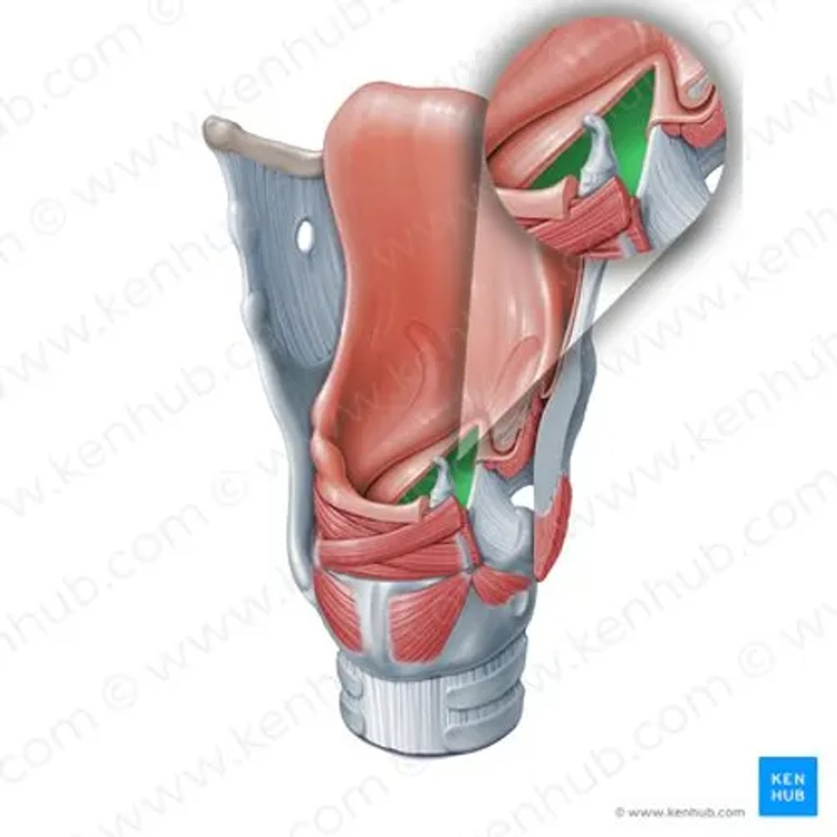
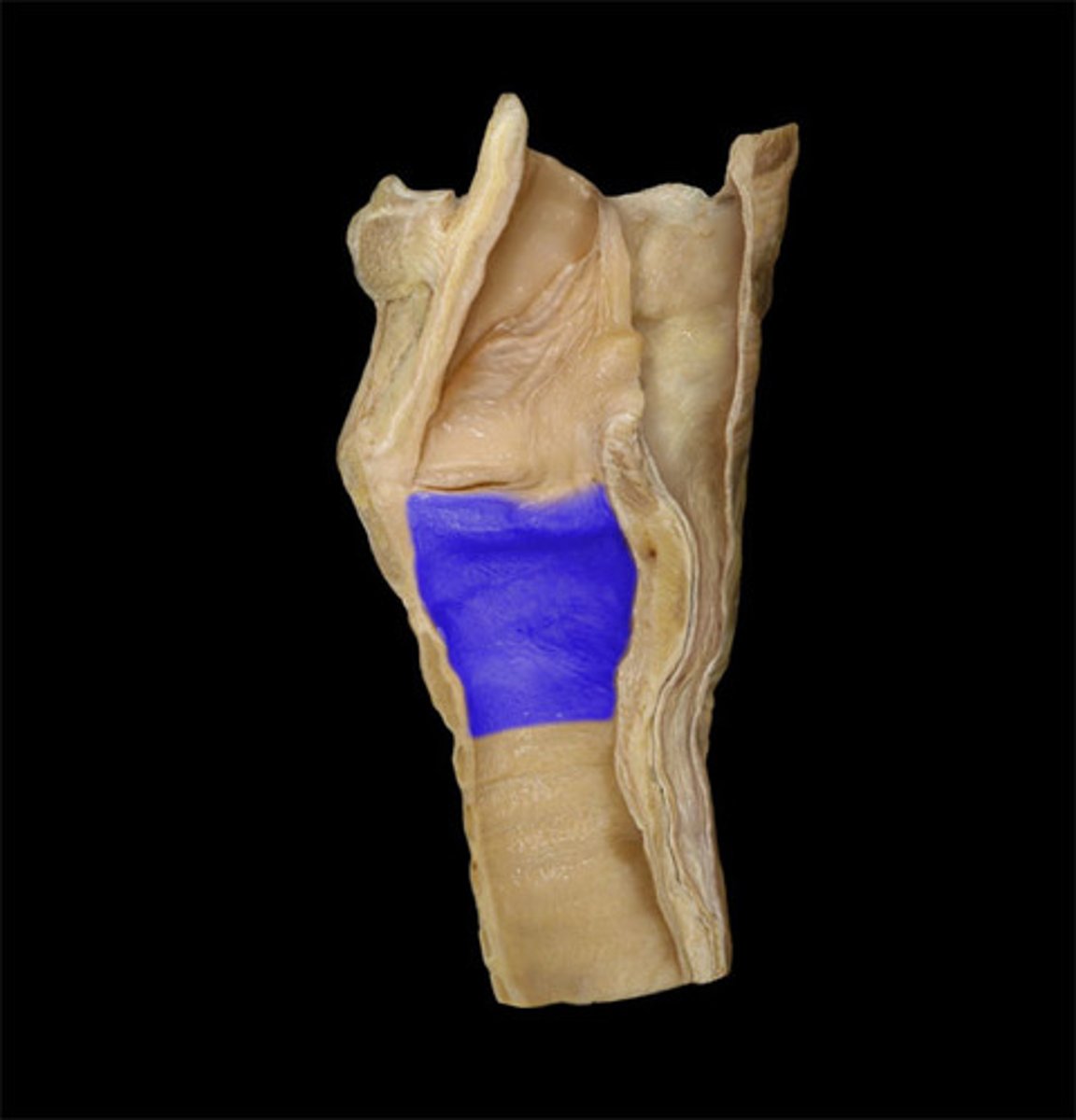
inlet of larynx
TA will lay probe across front
2D opening
aryepiglottic folds
ridges that make epiglottis slide tracts
ridges
vestibule of larynx
TA will insert the probe into the larynx
vestibular fold
the top triangle on larynx inside
strucutre
ventricle of larynx
the crease between triangle folds
glottis of the larynx
BQ ONLY: part of the larynx that produces sound
vocal folds of larynx
underneath ventricle
structure
Rima Glottidis of the larynx
TA will look through the bottom of larynx, a teardrop shape
changing space
infraglottic cavity
space underneath vocal fold
space
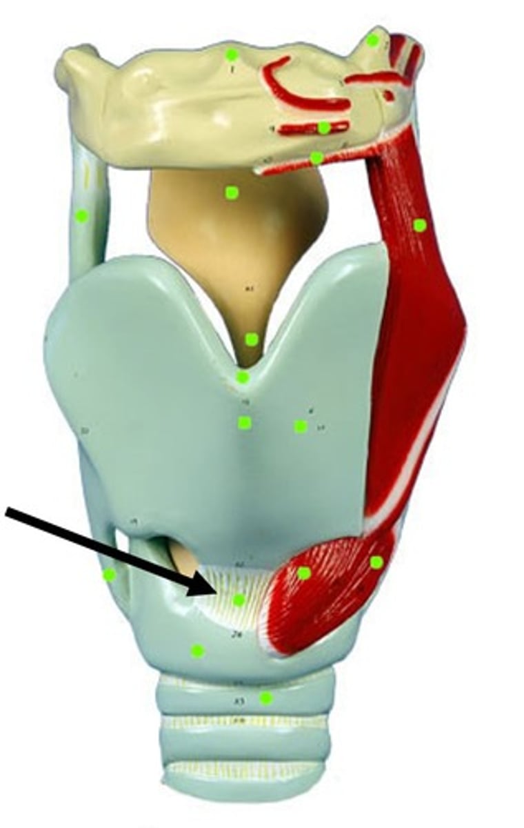
posterior cricoarytenoid muscle
the butt cheeks on the posterior side of the larynx
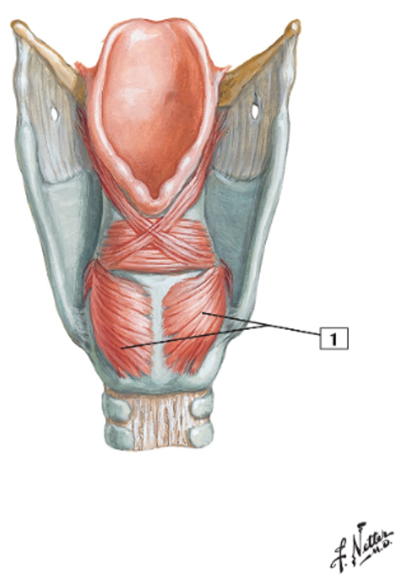
lateral cricoarytenoid muscle
5 in image
bottom wing of the butterfly
structure
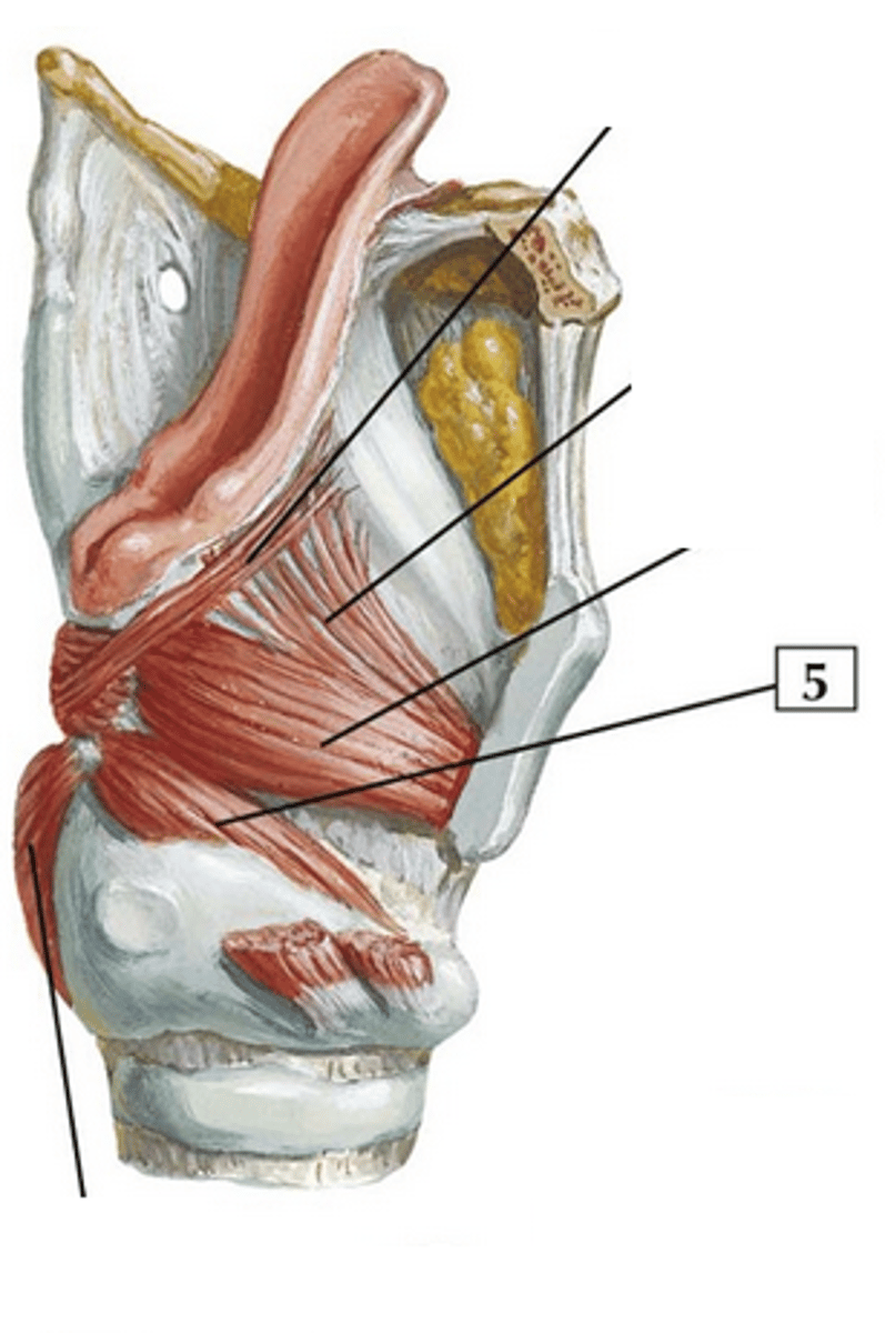
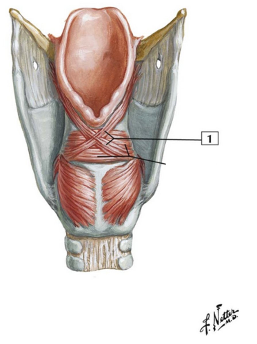
transverse arytenoid muscle
underneith the “X”
strucutre
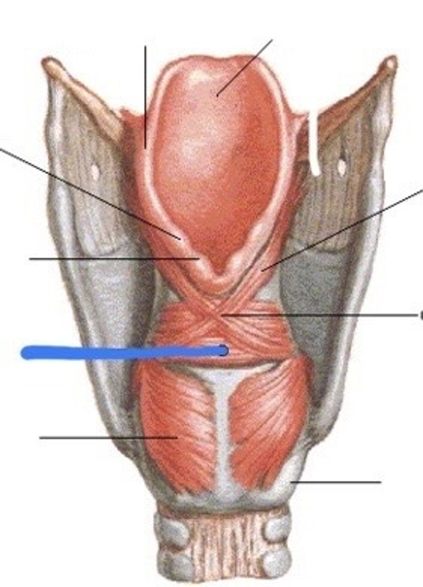
oblique arytenoid muscle
1 in image
the “X” itself
strucutre
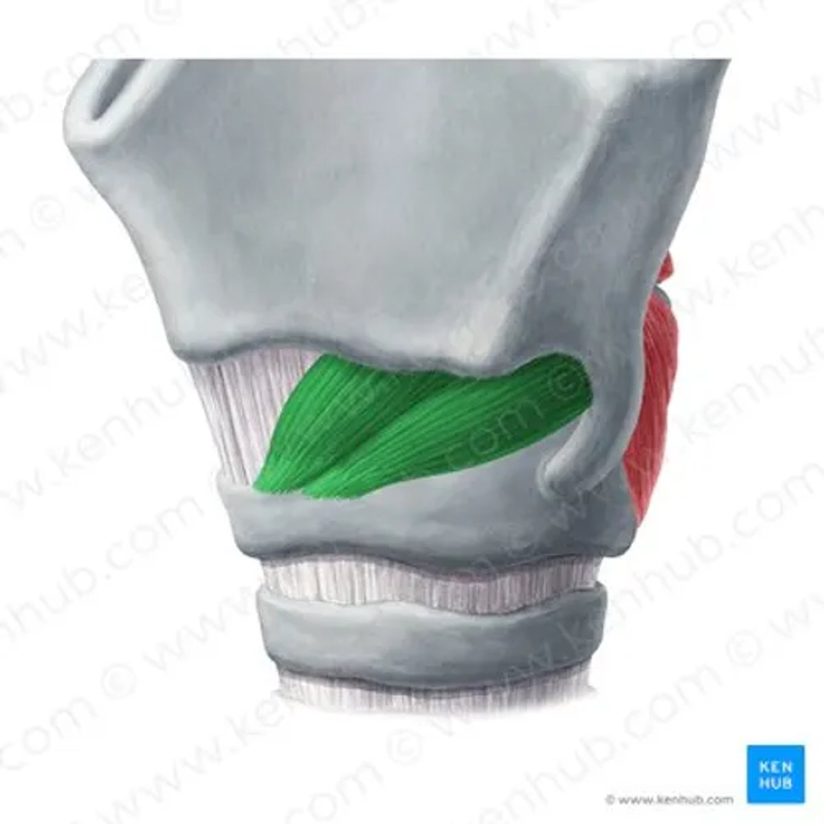
thyroarytenoid muscle
upper butterfly wing
structure
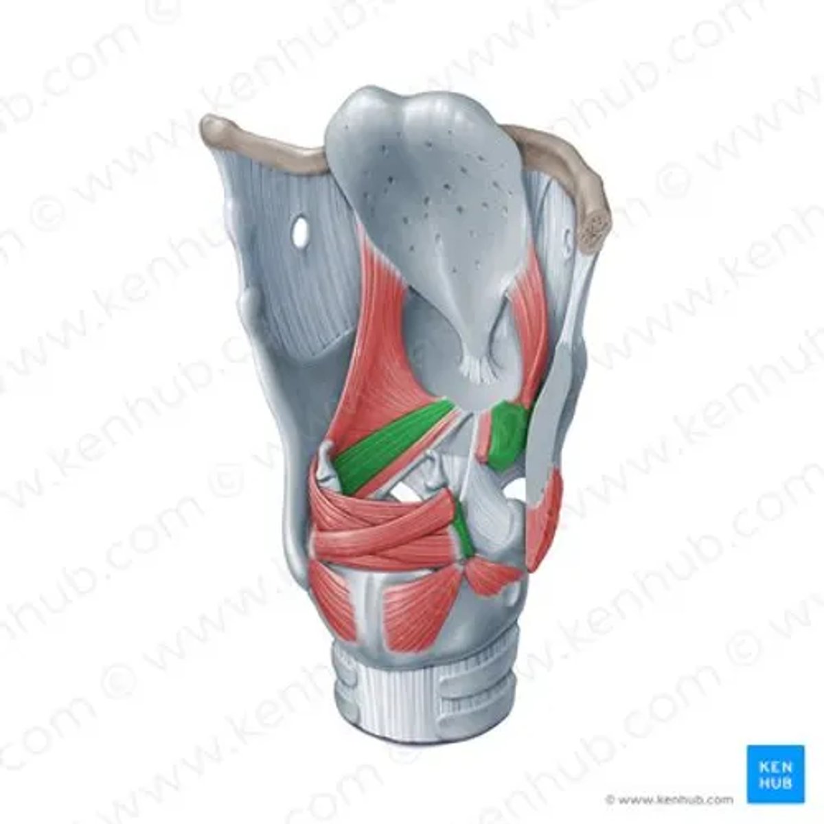
cricothyroid muscle
thick “M” on the anterior side
strucuture
innervation of intrinsic muscles of the larynx
BQ ONLY: vagus nerve
innervation of intrinsic muscles of the larynx
BQ: only vagus nerve
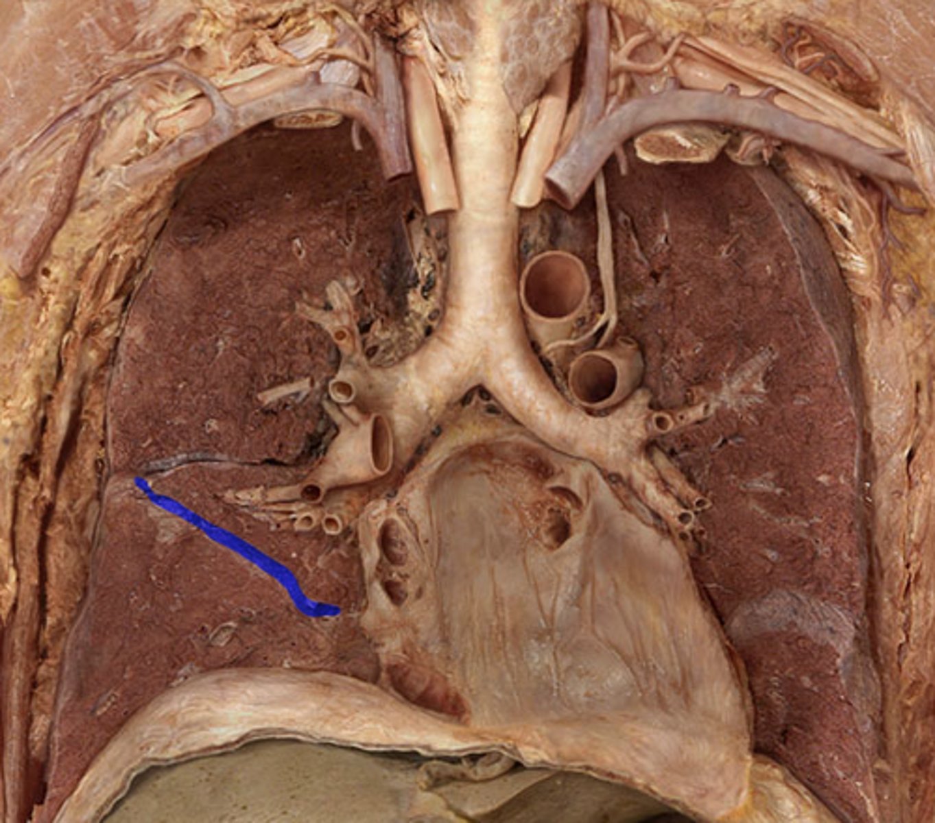
Trachea
the tube part of the windpipe before the split, TA will lift aorta and vena cava
entire strucutre
2352
tracheal cartilages
help make the individual C shaped rings on trachea, labeled C on image
individual structures
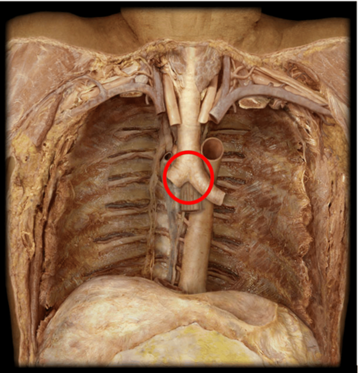
tracheal bifurcation
the division middle of the tracheal split
division
right main bronchus
structure
right main bronchus
BQ: more vertical, shorter, and wider than the left, an inhaled foreign body is more likely to enter the right main bronchus
left main bronchus
structure
segmental bronchi
BQ ONLY: third order bronchi, branch from the lobar bronchi, ten in each lung
R lung
larger lung, consists of three lobes: upper, middle, and lower, responsible for oxygen exchange.
L lung
smaller lung, consists of two lobes: upper and lower, also responsible for oxygen exchange.
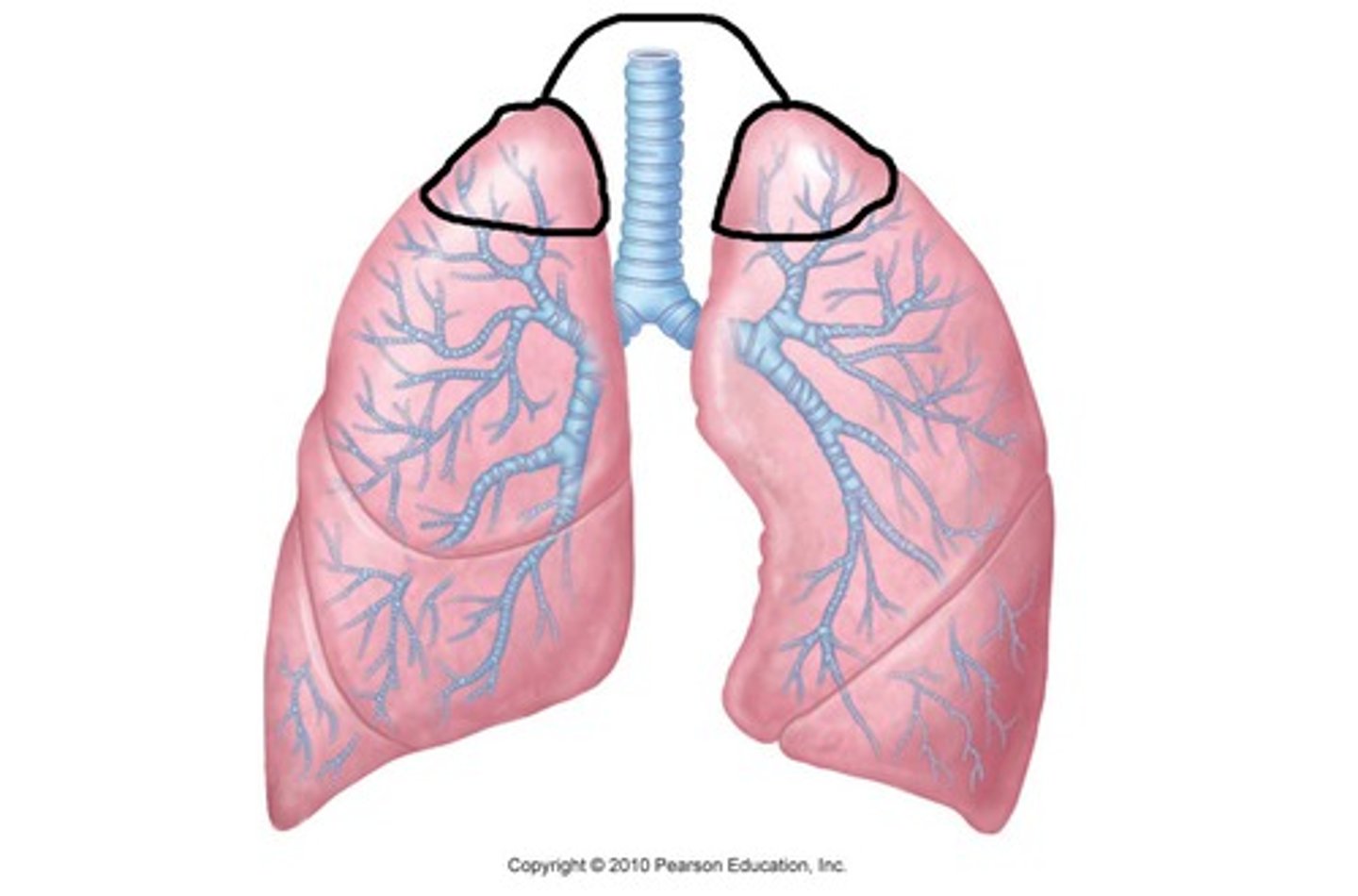
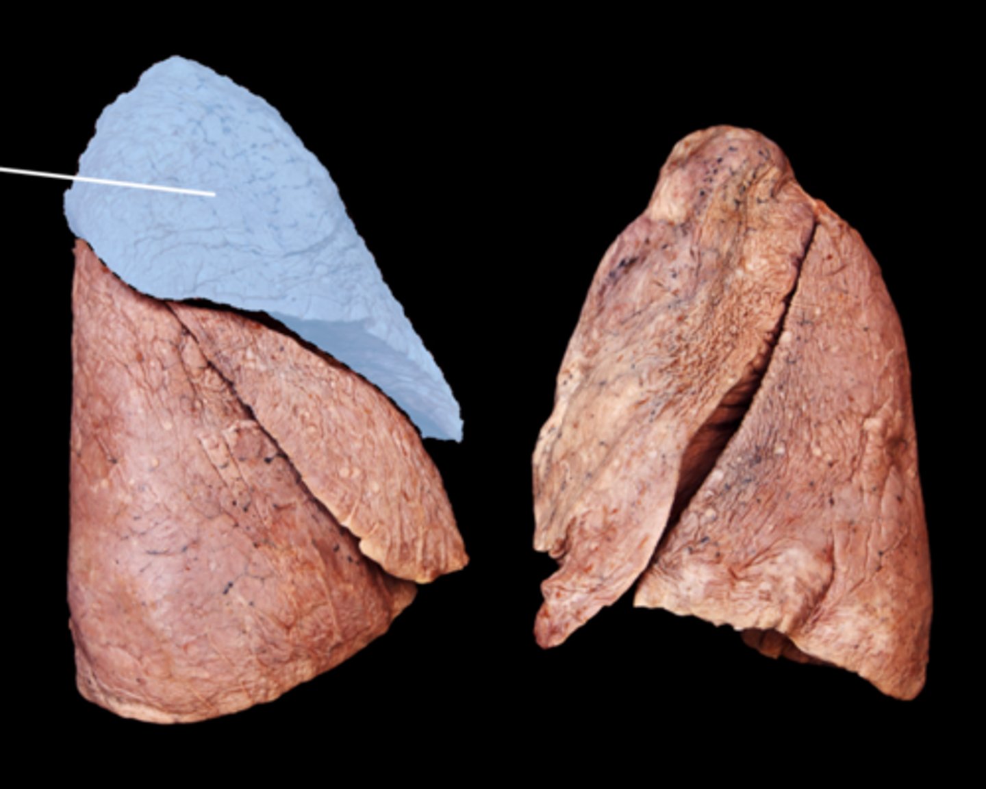
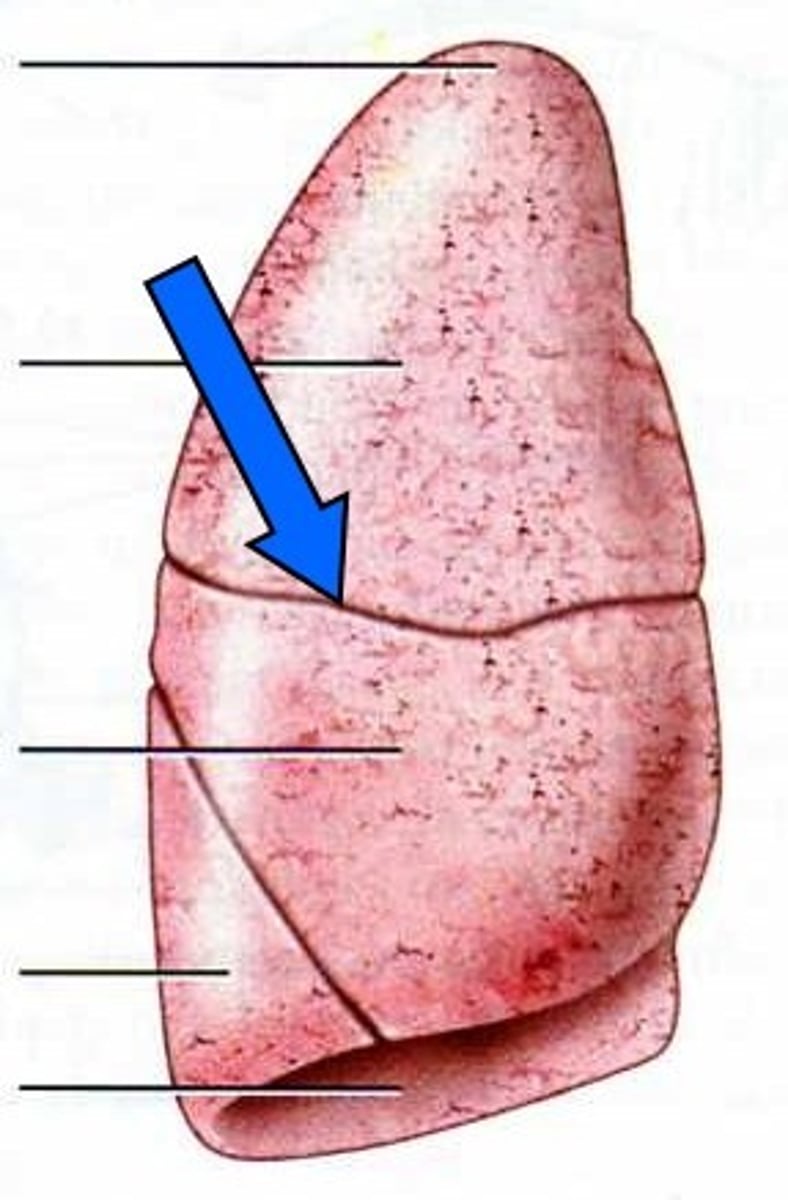
apex of the R/L lung
the top peak on the lungs
feature
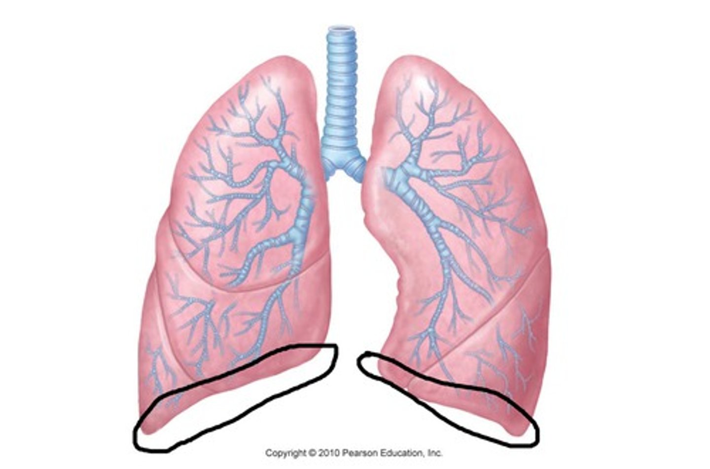
base of lung of the R/L lung
flat base of lung, TA will wave hand below lung
feature
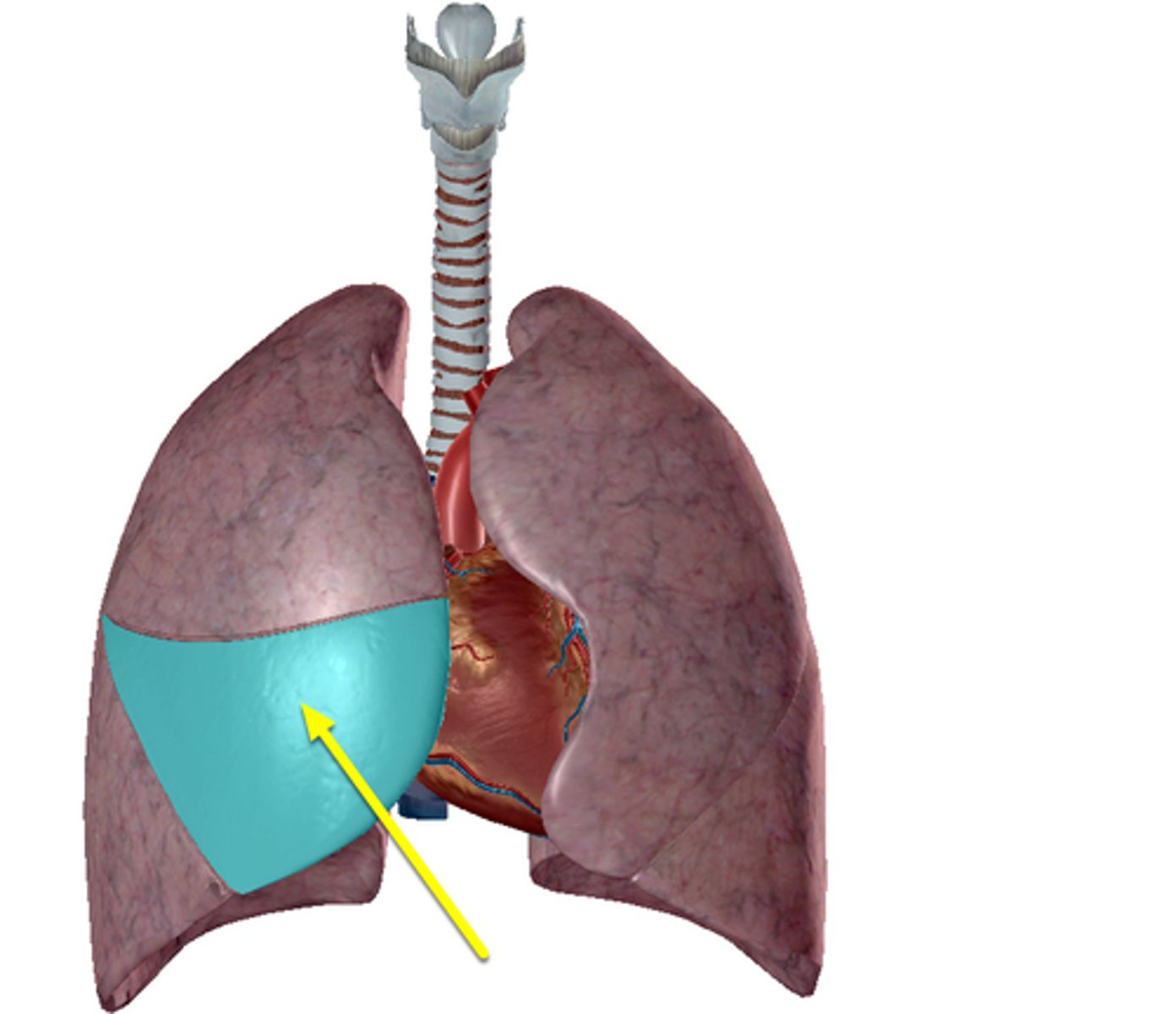
costal surface of the R/L lung
the surface of the lung that faces the rib cage
where blueish lines where the ribs are
layer
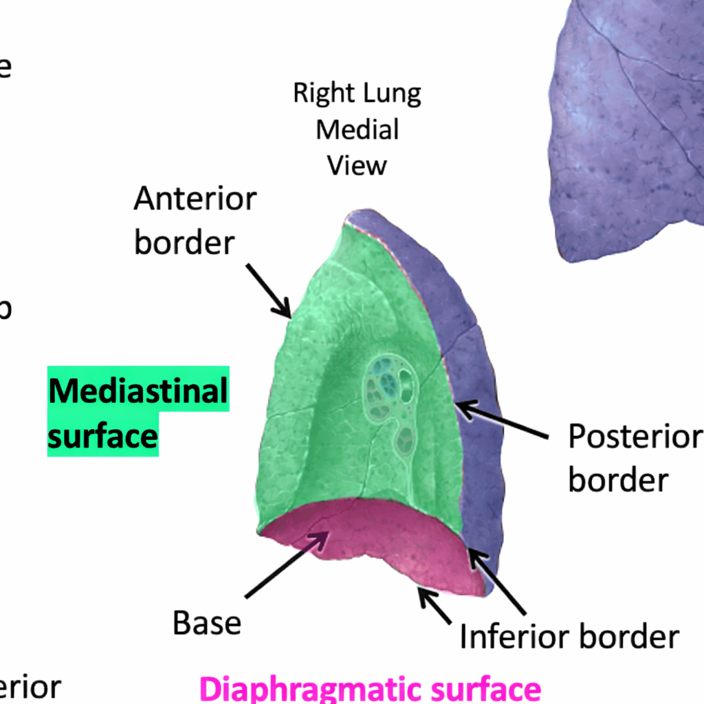
mediastinal surface of the R/L lung
the surface of the lung that faces the mediastinum, containing structures such as the heart and great vessels.
layer
diaphragmatic surface of the R/L lung
concaved portion of the bottom lung, TA will put fist in concave dome
anterior border of the R/L lung
this is the left lung
boundary
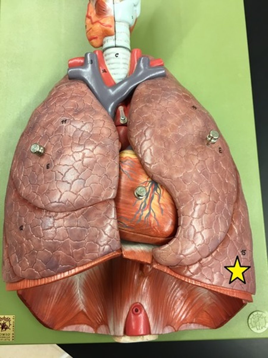
inferior border of the R/L lung
this is the lower edge of the lung, separating it from the diaphragm.
boundary
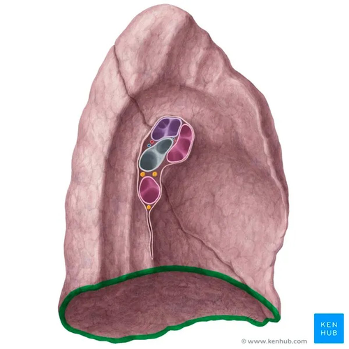
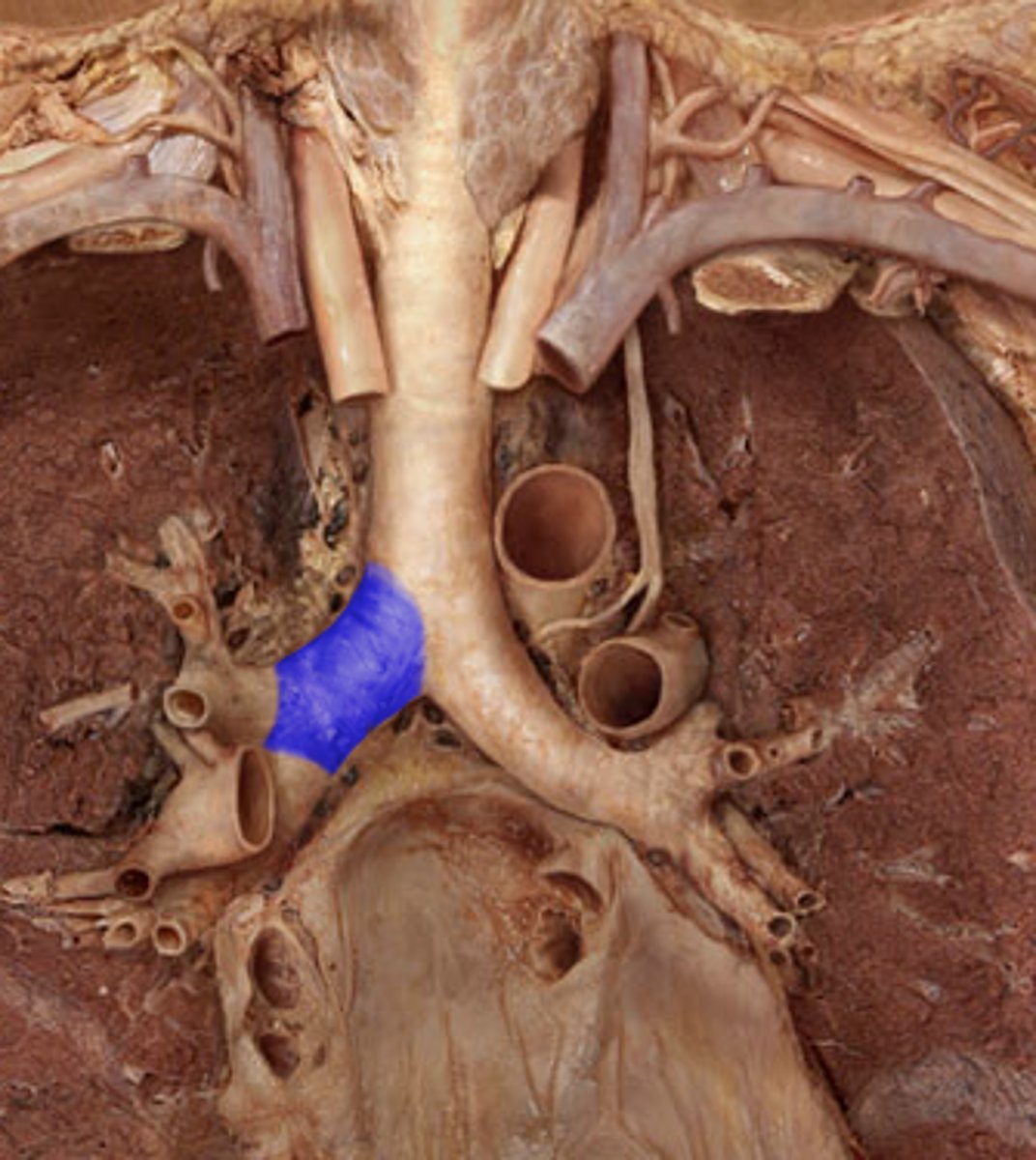
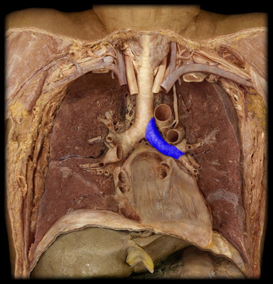
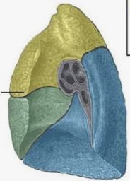
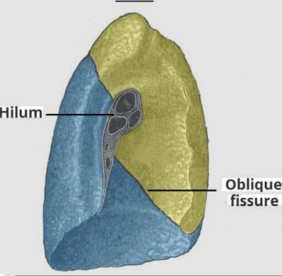
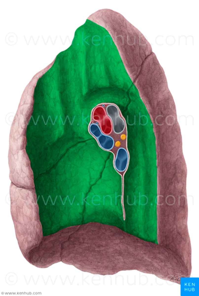
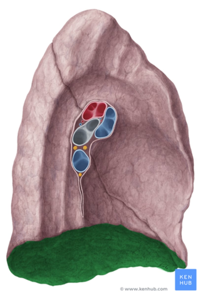
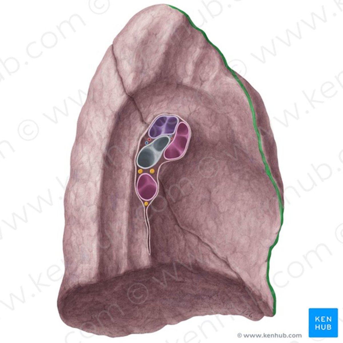
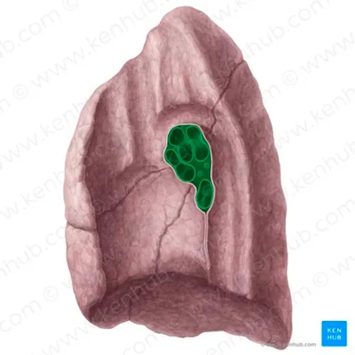
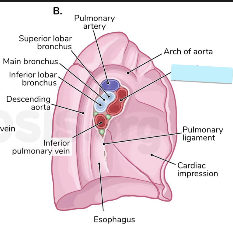
hilum of the R/L lung
all the blood vessels of the lung, TA will circle probe around
region
root of the R/L lung
the vessels within the hilum of the lung
TA will probably poke probe into all the holes
collective structure
lobes of right lung
superior, middle, inferior
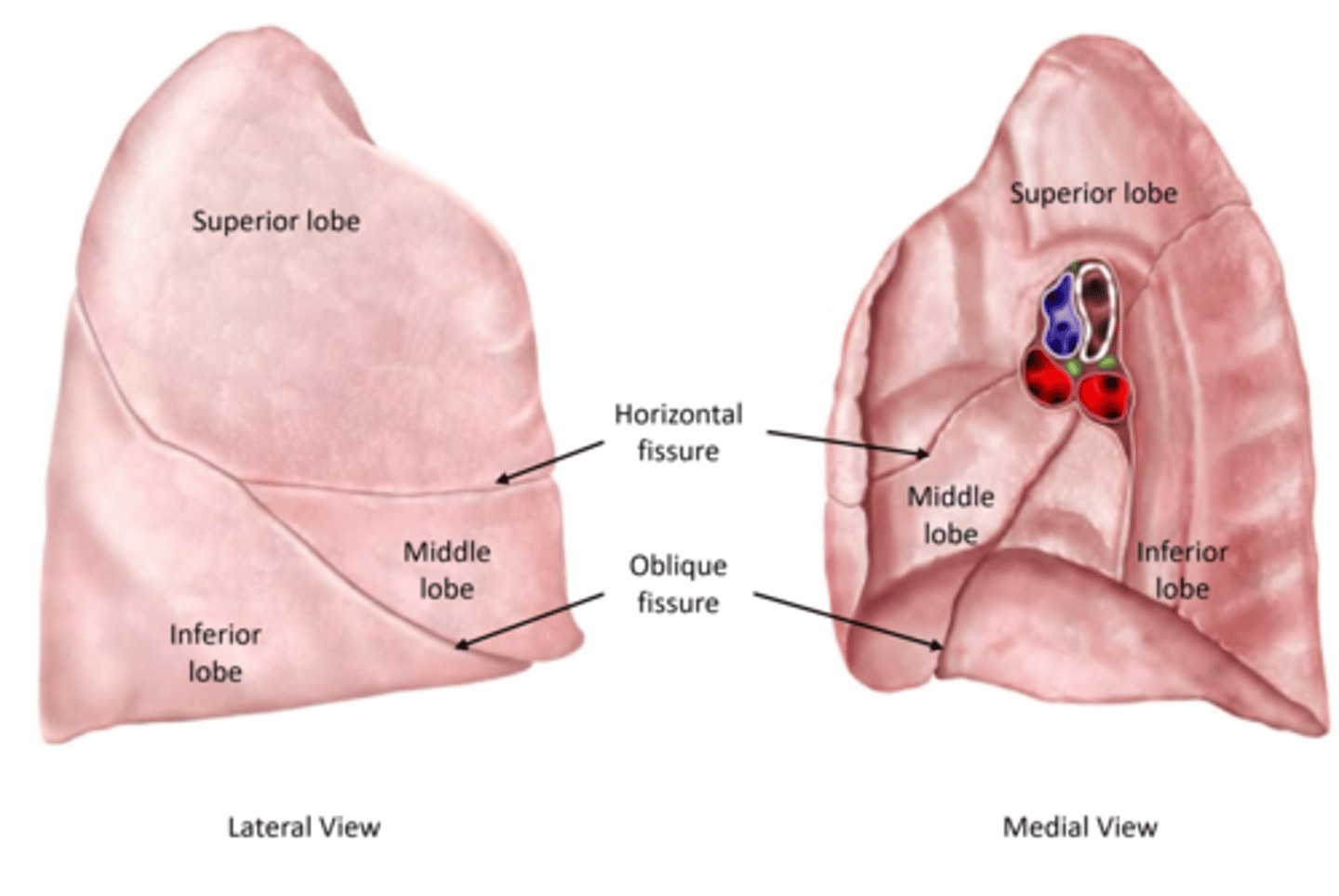
superior lobe of the right lung
top love of the three lobes
structure
middle lobe of the right lung
more anterior anatomically, more cubic in shape
strucutre
inferior lobe of the right lung
strucutre
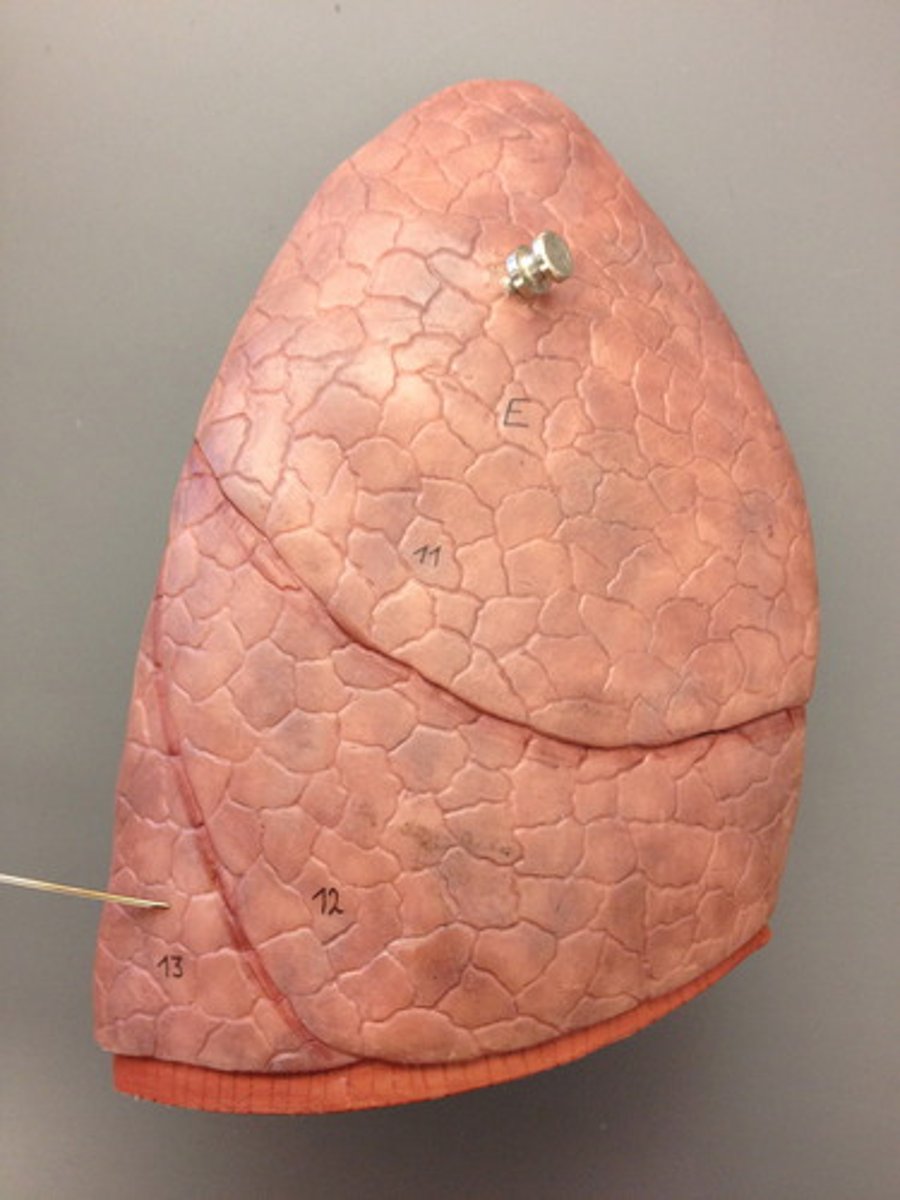
oblique fissure of the right lung
the “S” shaped fissure that goes through the superior, middle and inferior lobes
space
horizontal fissure of the right lung
goes straight across between the superior and middle lobes
space
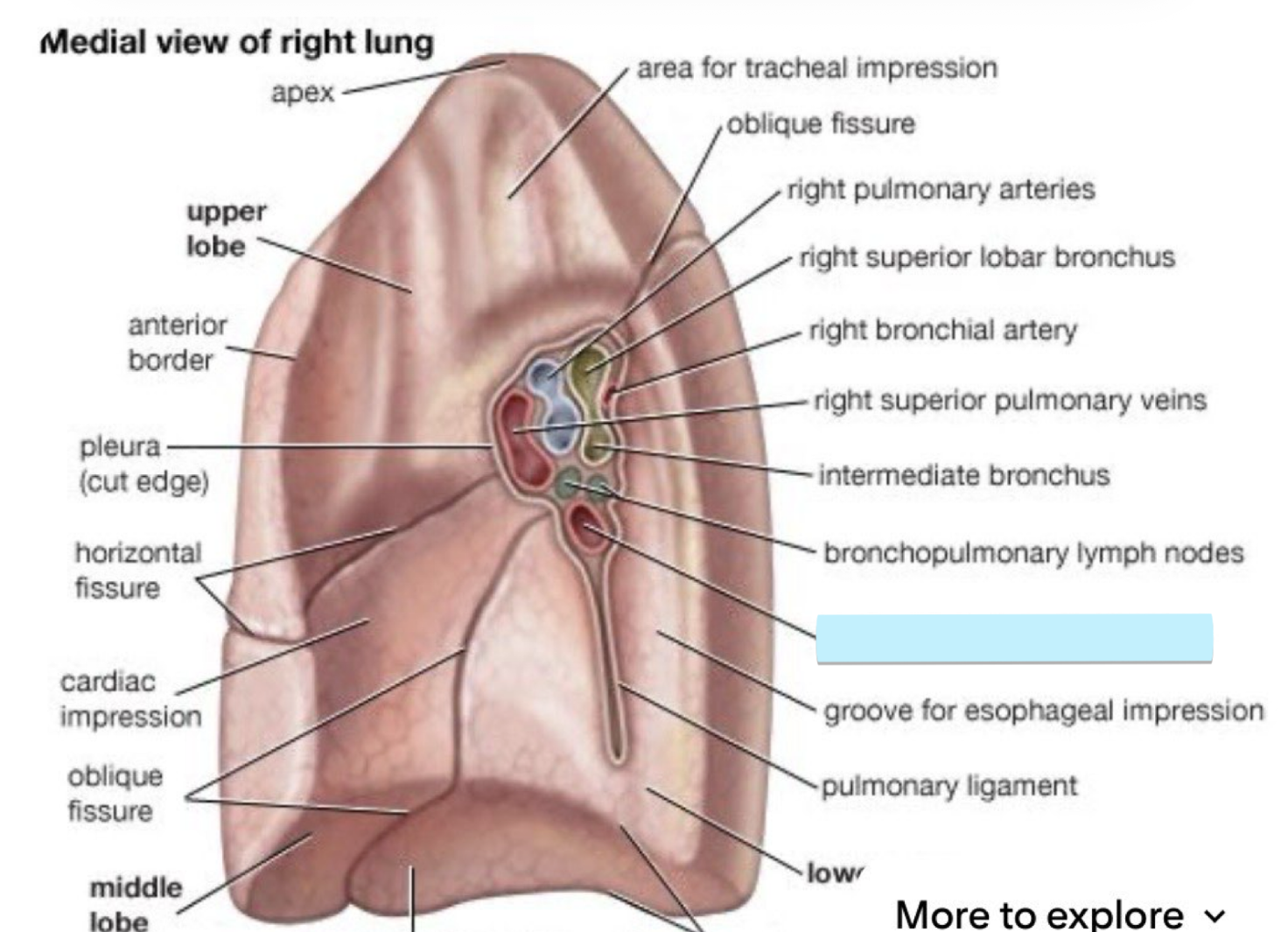
right superior lobar bronchus of the right lung
some cartilage, extends up to R superior lobe
structure
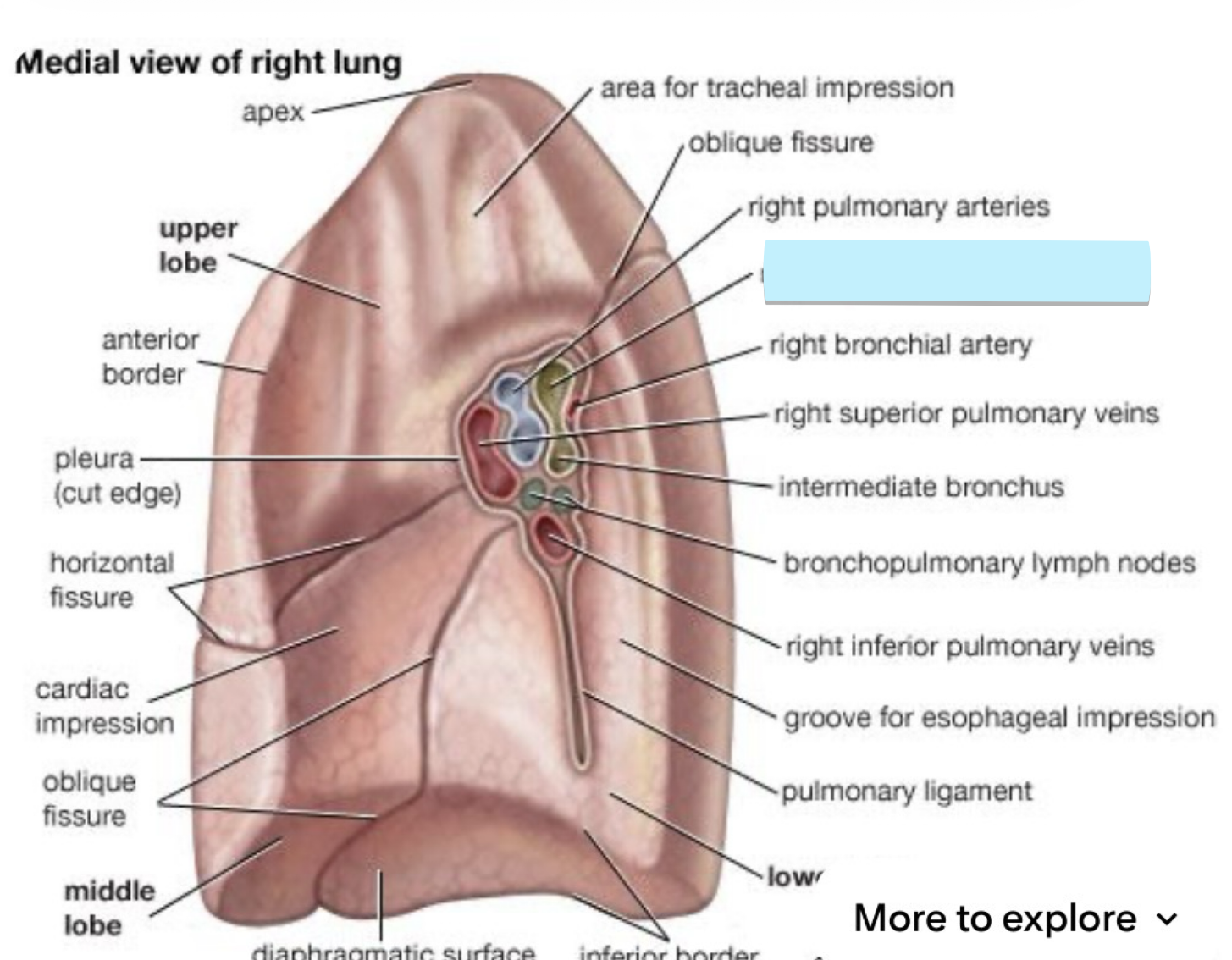
interlobar bronchus of the right lung
below the right superior lobar bronchus, thicker walls
strucutre
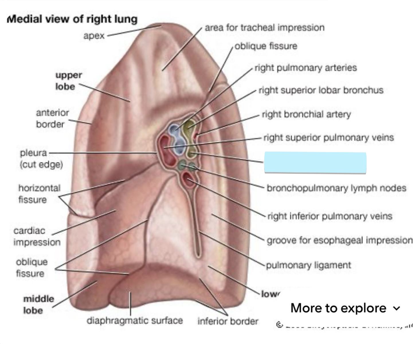
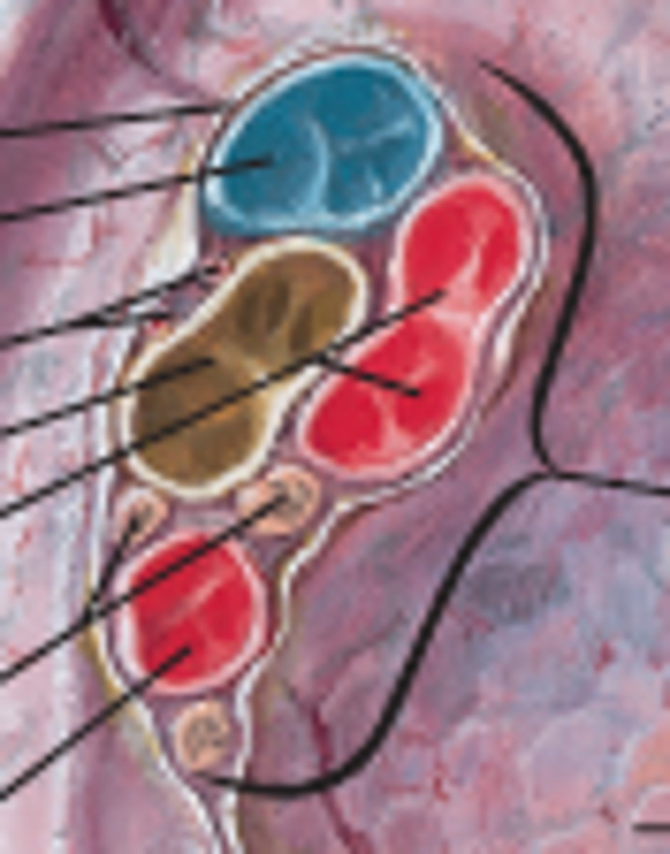
right pulmonary artery of the right lung
two vessels together on R side
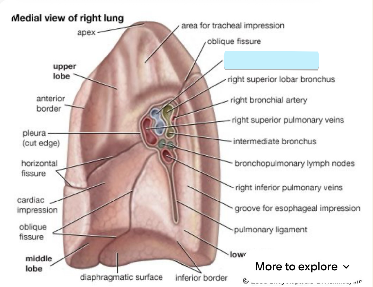
right superior pulmonary vein of right lung
below arteries
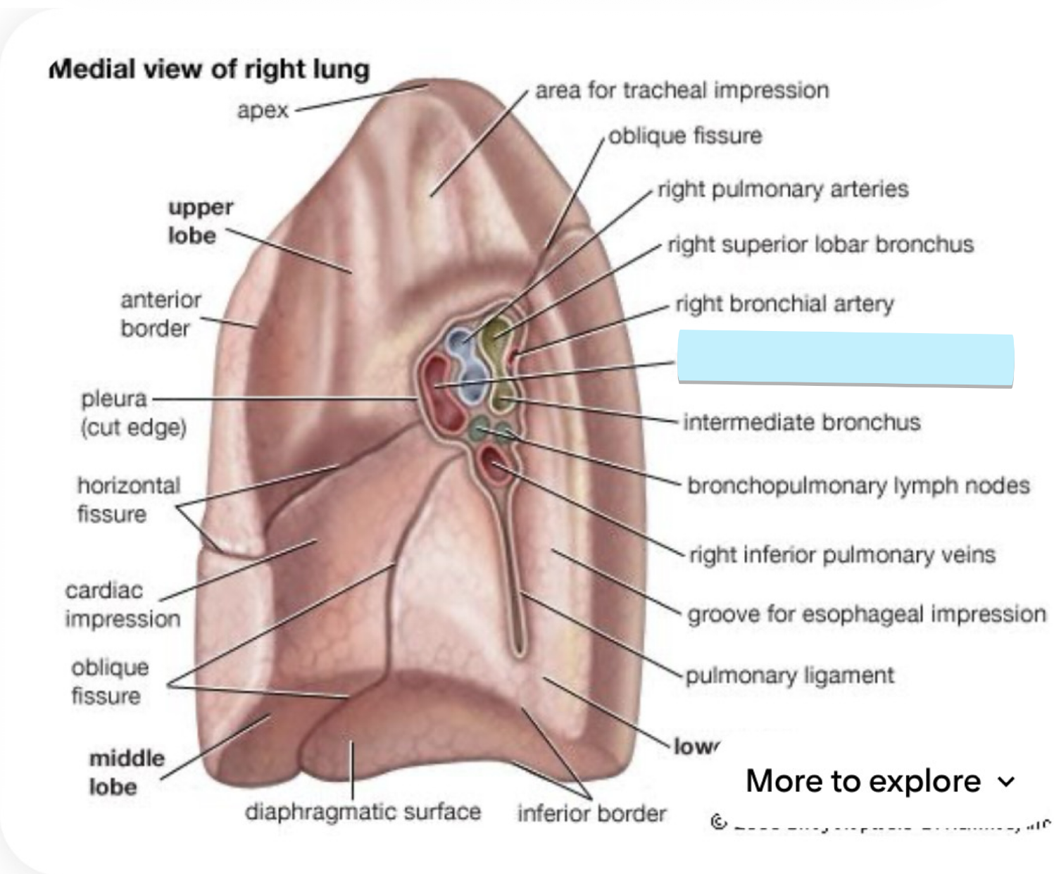
right inferior pulmonary vein of the right lung
below the bronchi
structure
superior lobe of left lung
the uppermost section of the left lung, looks like a pillar
strucutre
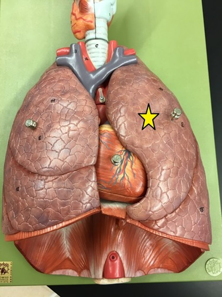
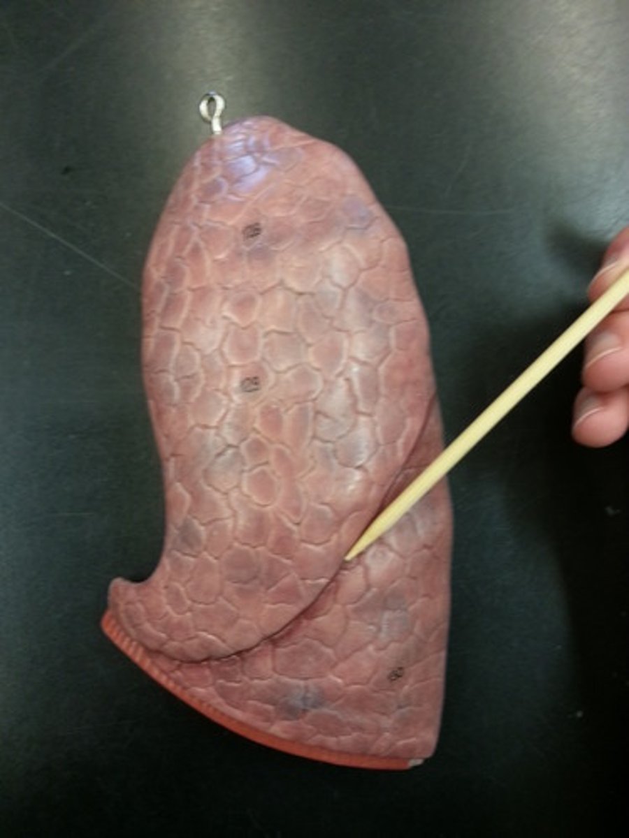
cardiac notch of left lung
looks kinda like a triangular shaped divot or a bite
on the mediastinal surface
depression
cardiac notch of left lung
BQ: like a bite taken out of the superior lobe, the pericardium is visible through the notch.
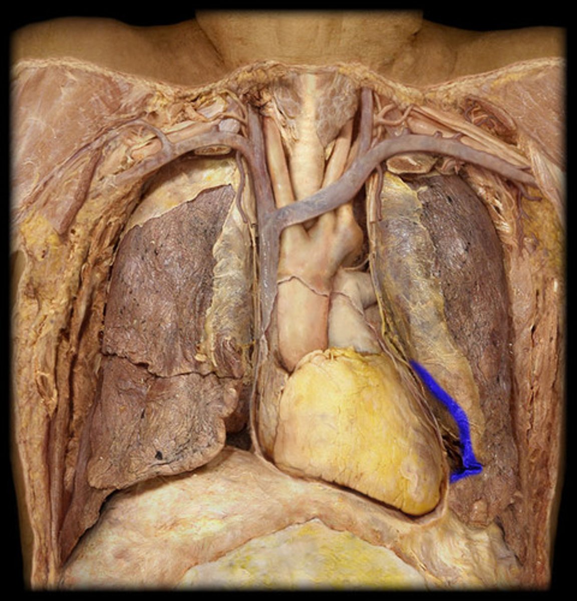
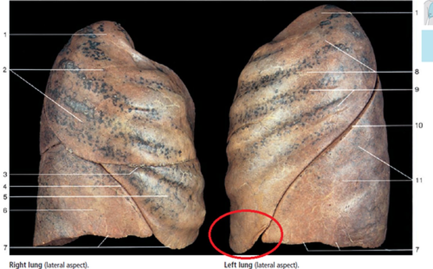
Lingula of left lung
flappy part at the bottom of the superior lobe
feature
inferior lobe of the left lung
bottom lobe
strucutre
oblique fissure of the left lung
space between the two lobes
space
root of left lung
collective structure
vessels that enter the left lung
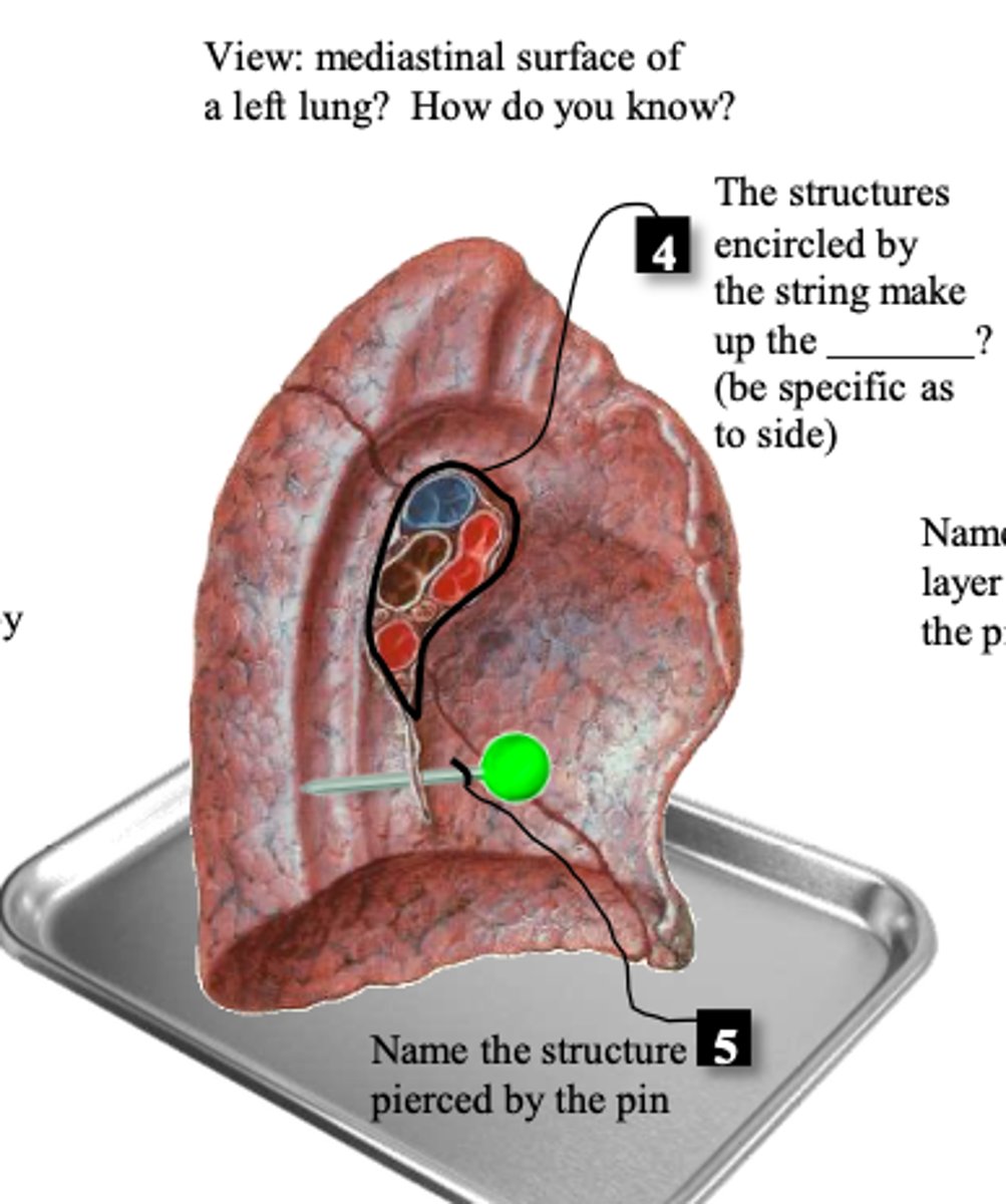
left main bronchus
kinda cut
structure
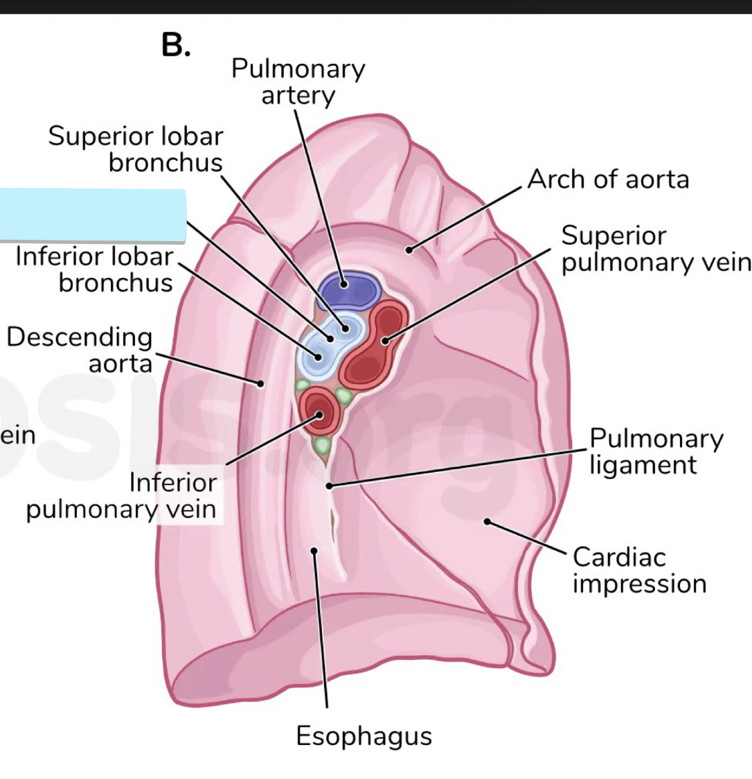
left pulmonary artery of left lung
the top and largest vessel, split up
strucutre
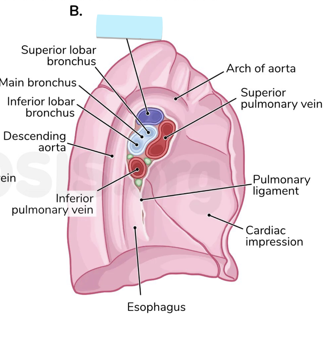
left superior pulmonary vein of left lung
strucutre
left inferior pulmonary vein of the left lung
structure
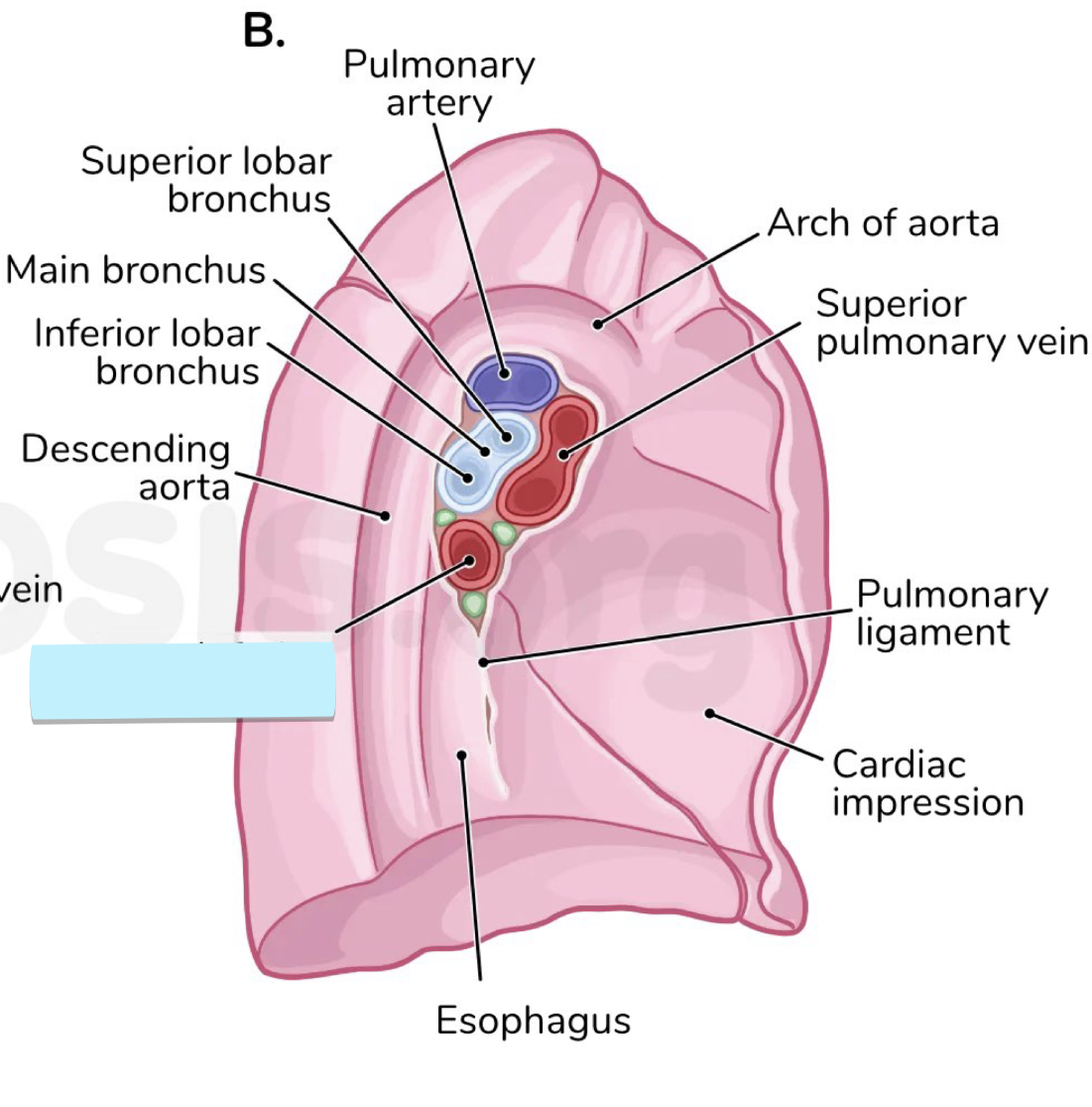
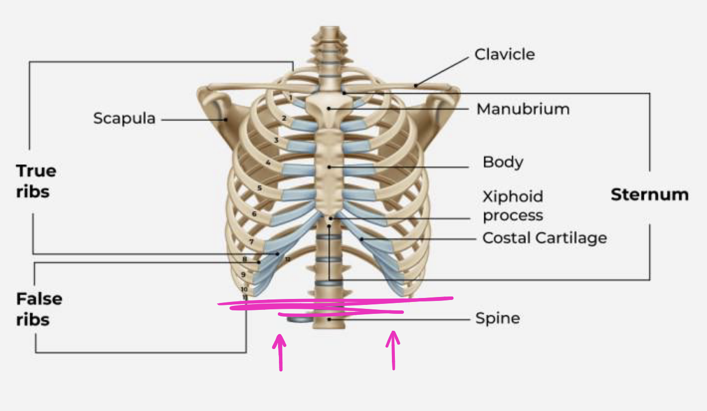
superior boundary of the mediastinum
ta will wave hand over clavicle
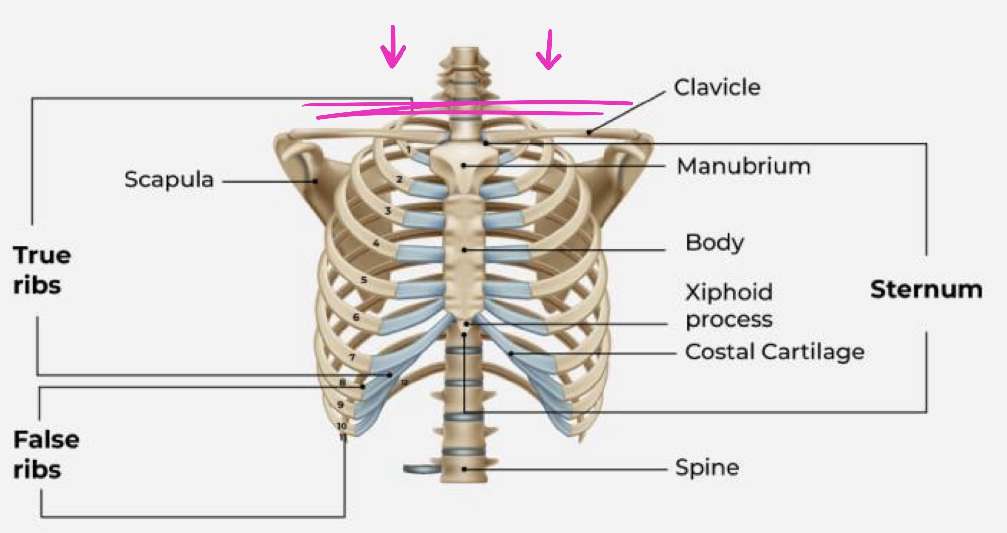
inferior boundary of mediastinum