SAM3.1: Optho Orbit
1/68
There's no tags or description
Looks like no tags are added yet.
Name | Mastery | Learn | Test | Matching | Spaced | Call with Kai |
|---|
No analytics yet
Send a link to your students to track their progress
69 Terms
Microphthalmos
Congenitally small globe +/- abnormalities.
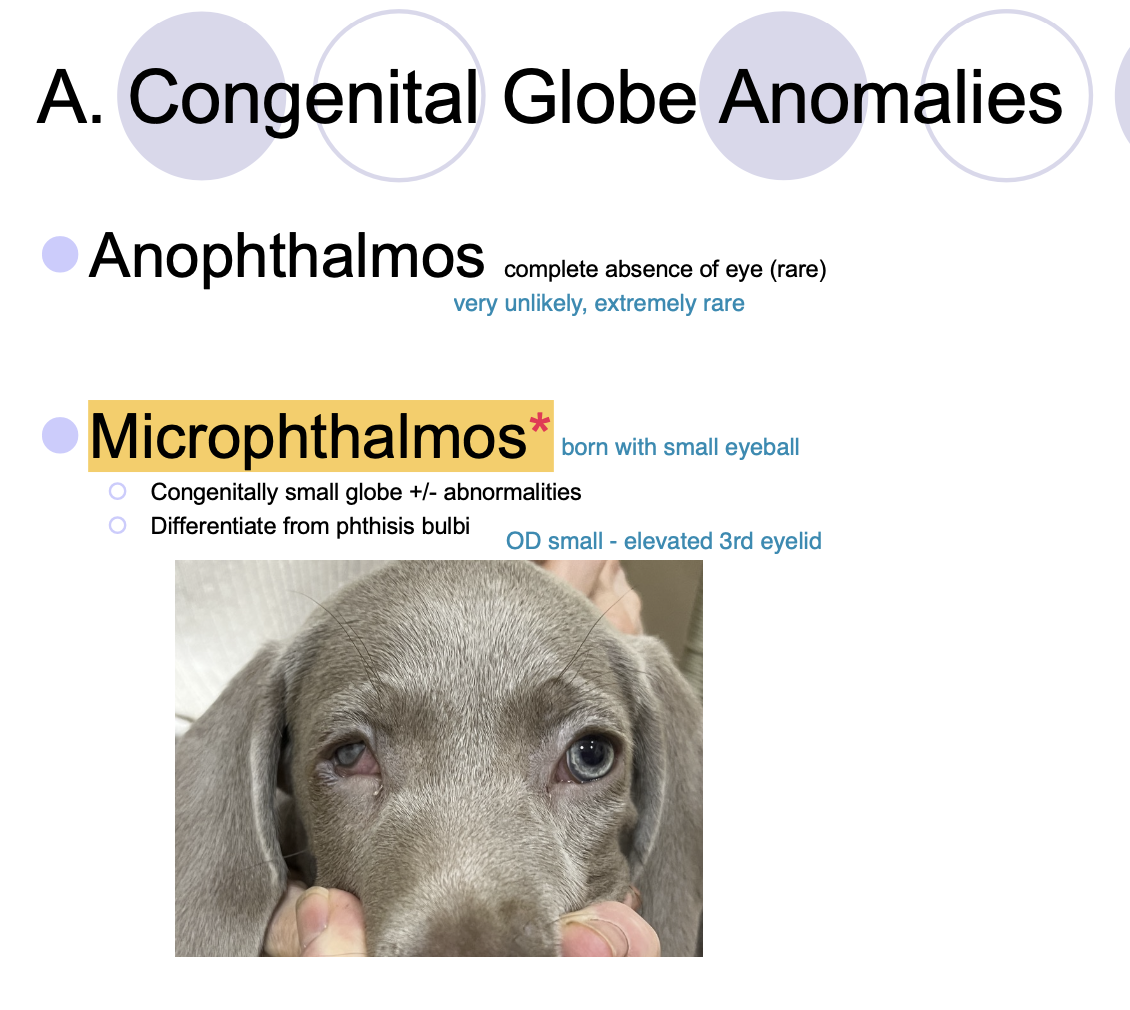
Microphthalmos must be differentiated from
phthisis bulbi.

True microphthalmos is association
without other ocular defects (cataracts, anterior segment dysgenesis, PHPV, retinal dysplasia).

Vision w/ microphthalmos
May or may not be present
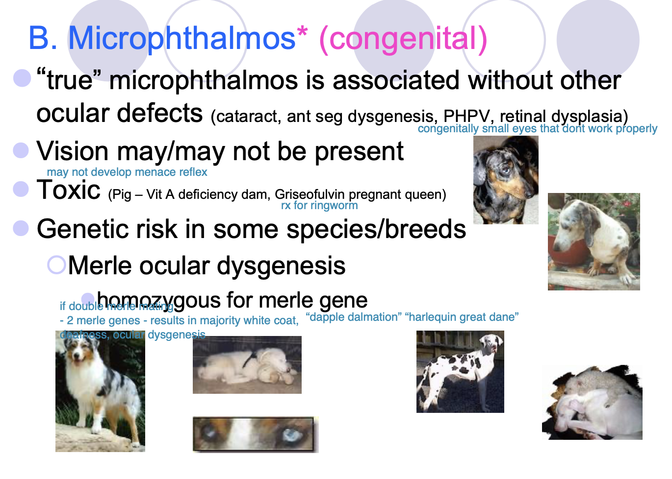
Toxic microphthalmos
Pigs: Vitamin A deficient dams.
Griseofulvin pregnant queen (not a pig... it's a cat).

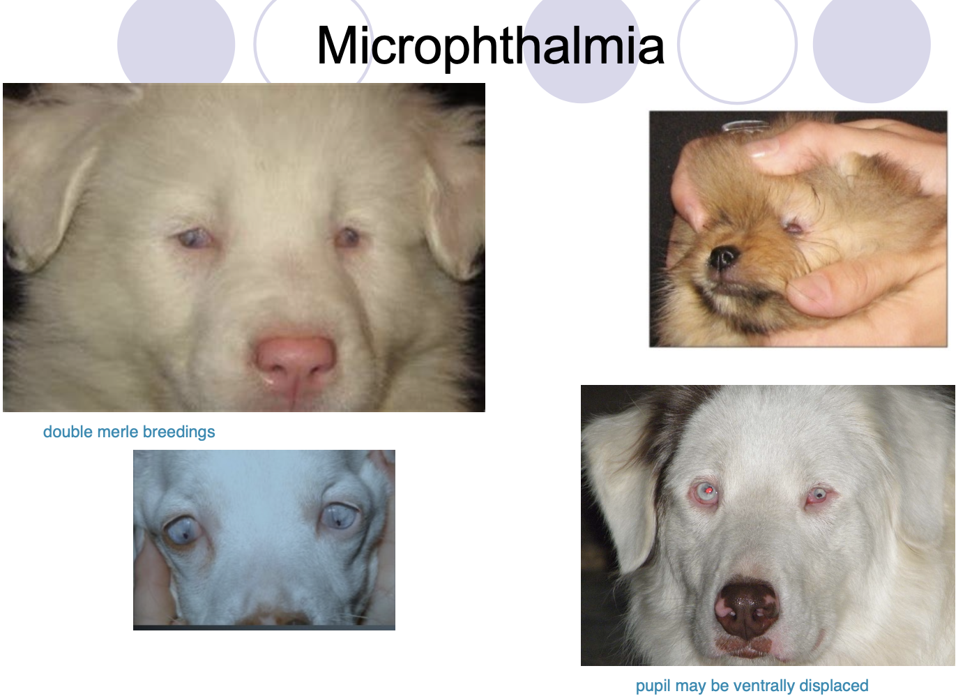
Genetic risk of microphthalmos in some spp/breeds includes
Merle ocular dysgenesis - homozygous for merle gene.
Throughbreds.
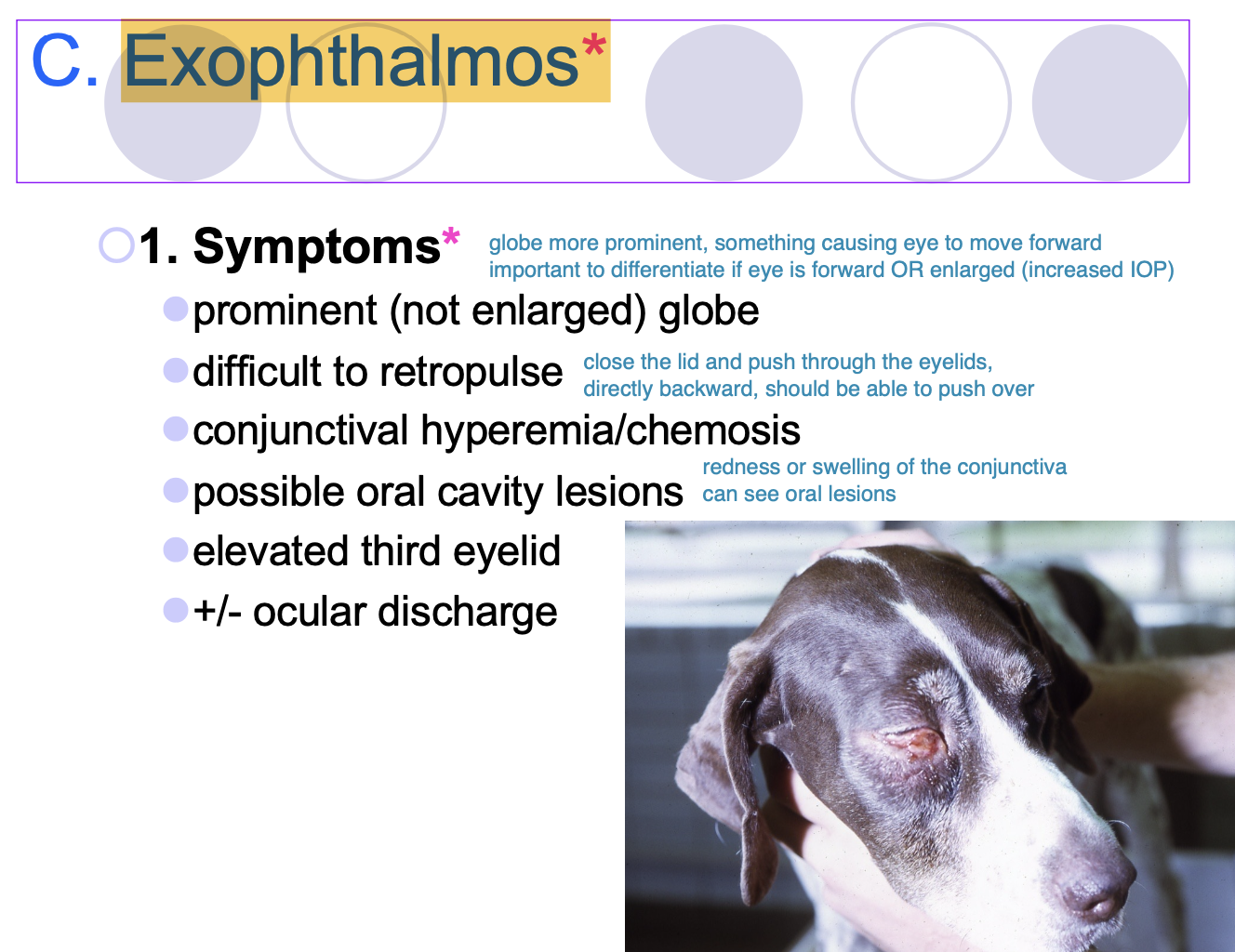
Exophthalmos
An eye that is more prominent or push forward w/in orbit.

Symptoms of exophthalmos (6)
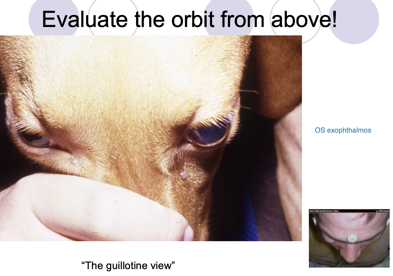
Prominent (not enlarged) globes.
Difficult to retropulse.
Conjunctival hyperemia/chemosis.
Possible PO cavity lesions.
Elevated third eyelid.
+/- ocular discharge.
Dx for exopthalmos (6)
Palpation of the orbit/oral exam.
Chest rads and bloodwork.
Skull/dental rads.
FNA for cytology/biopsy.
U/S.
CT/MRI.

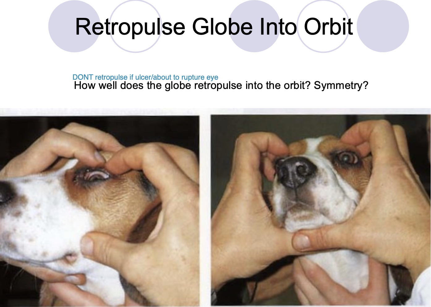
Palpation of the orbit/oral exam includes (2)
Retropulsion of the globe.
Examine PO cavity at the end of the exam (b/c it can be painful)
Chest rads and bloodwork should be done to
r/o disseminated neoplasia/infectious.
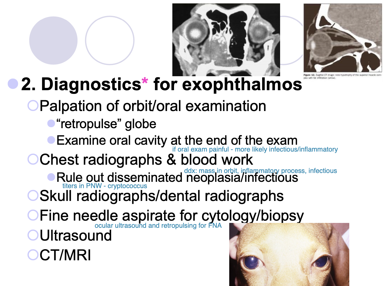
2 most common causes of exophthalmos are
masses or infections/inflammatory processes.
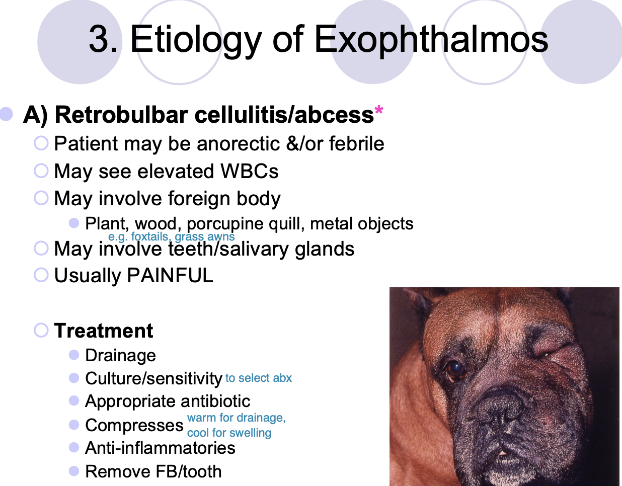
Etiology of Exophthalmos (12)
Retrobulbar cellulitis/abscess.
Neoplasia.
Crypto.
Eosinophilic myositis of masticatory mm.
Extraocular polymyositis.
Craniomandibular Osteopathy.
Cysts.
Hemorrhage.
Orbital fxs.
Vascular Anomalies.
Granulomas.
Tetanus.
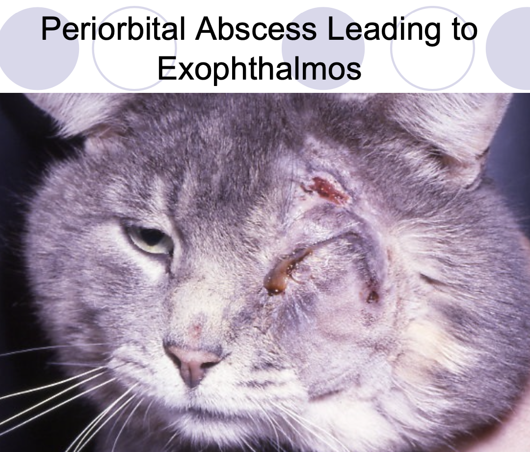
What may you see in a P' w/ retrobulbar cellulitis/abscess
anorectic and/or febrile.
elevated WBCs.
involve FB.
involve teeth/SG.

Retrobulbar cellulitis/abscess is usually
very painful.

Tx for retrobulbar abscess
Drain.
C&S.
Appropriate ABX.
compresses.
Anti-inflam.
Remove FB/tooth.
Most common etiology of exophthalmos
Neoplasia (generally older P's)

Neoplasia pain in the eye
usually painless and slowly progressive.

What may you see with exophthalmos from neoplasia (2)
globe depressed by mass.
Secondary inflam can develop.
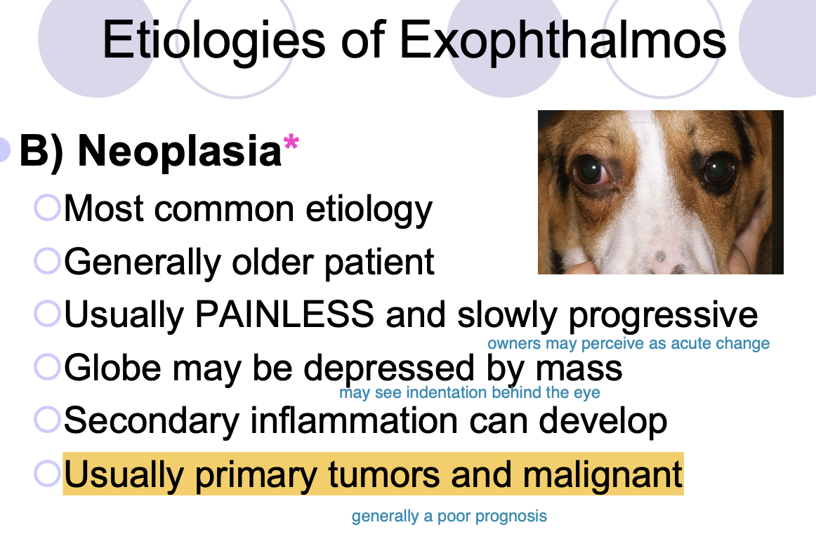
Trend for ocular neoplasia
usually primary tumors and malignant.

Ddx for primary retrobulbar tumors (6)
multilobular osteoma,
fibrosarcoma,
meningioma,
adenocarcinoma,
SCC,
myxoma

Ddx for secondary retrobulbar tumors
metastatic,
adenocarcinoma from nasal cavity,
lymphosarcoma
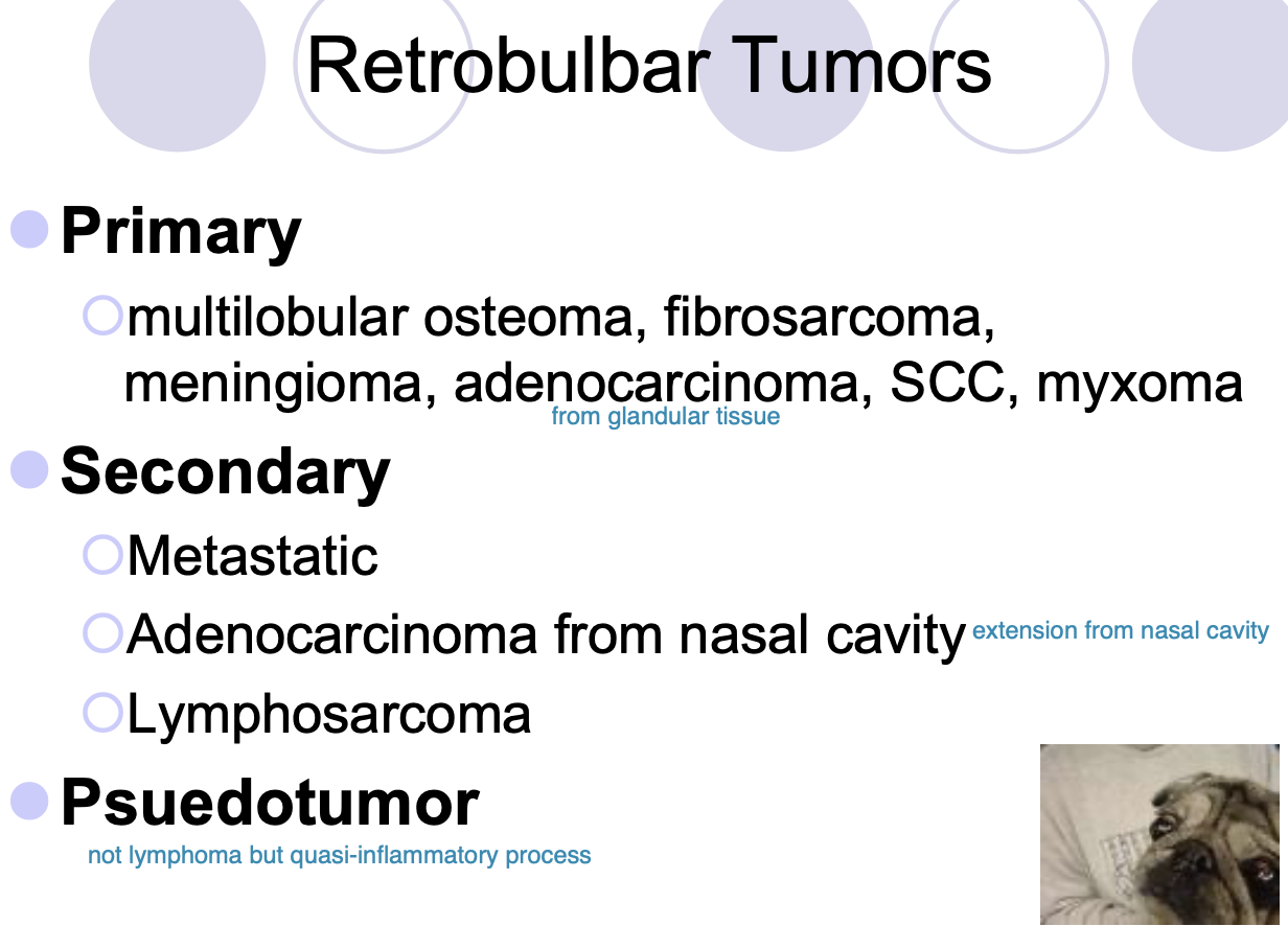
Pseudotumor
a retrobulbar tumor that is a quasi-inflammatory process (not lymphoma)

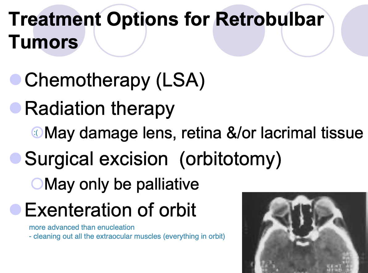
Tx for retrobulbar tumors
Chemotherapy (LSA)
Radiation therapy (may damage lens, retina, lacrimal tissue)
Surgical excision (orbiotomy, may only be palliative)
Exenteration of orbit
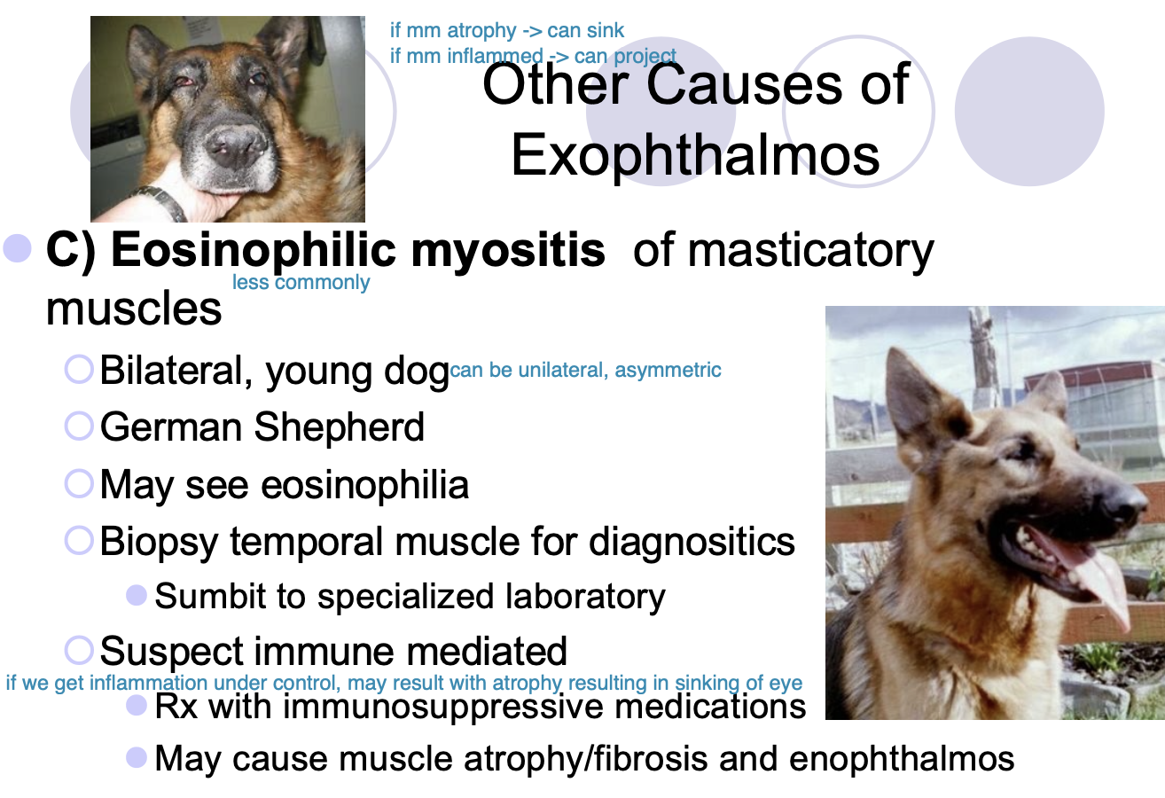
Eosinophilic myositis of masticatory muscles signalment
bilateral, young dog
GSD

Eosinophilic myositis of masticatory muscles effect on eye
exophthalmos (during inflammation),
may cause muscle atrophy/fibrosis and enophthalmos
Etiology of eosinophilic myositis of masticatory muscles
immune-mediated

Dx of eosinophilic myositis of masticatory muscles
Bx of temporal m, submit to specialized lab
May see eosinophilia

Tx of eosinophilic myositis of masticatory muscles
immunosuppressive medications
Extraocular polymyositis signalment
bilateral, usually young, large breed dog
golden retriever may be predisposed

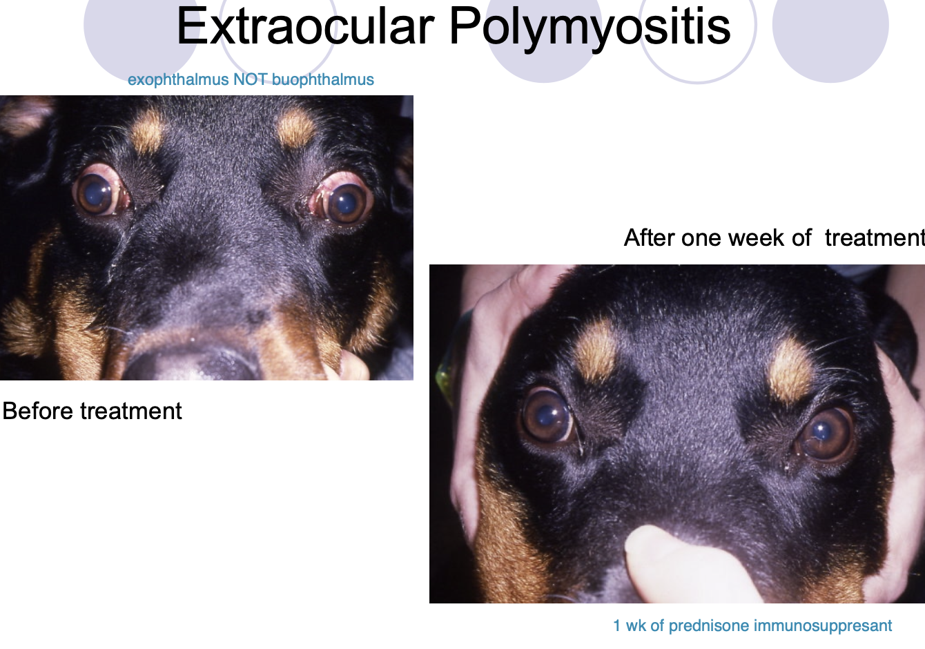
Extraocular polymyositis effect on eye
exophthalmos
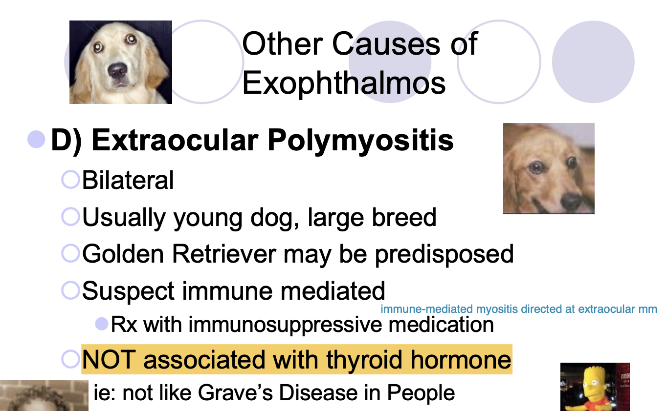
T or F? Extraocular polymyositis is associated with thyroid hormone
False. NOT assoc with thryoid hormone, not like Grave’s disease in people

Tx of extraocular polymyositis
immunosuppressive medication (e.g. prednisone)
Craniomandibular osteopathy signalment
usually 3-6mo old,
scottish terrior/WHWT predisposed
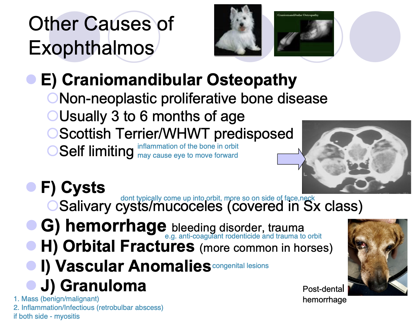
Craniomandibular osteopathy etiology
self-limiting, non-neoplastic proliferative bone disease

What condition must be differentiated from exopthalmos
buphthalmos
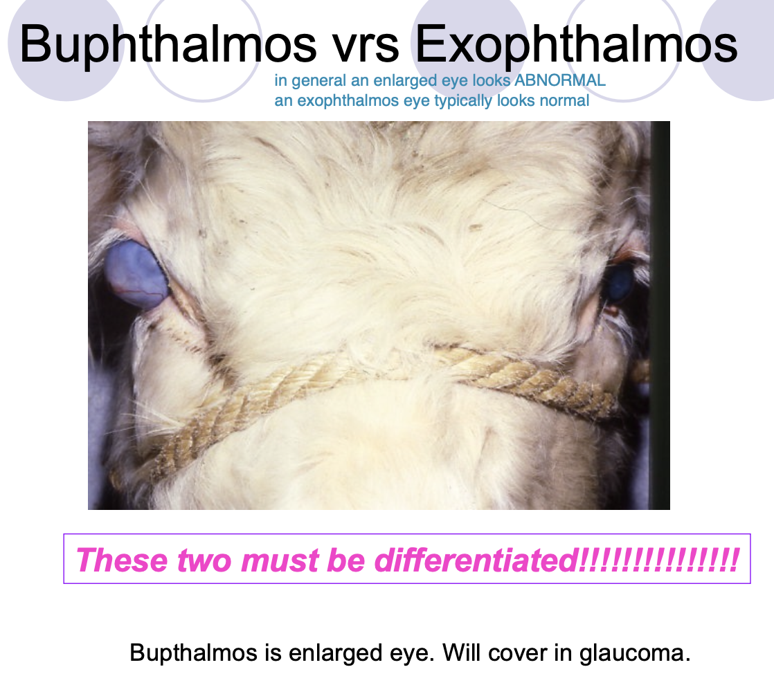
Buphthalmos is
enlarged eye

How do you differentiate Buphthalmos and Exophthalmos
Buphthalmos eyes usually appear to have abnormalities.
Exophthalmos eyes are healthy eyes that look displaced.

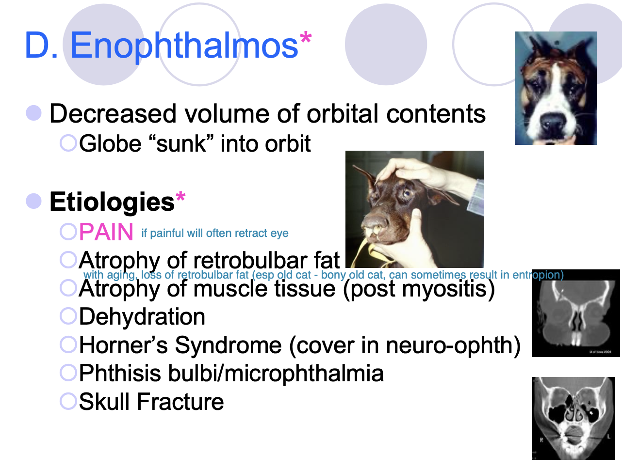
Enophthalmos is
decreased volume of orbital contents - globe sunk into orbit.

Etiologies of Enophthalmos (5)
Pain.
Atrophy of the retrobulbar fat or m. tissue.
Dehydration.
Horner's syndrome.
Phthisis bulbi/microphthalmia.
Skull Fx.
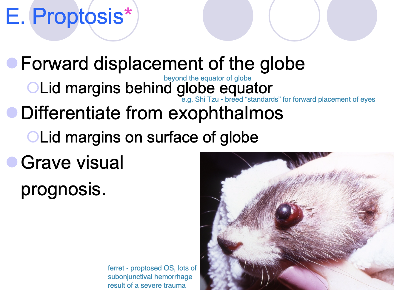
Proptosis def
Forward displacement of the globe. Lid margins are behind globe equators.

proptosis is differentiated from
exophthalmos - lid margins on surface of the globe.

Prognosis for Proptosis?
Grave for vision.
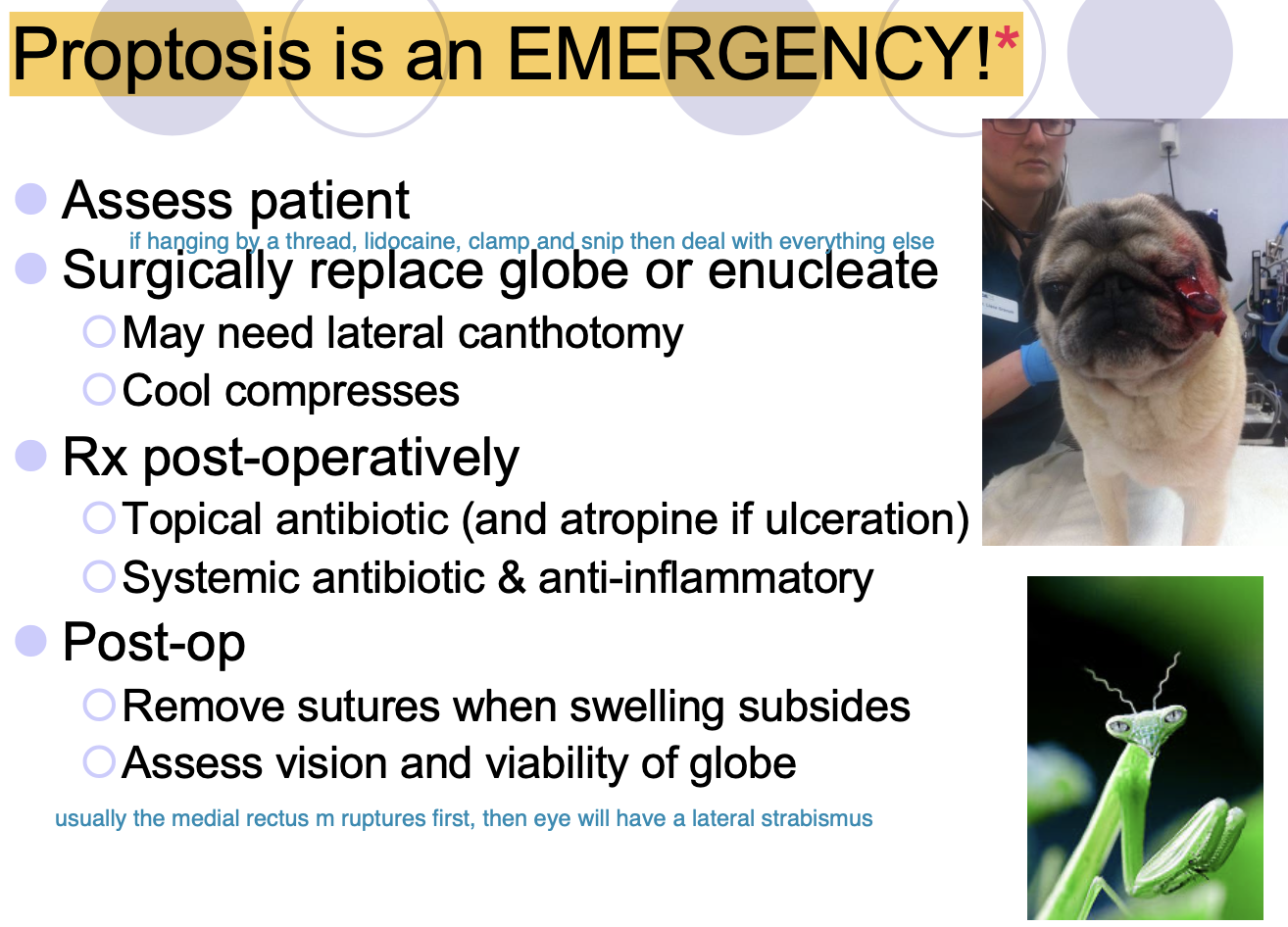
Proptosis is
an emergency
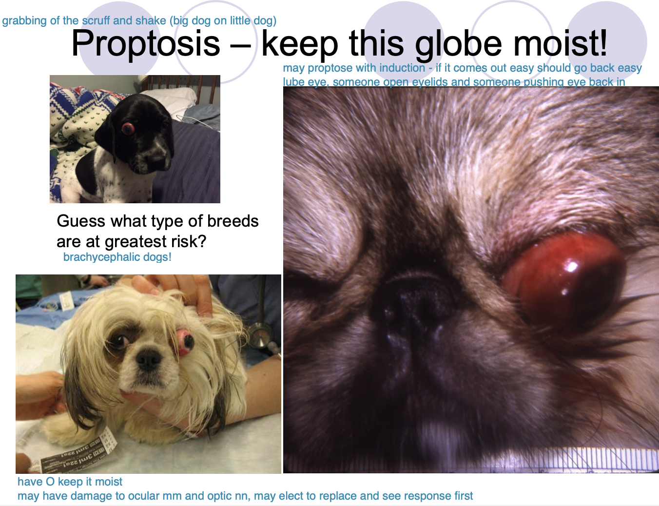
At risk breeds for proptosis
brachycephalic dogs

Proptosis Tx
Surgically replace globe or enucleate - cool compresses and may need lateral canthotomy

Post-op Rx for Proptosis (2)
Topical ABX (and atropine if ulceration).
Systemic ABX and anti-inflammatory.

Post-op Sx for Proptosis (2)
Remove sutures when swelling subsides.
Assess vision and viability of globe.
Describe prolapse retrobulbar fat
herniation of retrobulbar fat through Tenon’s capsule
usually dorsal-lateral and presents as conjunctival swelling

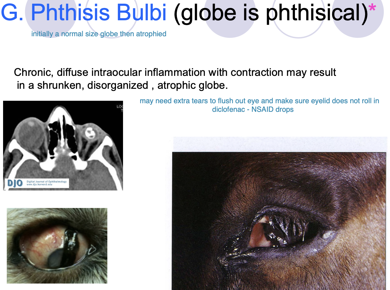
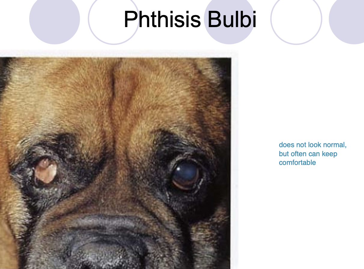
Phthisis Bulbi (3)
Globe is phthisical.
was normal at one point.
chronic, diffuse intraocular inflammation w/ contraction may result in shrunken, disorganized, or atrophic globe.
Phthisis Bulbi is usually
comfortable (globe can remain until it becomes painful)
Globe position: Exotropia
Wall eyes - lateral strabismus
Globe position: Esotropia
Cross eyed (Siamese cats)

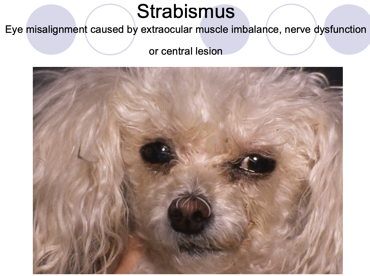
Strabismus can be the result of (3)
m. imbalance.
n. dysfxn.
central lesion.
Surgeries invoving the opthoorbit (6)
enucleation
exenteration
orbital prothesis
intraocular prosthesis
obitotomy
extrascleral (shell) prosthesis
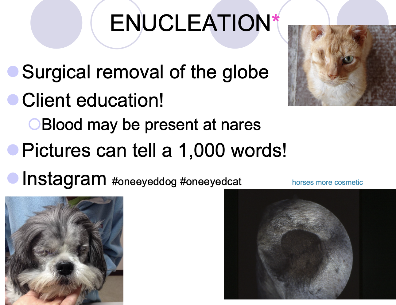
Enucleation def
Removal of the globe, third eyelid, lacrimal tissue and eyelid margins.
Enucleation is indicated for
blind/painful eyes or neoplasia

Enucleation techniques (2)
Subconjunctival approach.
Transpalpebral approach - harder to do and takes longer, but more ideal for containment of potential infection/neoplasia.

Post op warning to client for enucleation
blood from the nostril on the same side as the enucleated eye w/in a few days post-op is normal.
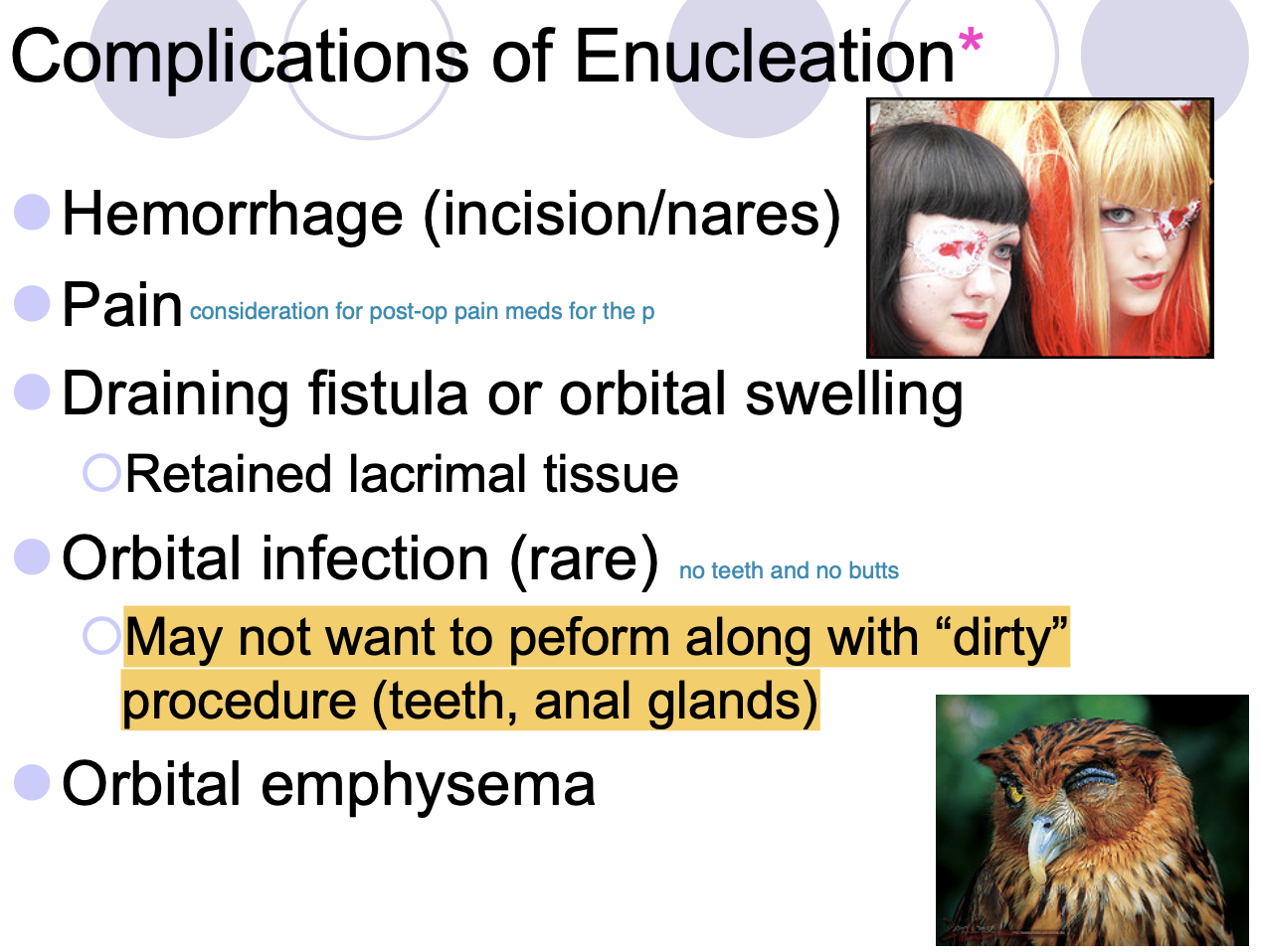
Complications of Enucleation (5)
Hemorrhage.
Pain.
Draining fistula or orbital swelling - retained lacrimal tissue.
Orbital infection (rare).
Orbital emphysema.

Orbital infections: You may not want to do enculeation
at the same time as dirty procedures (teeth and AG)
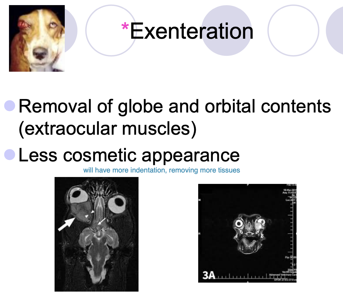
Exenteration def
Removal of globe and orbital contents (extraocular mm). Less cosmetic appearance.
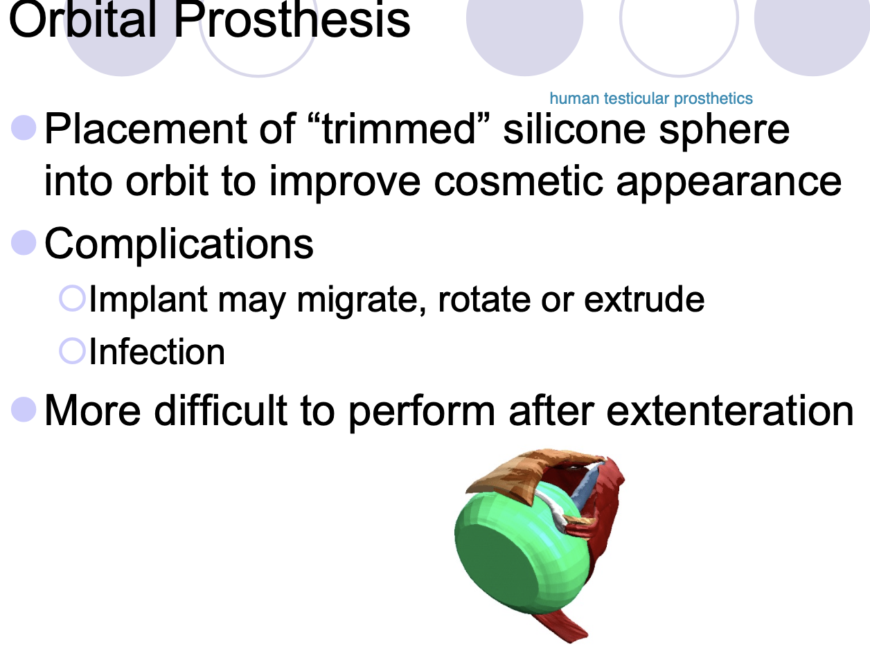
Orbital Prothesis placement of (2)
trimmed silicone sphere into orbit to improve cosmetic appearance.
More difficult to perform after exenteration.
Intraocular Prothesis has (2)
more cosmetic appearance.
dark silicone sphere.

Intraocular prothesis will allow
globe to move normally, but cornea can still be injured.
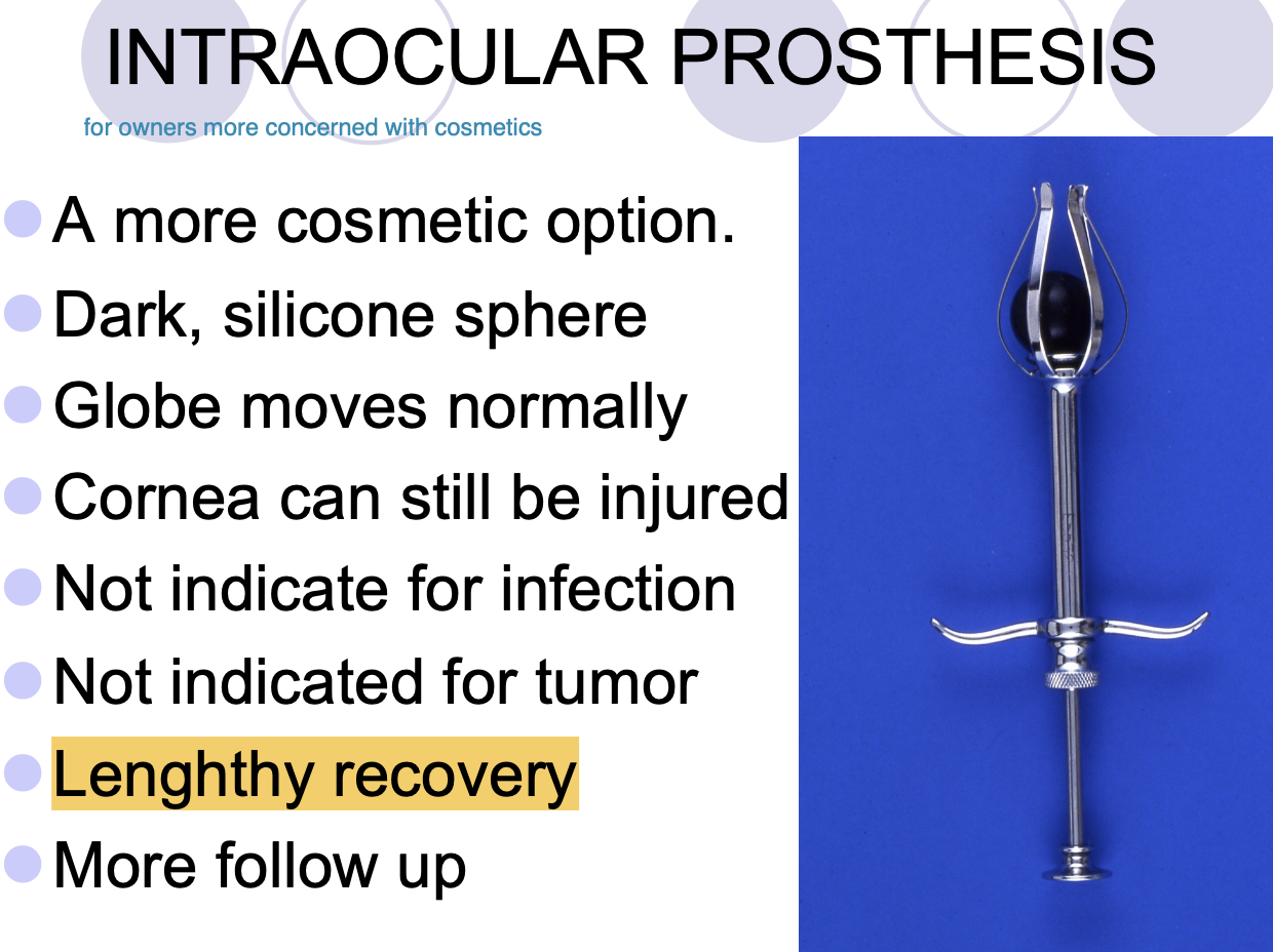
Intraocular prothesis is not indicated for
infection/tumor.

Intraocular prothesis has
lengthy recovery.
More follow up.

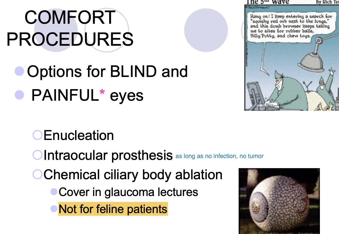
Comfort procedures as options for blind and painful eyes
Enucleation.
Intraocular prosthesis (only if no infection/tumor)
Chemical ciliary body ablation.

Chemical ciliary body ablation is not for
feline patients.