Chapter 18: eye anatomy
1/60
There's no tags or description
Looks like no tags are added yet.
Name | Mastery | Learn | Test | Matching | Spaced | Call with Kai |
|---|
No analytics yet
Send a link to your students to track their progress
61 Terms
State the accessory structures of the eye?
eye brows
eye lashes
eye lids (palpebrae)
conjunctiva
lubricating lacrimal apparatus
extraocular muscles (move the eyes in the socket)
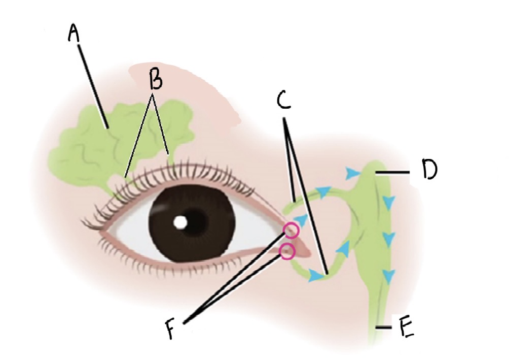
Label the lacrimal apparatus of the eye
A: lacrimal gland
B: excretory ducts
C: lacrimal canaliculi
D; Lacrimal sac
E: nasolacrimal duct
F: lacrimal puncta
Describe how tears are produced and drained by the lacrimal apparatus
After exiting the lacrimal gland through the excretory ducts, tears wash across the eye to the medial corner
Tears drain through tiny openings called lacrimal puncta into narrow tubes called lacrimal canaliculi
The superior and inferior lacrimal canaliculi drain into the lacrimal sac
Once in the lacrimal sac, the tears wait for your next sniff, then when you inhale forcefully through the nose the tears are sucked out of the lacrimal sac into the nasal cavity though the nasolacrimal ducts
tears are then swallowed to be recycled by the body
Name the 6 extraocular muscles
superior rectus
inferior rectus
medial rectus
lateral rectus
inferior oblique
superior oblique
What does the superior rectus do and what is it innervated by?
moves the eye: up
innervated by: the oculomotor nerve
What does the inferior rectus do and what is it innervated by?
moves the eye: down
innervated by: the oculomotor nerve
What does the medial rectus do and what is it innervated by?
moves the eye: toward the nose
innervated by: the oculomotor nerve
What does the lateral rectus do and what is it innervated by?
moves the eye: toward the ears
innervated by: abducens nerve
What does the inferior oblique do and what is it innervated by?
moves the eye: rotate toward the nose from below
innervated by: oculomotor nerve
What does the superior oblique do and what is it innervated by?
moves the eye: rotate toward the ear from above
innervated by: trochlear nerve
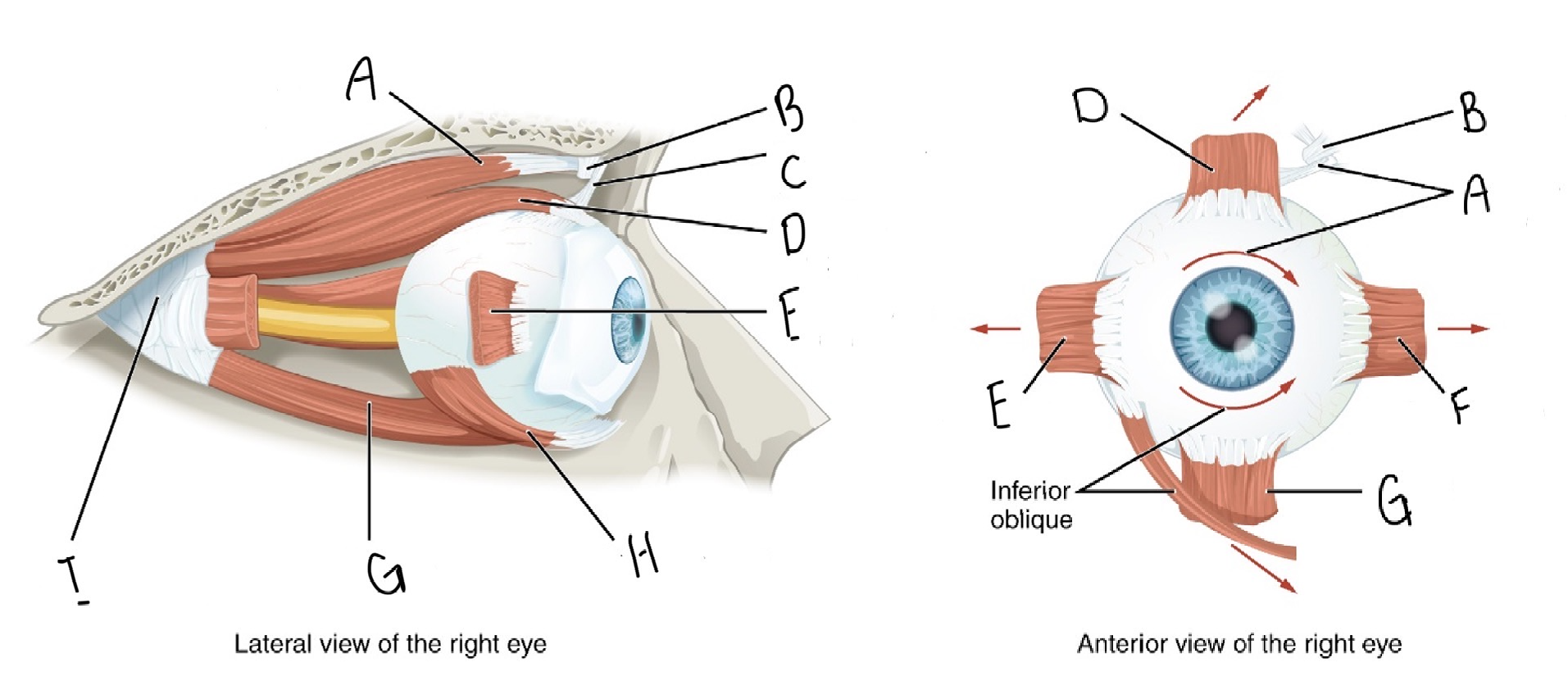
Label the external muscles of the eye
A: Superior oblique muscle
B: trochlea
C: superior oblique tendon
D: superior rectus muscle
E: lacteral rectus muscle
F: medial rectus
G: inferior rectus
H: inferior oblique muscle
I: common tendinous ring
Label the transverse section of the eye
A: Lens
B: Cornea
C: Iris
D; Pupil
E: Ciliary muscle
F: Lateral rectus muscle
G: Sclera
H: Choroid
I: Retina
J: Macula lutea
K: Fovea centralis
L: Optic nerve
M: Optic disc
N: Medial rectus muscle
Label, describe, and give the function of the internal and external structures of the eye
Internal structures
Choroid
Description: darkly pigmented layer deep to the sclera
Function: absorbs stray light and provides nourishment for the retina
Ciliary muscle
Description: muscle deep to the junction between the sclera and cornea
Function: alters the shape of the lens for focusing
Lens
Description: crystalline, disc-shaped structure centered behind the pupil
Function: focuses light for sharp vision
Retina
Description: deepest layer of the eyeball
Function: nervous layer that receives light for vision
Macula lutea
Description: yellow spot lacking blood vessel coverage
Function: area of very good vision
Fovea centralis
Description: indentation at the center of the macula lutea
Function: Area of sharpest color vision
Optic disc
Description: location where the optic nerve enters the eyeball
Function: this area lacks photoreceptors (blind spot)
External Structures
Sclera
Description: The white of the eye
Function: Protective, tough outer layer
Cornea
Description: the clear, solid structure over the iris and pupil
Function: Allows light to enter the eye. This is where most of the light bending takes place for focusing
Iris
Description: the two smooth muscles covered in pigmented cells
Function: controls how much light enters the eyes and inspires poems to its color
Pupil
Description: opening in the center of the iris
Function: allows light to enter the eyeball
Optic nerve
Description: cranial nerve that innervates the posterior, medial side
Function: transmits visual information to the brain
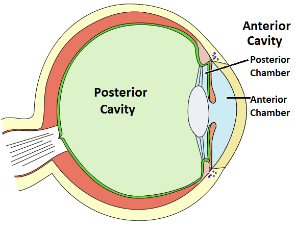
What are the two fluid chambers of the eye? describe them
anterior cavity: filled with aqueous humor (watery fluid that is constantly produced and reabsorbed)
Anterior chamber: space enclosed by the cornea and iris
Posterior chamber: behind the iris and around the lens
posterior cavity: filled with substance called vitreous humor
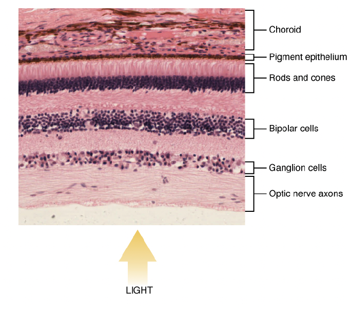
What is the role of the retina?
Layer that transduces light information into action potentials
What is the retina made up of?
Many layers of neurons and one layer of photoreceptors
What is this?
Retina
What is refraction?
Eyes ability to focus light sharply on the retina, particularly on the fovea centralis
Refractive defects are all of the problems related to focusing the eye
Can be caused by problems with any part of the refractive structure
What is emmetropia?
The normal focusing of light on the retina of the eye
When the eye is relaxed, or looking at very distant objects, the light entering the eye focuses sharply
normal vision
What is presbyopia?
Means ‘old eyes’
Eye can focus correctly at a distance, the eye can no longer focus up close due to stiffening of the lens
What is hyperopia?
Farsightedness occurs when the focal point of the light entering the eye occurs behind the retina
when light reaches the retina it is not yet in focus
can be caused by under-curvature of the cornea or lens, or much more commonly, by the foreshortened growth of the eyeball
What is Myopia?
Nearsightedness occurs when the focal point of the light entering the eye occurs in front of the retina
by the time the light reaches the retina, it is back out of the focus
can be caused by over-curvature of the cornea or lens, or by the oblong growth of the eyeball
What is the difference between nearsightedness and farsightedness?
Nearsightedness (myopia) means you see near objects clearly but distant objects are blurry because light focuses in front of the retina.
Farsightedness (hyperopia) means you see far objects clearly but near objects are blurry because light focuses behind the retina
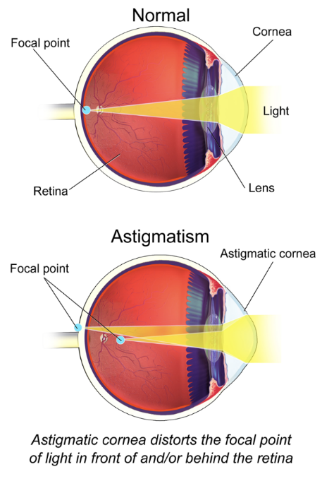
What is astigmatism?
Occurs when light is not focused in a single spot on the retina, but has multiple focal points that may or may not be on the retina.
It is a result of irregular curvature of one or more of the focal structures of the eye, but mostly irregular curvature of the cornea
What are the three types of vision tests?
Snellen Eye chart
Radial astigmatism chart
near point of accommodation
Explain the snellen eye chart
Traditional method of identifying the presence of a refractive error but can not distinguish between types of refractive errors —> can only give a general idea of the severity of a deficit
Is the source of the 20/20 vision
Procedure:
standing 20 feet away from the chart
subject then reads the smallest line of text that they are able to distinguish, any error is considered a failure to read the line
each line is assigned a distance —> the assigned distance is how far away a person with emmetropic (normal) vision would be able to stand and read the line clearly
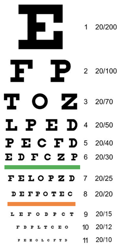
What does 20/20 vision mean?
The patient can, at 20 feet from the chart, see what a person with emmetropic vision could read from 20 feet away from the chart
What does 20/60 vision mean?
The patient can, at 20 feet from the chart, see what a person with emmetropic vision could read from 60 feet away from the chart (patient must stand much closer than average to see the line of text - patient has poor vision
Explain the radial astigmatism chart
The result of irregularity is that parts of the visual field may be in focus while others are out
a radial astigmatism test places identically thick, straight dark lines in the planes in which typical irregularities happen
a patient with astigmatism will not see the lines as identical, some may appear narrow, dark, wide fuzzy, or curved —> any observation indicates the presence of astigmatism
What is accommodation?
The process of focusing on objects as they approach the face
What is the near-point of accommodation?
The closest distance at which you can hold an object and still focus on it sharply
What can alter the near-point of accommodation
All types of refractive errors
What does a consistently long near-point distance indicate?
Hyperopia (farsightedness)
What condition is indicated by an increasing near-point distance with age?
Presbyopia
What happens in the eye to focus on a nearby object?
The ciliary body contracts, allowing the lens to assume its natural, curved shape
How does aging affect the lens?
The lens stiffens and becomes flatter, making it harder to focus on close objects
How is myopia related to near-point distance?
Individuals with myopia often have a shorter near-point distance
What are photoreceptors?
Sensory cells of vision that convert light information into action potentials
What does it mean that photoreceptors “transduce” light?
They convert light energy into action potentials
What happens when a photo hits a photoreceptor? what happens after that?
A pigment chemical inside the receptor changes shape causing an action potential in the optic nerve
What are the two types of photorecptors?
Rods and cones
What are rods most sensitive to?
Low light and a wide range of wavelengths
What kind of vision do rods provide and when are they used?
brightness and shades of gray
in dim or low-light conditions
Why is color hard to distinguish in low light?
Because rods do not detect color well
What are cones responsible for and how many types are there?
colour vision
three
What colours do cones respond to?
blue, green, and red light
How does the brain perceive different colours?
By comparing the firing rates of the three cone types
Where are cones concentrated in the retina?
in the fovea centralis
Why is central vision good for detecting color?
Because it has high concentration of cones
Define pigment bleaching
What is retinal disparity, what causes it, and why is it important
the difference between images seen by each eye
caused by the distance between the two eyes
it allows for depth perception (3D vision)
What is optic chiasm?
A structure where some optic nerve fibers cross to the opposite side of the brain
How is visual information processed in the brain?
Visual input from the left visual field is processed in the right occipital lobe (and vice versa)
Does it matter which eye receives the visual information?
No, processing depends on the visual field, not the eye
What is the optic disc?
The point where the optic nerve exits the eye and contains no photoreceptors
Why is the optic disc called the blind spot?
Because it cannot detect light due to the absence of photoreceptors
Why don’t we normally notice our blind spot?
The brain fills in missing information using input from the other eye
How does the brain compensate for the blind spot when one eye is closed?
It fills in the gap using surrounding visual information and patterns
How does the brain perceive depth?
By comparing the differences between the images from each eye.
How do nearby vs. distant objects differ in retinal disparity?
Nearby objects have large differences; distant objects have small differences.
What is the purpose of this visual difference processing?
To estimate distance accurately (depth perception).