Deep Structures Of The Forebrain
1/15
There's no tags or description
Looks like no tags are added yet.
Name | Mastery | Learn | Test | Matching | Spaced | Call with Kai | Chat |
|---|
No analytics yet
Send a link to your students to track their progress
16 Terms
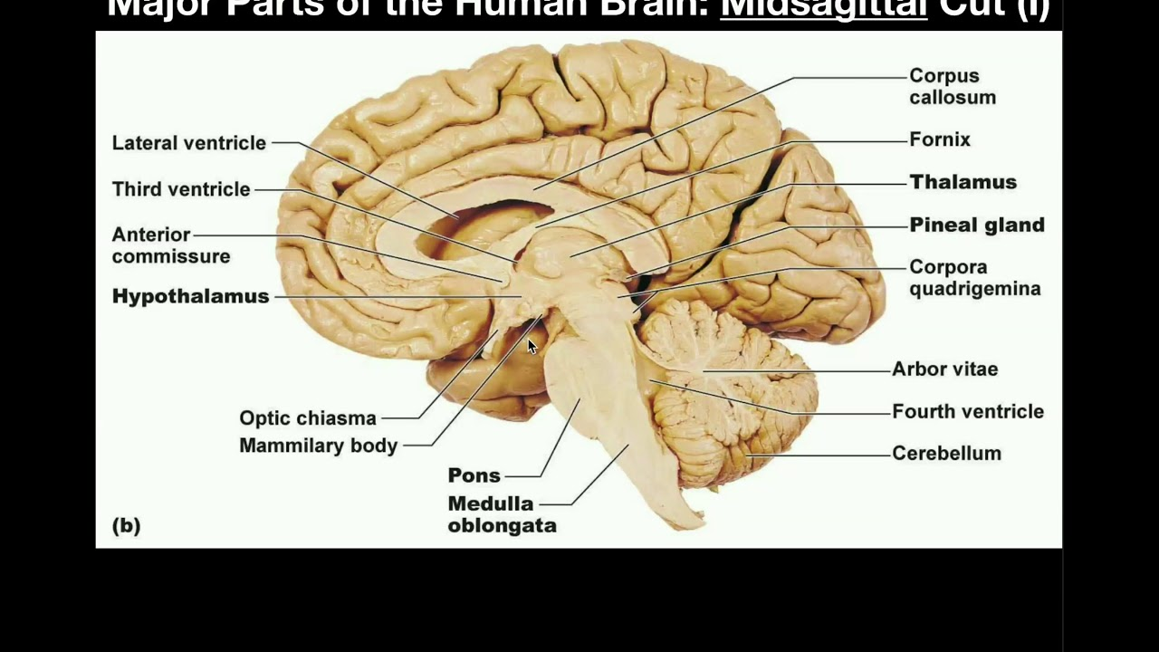
Name the major deep forebrain structures that are revealed by a mid-sagittal section.
A mid‑sagittal cut reveals the key midline deep structures:
Corpus callosum (all four parts)
Thalamus
Hypothalamus
Infundibular stalk (to the pituitary)
Optic chiasm
Mammillary bodies
Pineal gland (epithalamus)
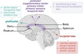
Name and describe the four named parts of the corpus callosum in order from anterior to posterior.
Genu — the front end
Body — the large middle section
Splenium — the rounded back end
Rostrum — a thin projection from the genu, running down and back toward the lamina terminalis
What is the splenium of the corpus callosum?
The splenium is the rounded back end of the corpus callosum
It’s important because the cingulate gyrus wraps around it before becoming the parahippocampal gyrus via the isthmus
This makes the splenium a key landmark for both white‑matter anatomy and the limbic lobe pathway
What is the thalamus and what is its broad function?
The thalamus is one of the two parts of the diencephalon (the other is the hypothalamus)
Its broad job is to act as the brain’s major sensory relay station
It receives incoming sensory signals and sends them to the correct cortical areas for conscious processing
Exception: smell does not relay through the thalamus
Describe the thalamus's role as a "relay station" for sensory information.
The thalamus receives sensory signals coming from the body and brainstem
It then routes each modality to the correct cortex:
LGN → occipital lobe for vision
MGN → temporal lobe for hearing
VPL/VPM → parietal lobe for touch, pain, temperature, proprioception
A patient suffers a thalamic infarct. Based on the thalamus's broad function, which type of deficit would be most expected?
Impaired conscious sensory perception (sensory loss)
What is the hypothalamus and what is its broad function?
The hypothalamus is the second part of the diencephalon (with the thalamus)
Its broad job is to act as the body’s master homeostasis regulator
It controls:
Autonomic functions
Hormone release via the pituitary
Temperature
Fluid balance
Hunger and satiety
Circadian rhythms
How is the hypothalamus connected to the pituitary gland, and what is this connecting structure called?
The hypothalamus connects to the pituitary via the infundibular stalk
This stalk carries:
Axons to the posterior pituitary (for direct hormone release)
Portal blood vessels to the anterior pituitary (for hormone control)
Describe the precise anatomical position of the infundibular stalk in relation to the optic chiasm and the mammillary bodies.
The infundibular stalk sits between two key landmarks:
Optic chiasm → infundibular stalk → mammillary bodiesSo it is posterior to the optic chiasm and anterior to the mammillary bodies
What is the optic chiasm and why is its position adjacent to the infundibular stalk clinically significant?
The optic chiasm is the X‑shaped crossing where nasal retinal fibres decussate
It sits just in front of the infundibular stalk
Because of this position, pituitary tumours growing upward can compress the chiasm
Classic result: bitemporal hemianopia (loss of both temporal visual fields)
A pituitary macroadenoma expands superiorly and compresses the structure immediately anterior to the infundibular stalk. Which visual field defect would you expect?
A pituitary macroadenoma growing upward compresses the optic chiasm (which sits just in front of the infundibular stalk)
Compression of the crossing nasal fibres causes loss of both temporal visual fields
This produces bitemporal hemianopia — the classic “tunnel vision” of pituitary tumours
Where is the pineal gland located anatomically, and to which part of the diencephalon does it belong?
The pineal gland sits in the epithalamus, which is the dorsal/posterior part of the diencephalon
It is a midline structure located posteriorly, near the posterior commissure
What is the known function of the pineal gland?
The pineal gland’s overall function is largely unknown
What is known: it secretes melatonin
Melatonin helps regulate the body’s circadian (24‑hour) rhythm, especially the sleep–wake cycle in response to light and dark
What is melatonin, which structure produces it, and what is its physiological role?
Melatonin is a hormone
It is produced by the pineal gland (in the epithalamus of the diencephalon)
Its job is to regulate circadian rhythms — especially the sleep–wake cycle
Darkness increases melatonin release; light suppresses it, helping the body stay synced with the day–night cycle
A patient works night shifts and has severe disruption of their sleep-wake cycle. Dysfunction of which diencephalic structure is most directly implicated?
The structure most directly involved is the pineal gland (in the epithalamus)
It makes melatonin, which regulates the sleep–wake cycle based on light–dark cues
Night‑shift work disrupts those cues → melatonin secretion is suppressed → circadian rhythm becomes disordered
What is the clinical syndrome associated with disruption of the hypothalamic-pituitary axis via the infundibular stalk?
Damage to the infundibular stalk cuts off hypothalamic control of the pituitary
This causes panhypopituitarism — loss of all pituitary‑dependent hormones
Key hormone losses:
TSH → hypothyroidism
ACTH → adrenal insufficiency (life‑threatening)
LH/FSH → gonadal failure
GH → growth failure in children
Prolactin dysregulation