Speech Anatomy & Physiology-Resonance system
1/31
There's no tags or description
Looks like no tags are added yet.
Name | Mastery | Learn | Test | Matching | Spaced | Call with Kai |
|---|
No analytics yet
Send a link to your students to track their progress
32 Terms
Bony framework of VPN system
Skull
Facial skeleton
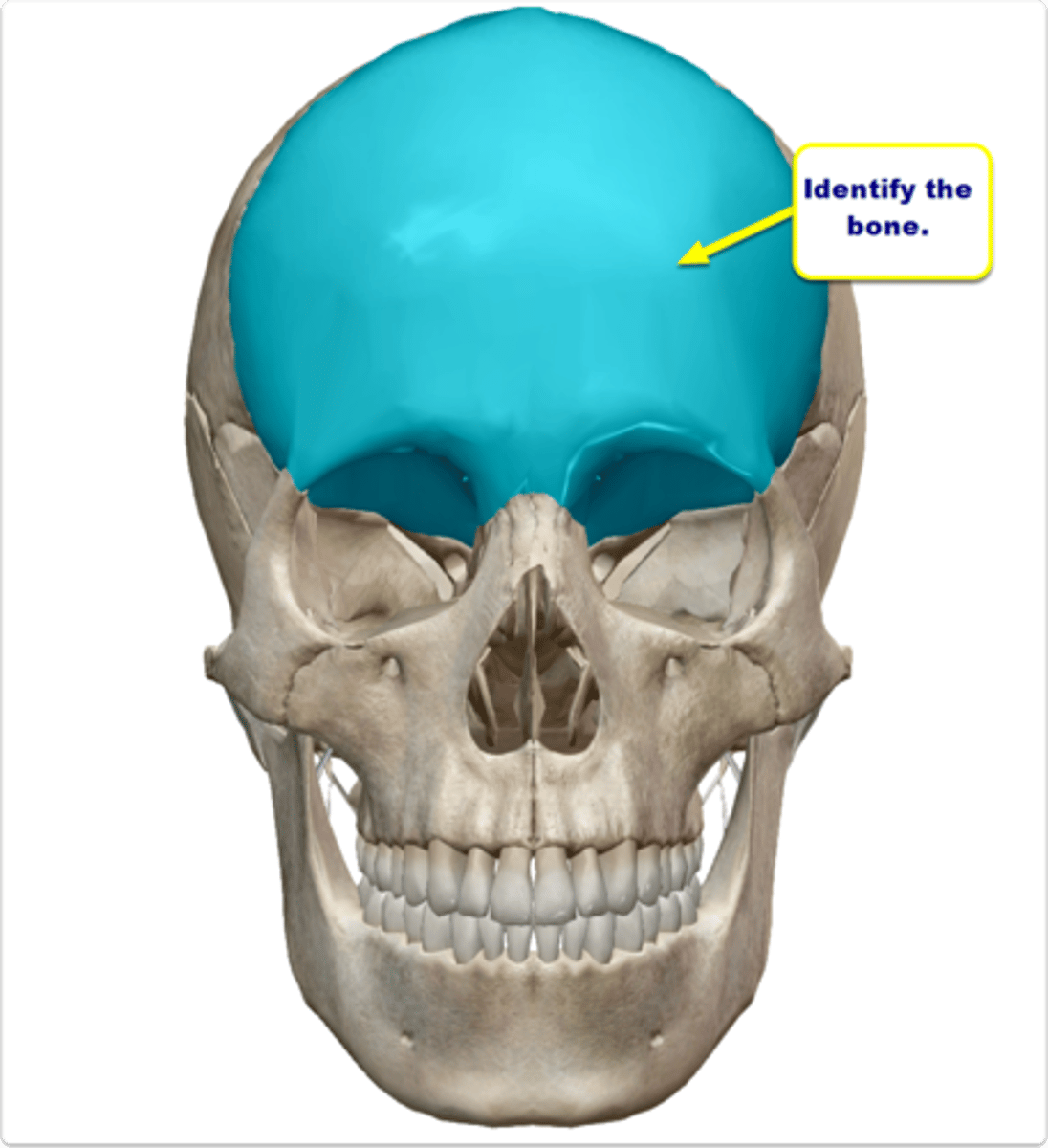
Frontal Bone
forms the forehead the roof of the nasal cavity.
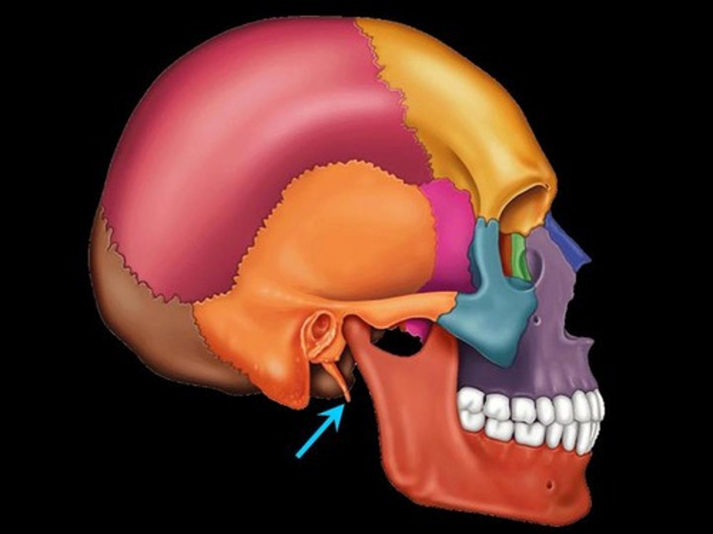
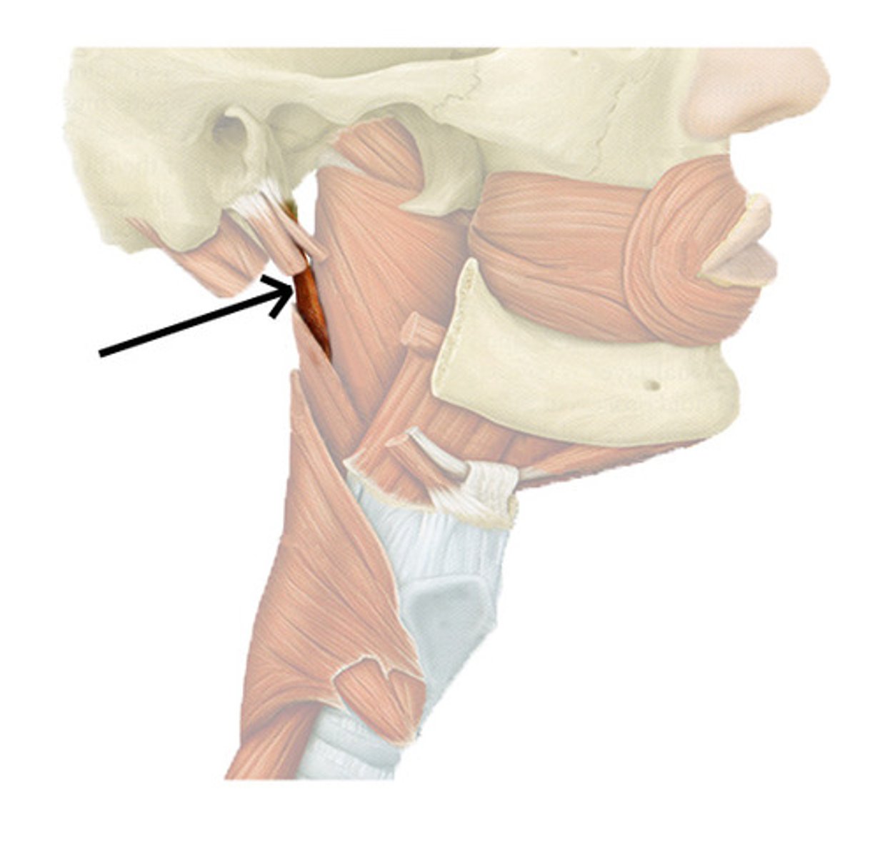
styloid process of temporal bone
serves as an anchorage for muscles associated with the tongue and pharynx.
sphenoidal crest
forms the back wall of the nasal cavities
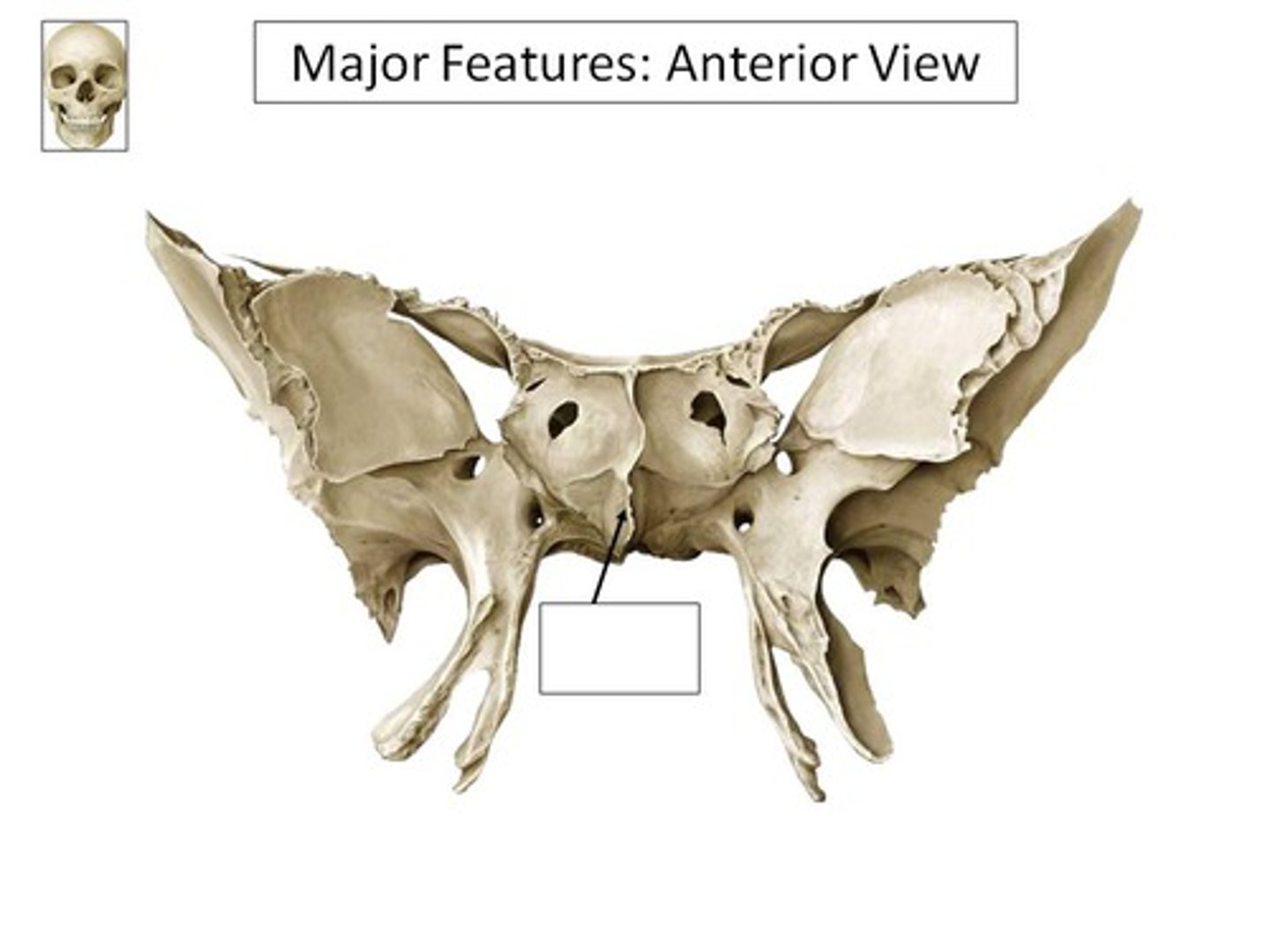
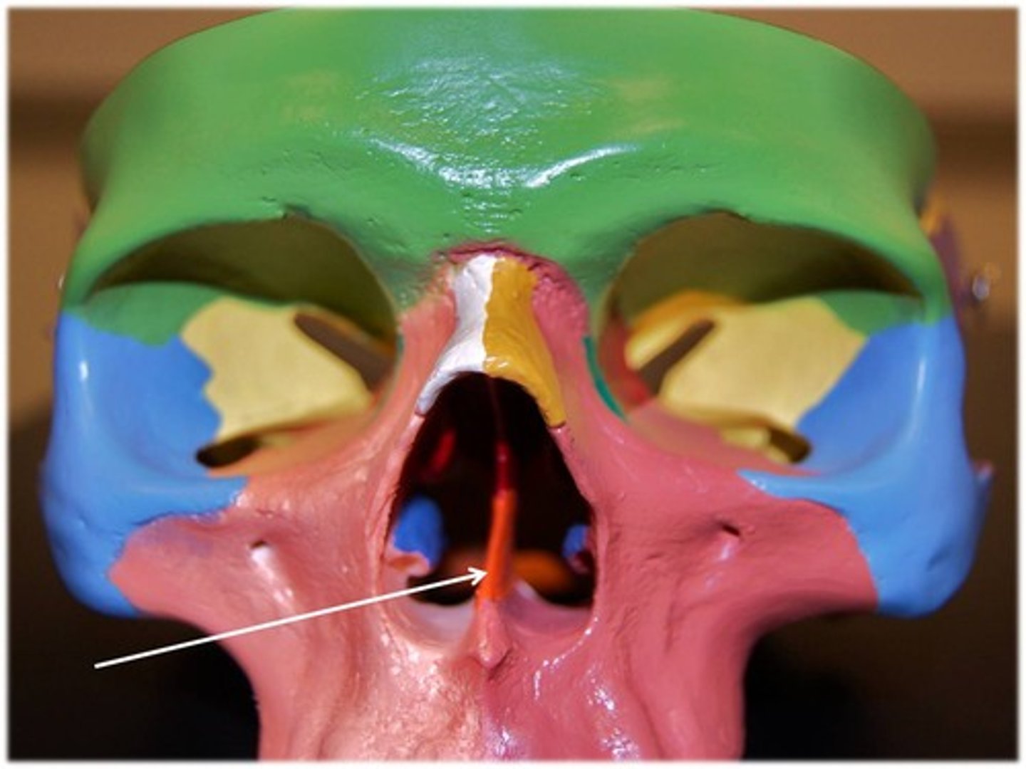
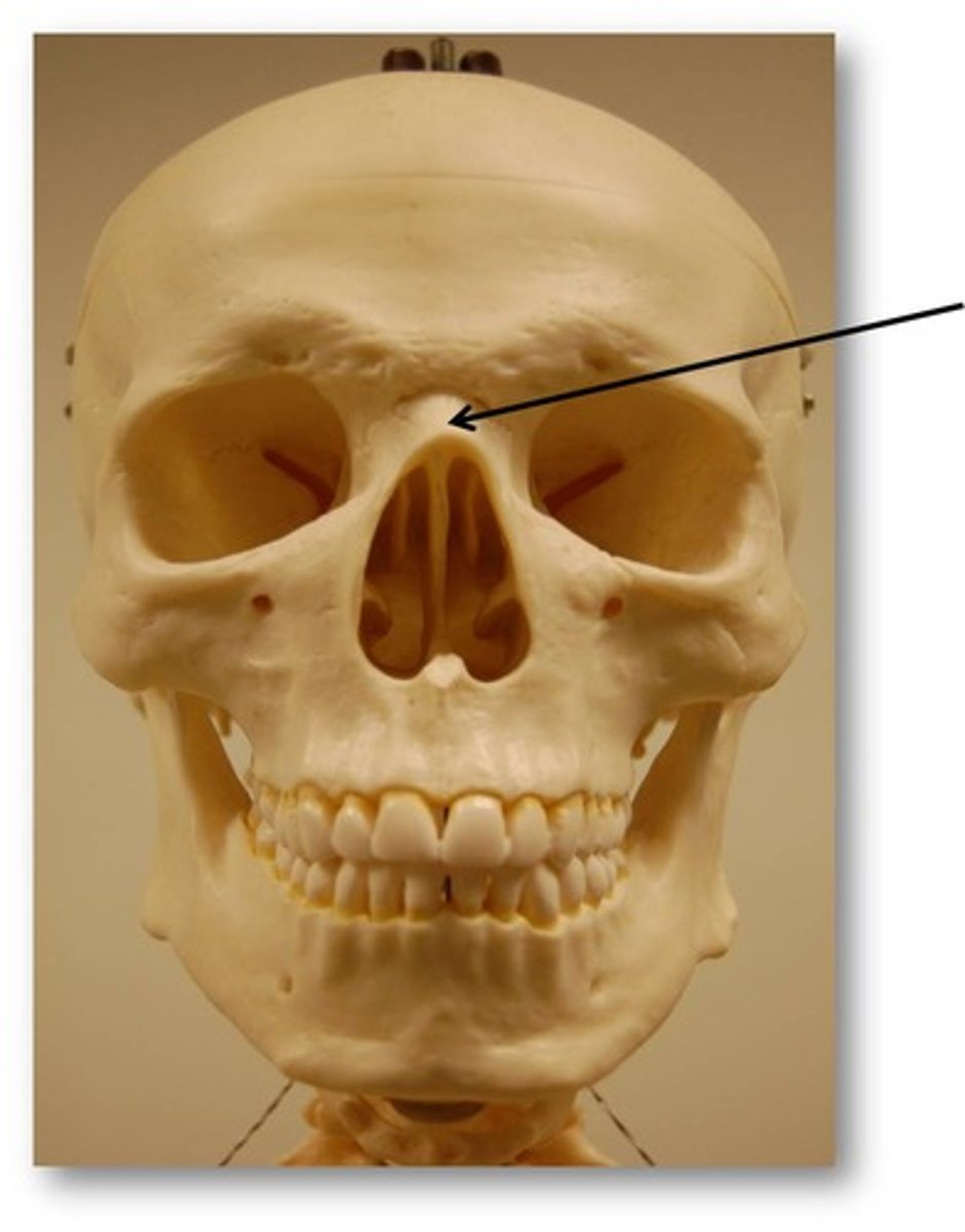
ethmoid bone
forms the upper part of medial wall (Septum), the roof, and upper side walls of the nasal cavities.
The superior and the middle concha from the lateral wall of the nasal cavities.
palatine bone
Forms the third posterior part of the floor of the nasal cavities.
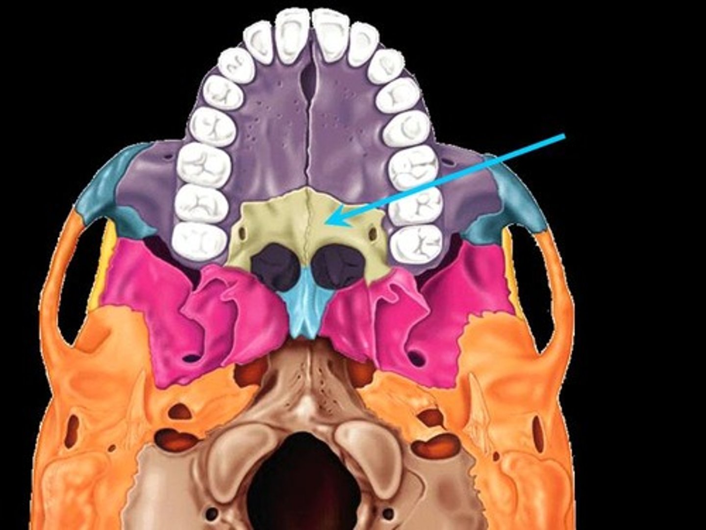
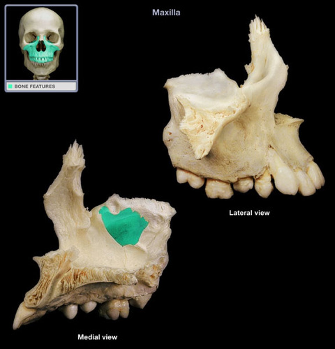
maxillary bone
Forms the anterior two thirds of floor of the nasal cavities.
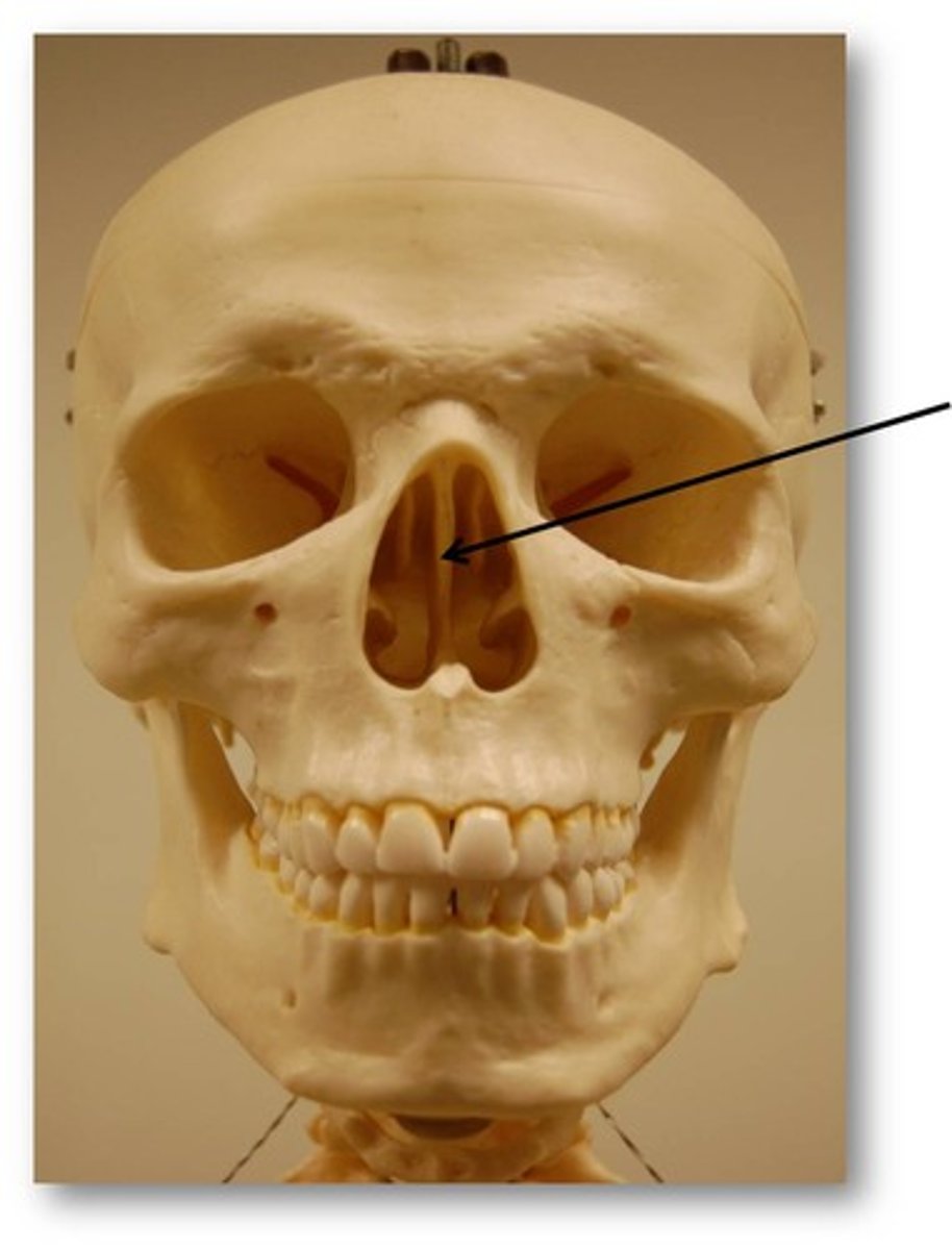
vomer bone
forms the base for the nasal septum
Inferior conchae
Located on the lower side walls of the nasal cavities

Nasal bones
form the bridge of the nose (part of the lateral wall)
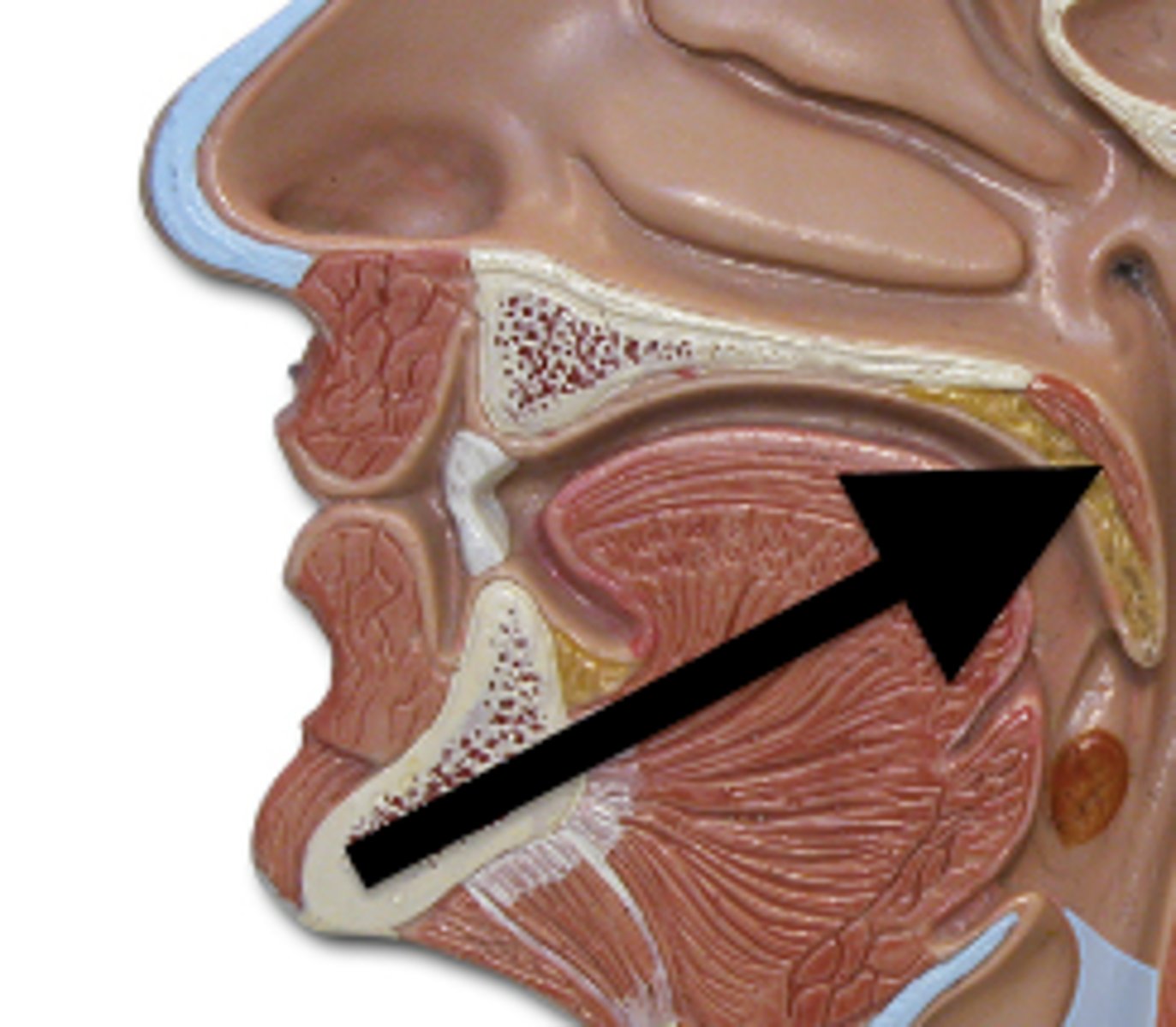
Pharynx
the membrane-lined cavity behind the nose and mouth, connecting them to the esophagus.
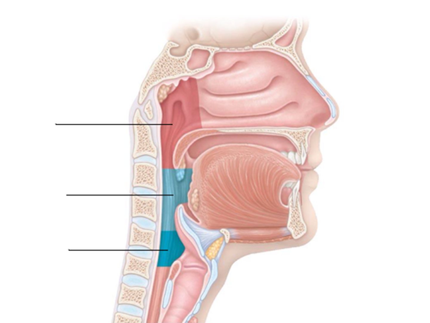
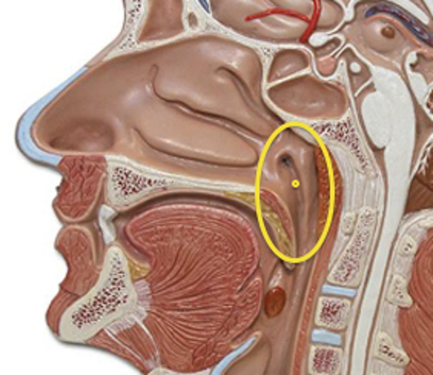
nasopharynx
region of the pharynx at the back of the nose and above the soft palate
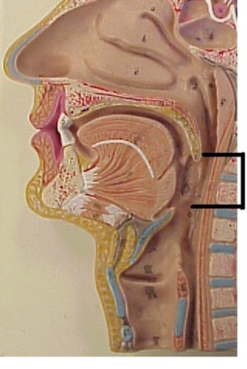
oropharynx
central portion of the pharynx between the roof of the mouth and the upper edge of the epiglottis
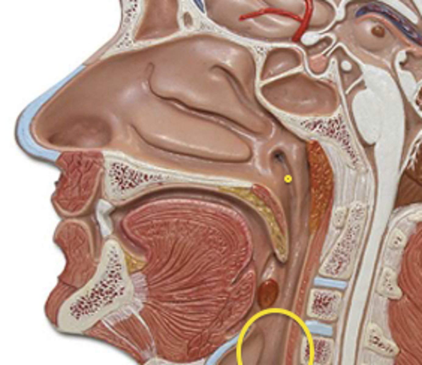
laryngopharynx
lower part of the pharynx, just below the oropharyngeal opening into the larynx and esophagus
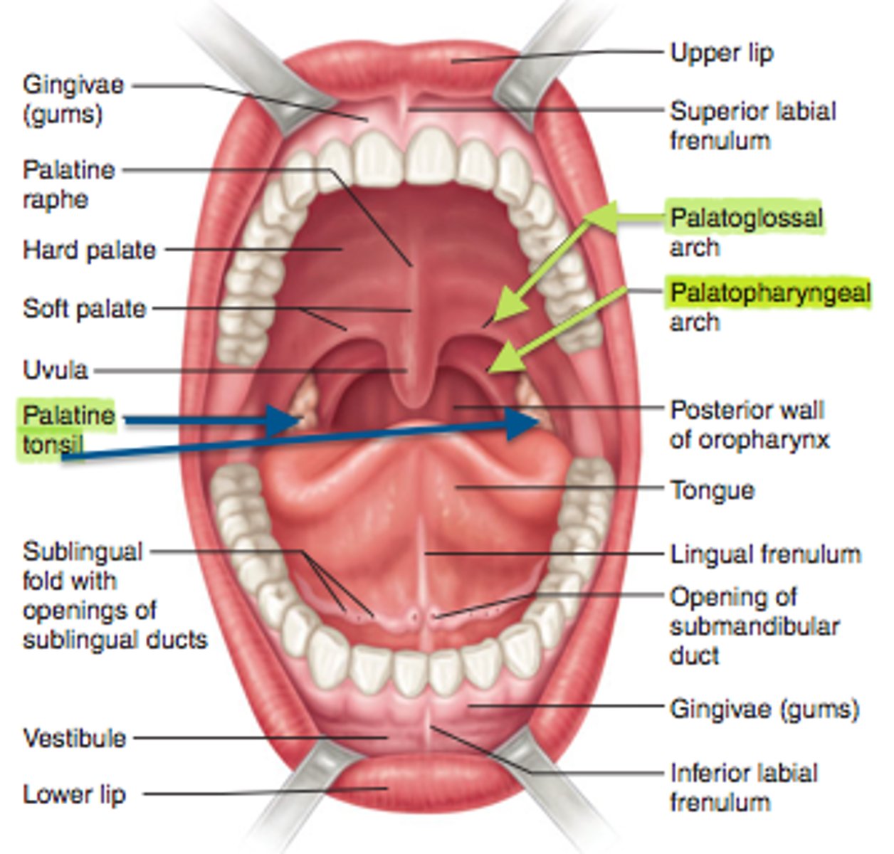
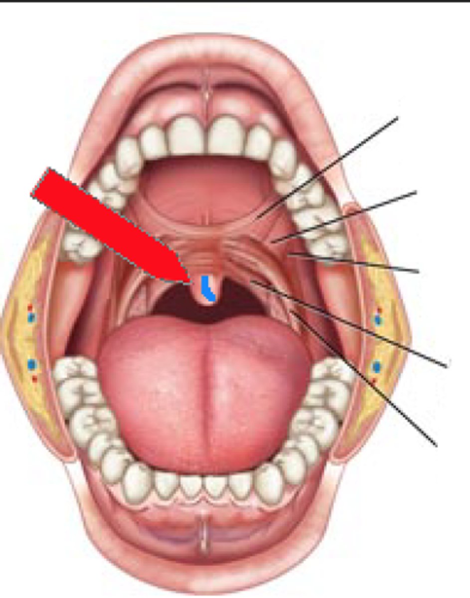
velum
soft palate+uvula
palatoglossal arch and palatopharyngeal arch
palatal levator (levator veli palatini)
forms much of the bulk of the velum; when contracts, velum moves upward and backward
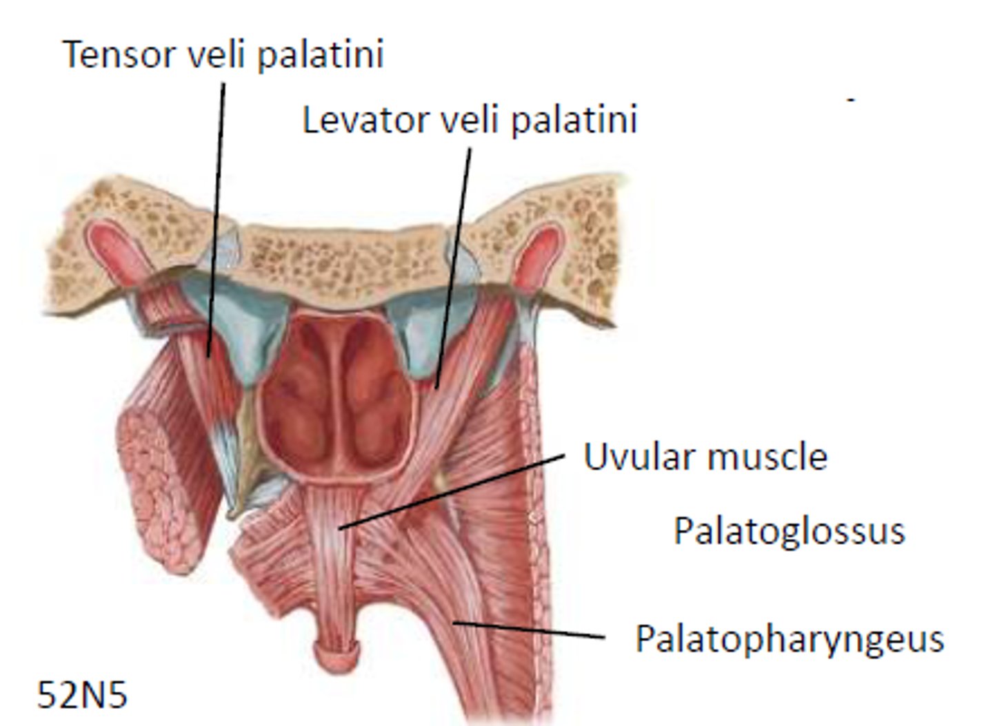
palatal tensor (tensor veli palatini)
opens eustachian tubes

Uvula muscle
to shorten and lift the soft palate
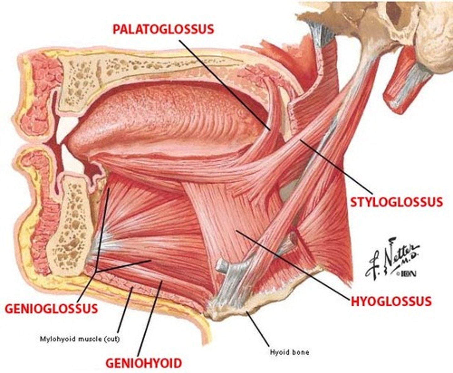
palatoglossus muscle
(muscular structure of anterior faucial pillar) - elevates back of tongue and depresses soft palate
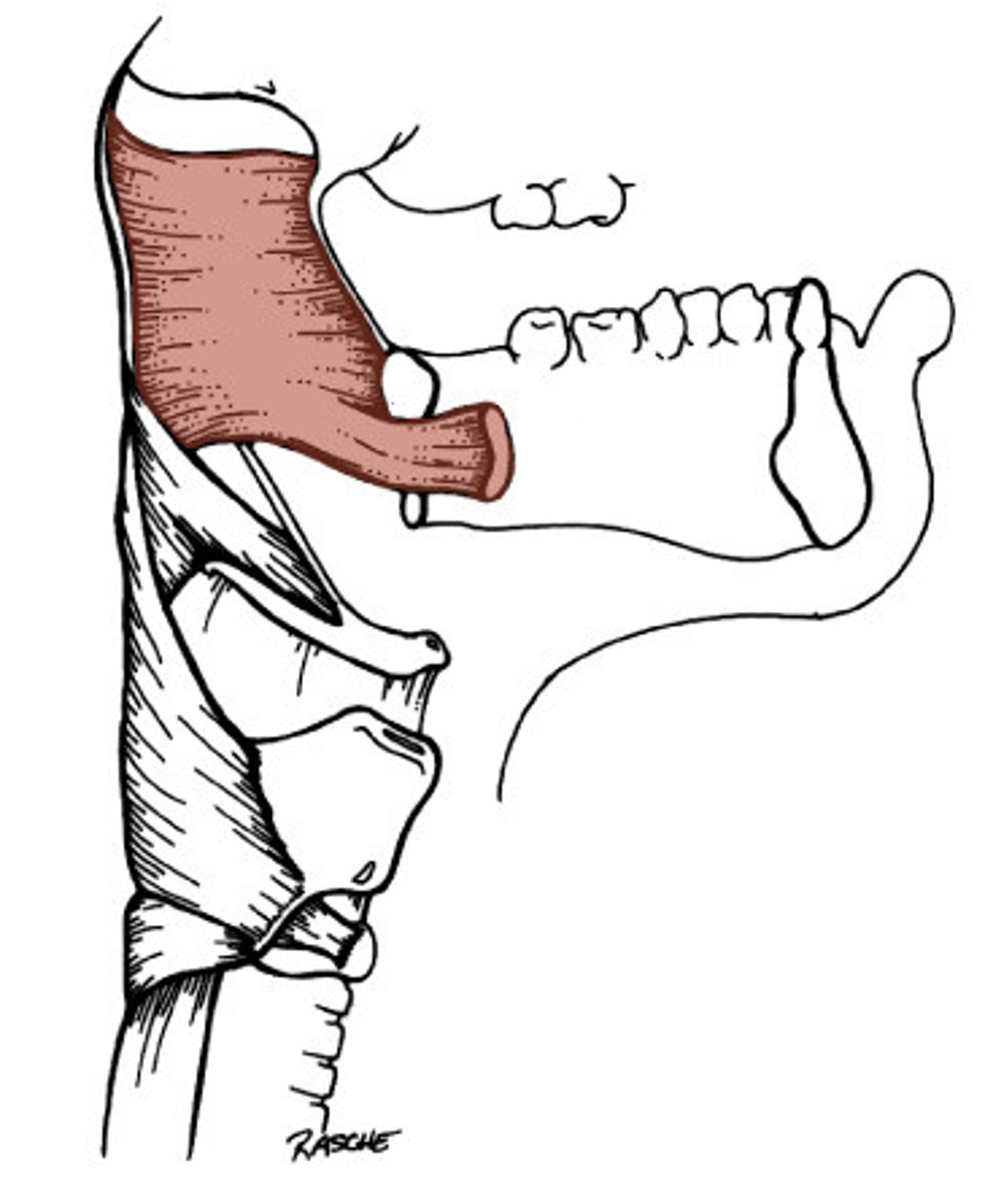
superior constrictor muscle
closes upper part of the pharyngeal tube in the manner of a sphincter
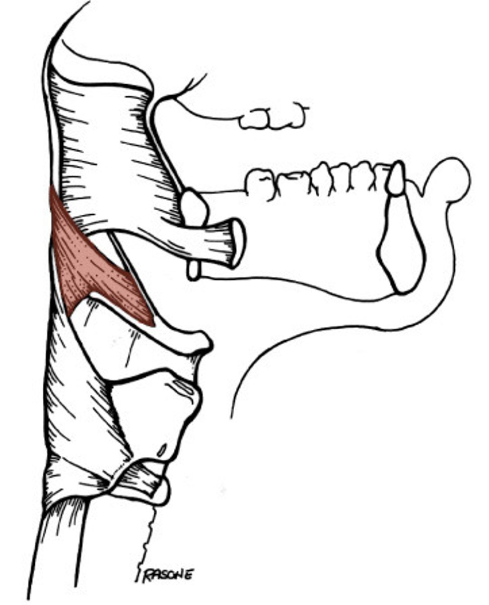
middle pharyngeal constrictor
Narrows middle part of the pharyngeal tube in the manner of a sphincter
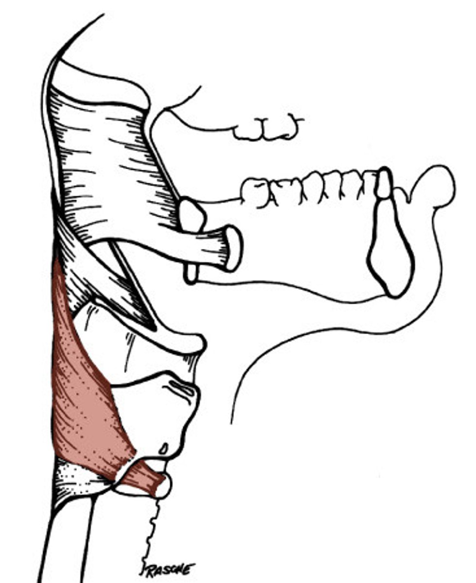
inferior pharyngeal constrictor
constrict the lower pharynx
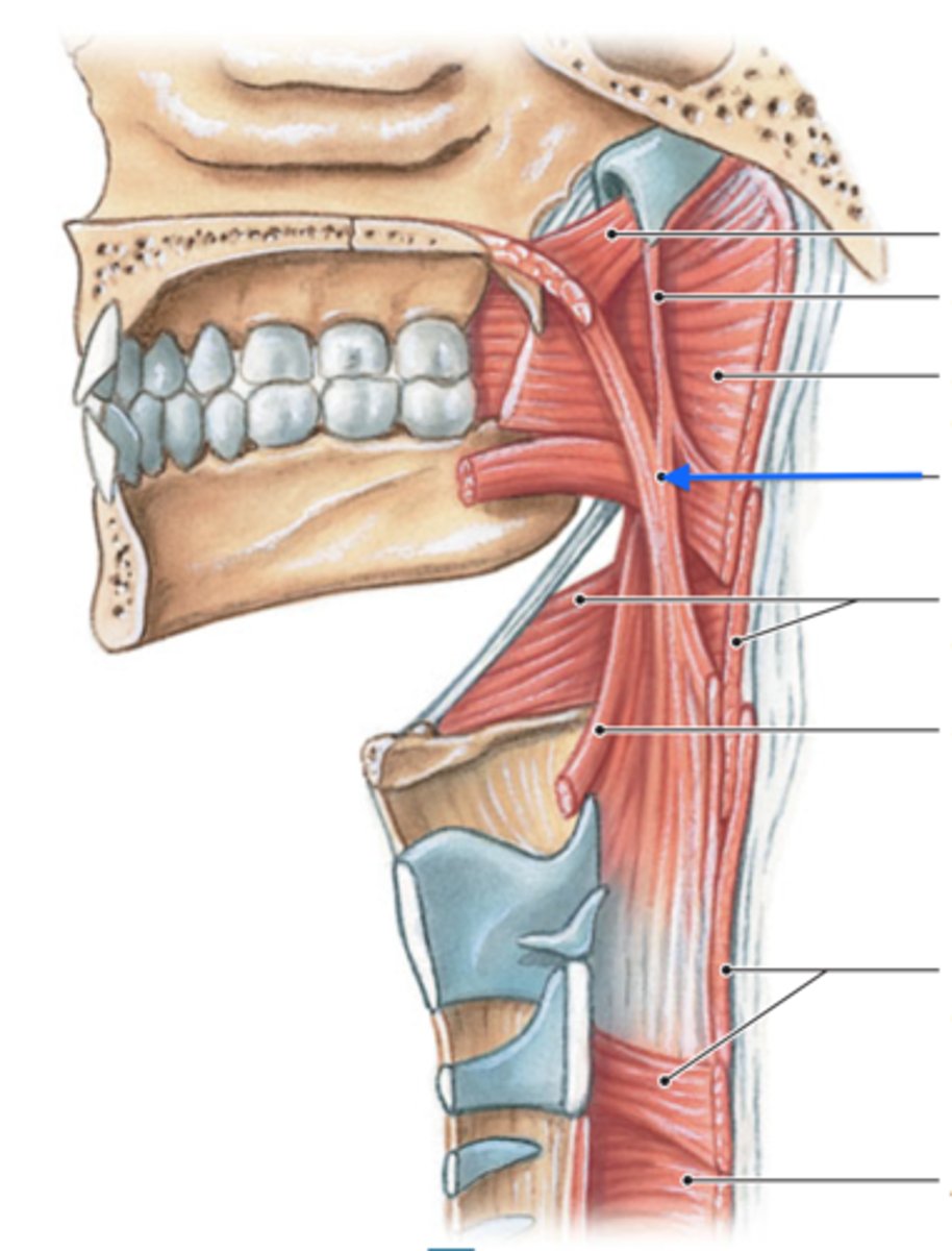
Salpingopharyngeus muscle
elevates lateral pharyngeal wall (shorten the pharynx)
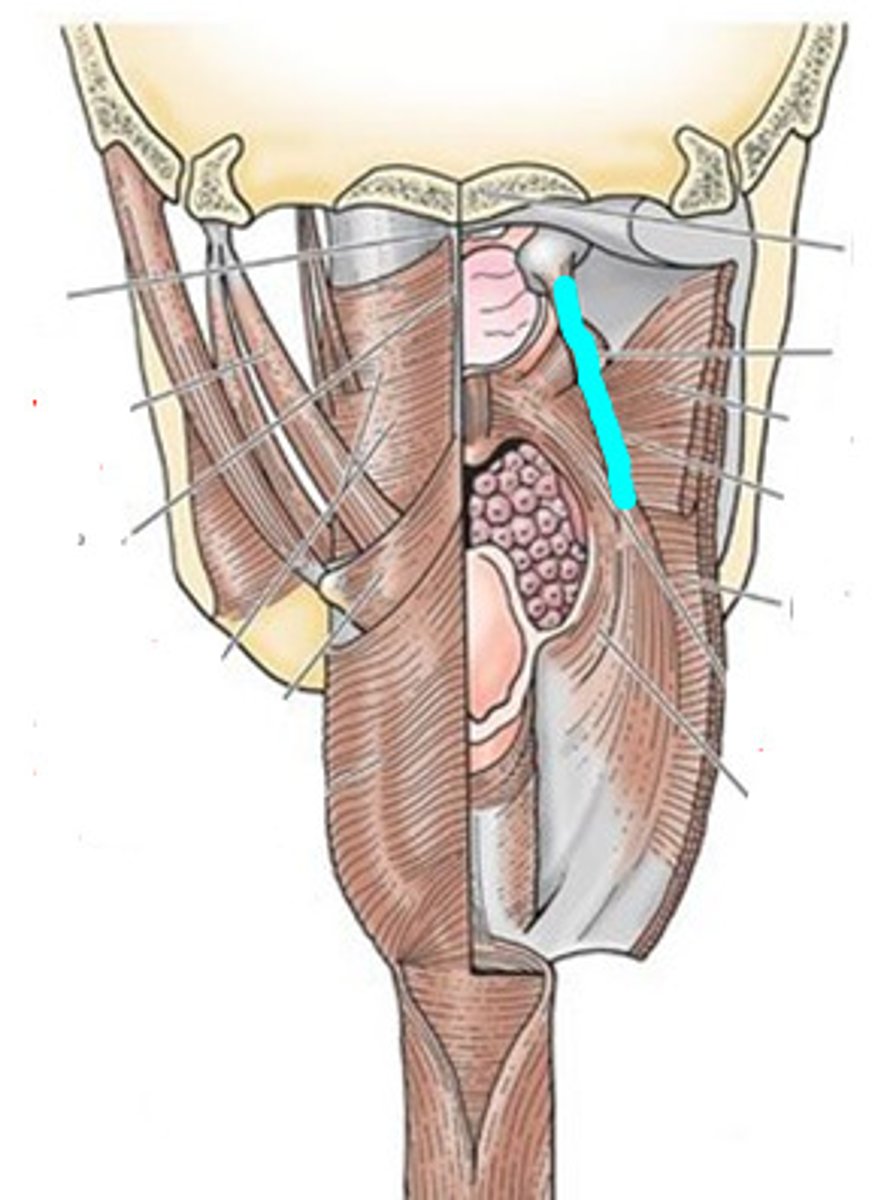
stylopharyngeus muscle
widen the pharynx
Palatopharyngeus muscle
The upper most fibers of the muscle draw the lateral pharyngeal wall inward to complement the action of the sup constrictor muscle while the lowermost fibers of the muscle pull upward the lateral pharyngeal wall and elevate (shorten) the pharynx
VPN airway resistance
opposition to the mass flow of air through structures of the VPN airway.
Factors that can change VP-nasal airway resistance
length of constriction
speed of airflow
VP port size
length of VP port
obstruction of nasal cavities
VP Sphincter Compression
compressive muscular pressure exerted to maintain the VP sphincter in a closed configuration
VPN acoustic impedance
opposition to passage of sound waves, being the product of the density of a substance and the velocity of sound in it.
Evaluation of velopharyngeal
Look in the mouth
X-ray or fluoroscopy
MRI
Pathologies that can impact on resonance
Cleft palate
Velopharyngeal insufficiency/incompetance
Englarged pharyngeal tonsils