Prostate & Penis
1/41
There's no tags or description
Looks like no tags are added yet.
Name | Mastery | Learn | Test | Matching | Spaced | Call with Kai |
|---|
No analytics yet
Send a link to your students to track their progress
42 Terms
What is the prostate?
Cone shaped retroperitoneal organ
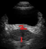
Identify this image.
Prostatic apex
Prostatic base
What is the transitional zone of the prostate?
Central zone composed of two lobes of glandular tissue surrounding proximal prostatic urethra
What is the verumontanum?
Part of prostate located near transitional zone where ejaculatory ducts enter urethra
What is the central zone of the prostate?
Glandular tissue located at base of gland posterior to urethra
Which prostatic zone is the largest?
Peripheral zone (70%)
What is the peripheral zone of the prostate?
Glandular tissue located posterolateral to distal prostatic urethra
What is the periurethral glandular zone of the prostate?
Glandular tissue that lines proximal prostatic urethra
What is the fibromuscular stroma or anterior region of the prostate?
Fibromuscular sheath covering entire anterior prostate surface
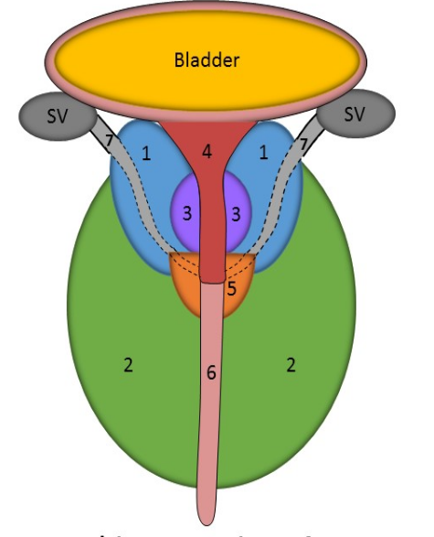
Identify this image.
Central zone
Peripheral zone
Transitional zone
Periurethral glandular zone
Verumontanum
Urethra
Ejaculatory duct
Fibromuscular region
What is prostate specific antigen (PSA)?
Protein used to assess increasing levels of benign or malignant tissue produced by acinar cells
What is the normal lab value for prostate specific antigen (PSA)?
0 - 4 ng/ml (> 10 ng/ml indicates malignancy)
What position is required for transrectal prostate exam?
LLD or lithomy
What is the normal sonographic appearance of the prostate?
Homogenous
Medium-level gray
Smooth capsule
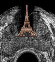
What is the Eiffel tower sign?
Shadowing created by dense tissues in area of urethra and verumontanum
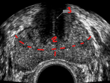
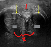
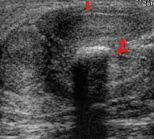
Identify this image.
Peripheral zone
Central and transitional zone
Urethra
What is benign prostatic hypertrophy (BPH)?
Enlargement of prostate that causes urinary infrequency and nocturia
Where is the most common location for benign prostatic hypertrophy (BPH)?
Transitional zone (95%)
What lab value is elevated in someone with benign prostatic hypertrophy (BPH)?
PSA
What is the sonographic appearance of benign prostatic hypertrophy (BPH)?
Diffuse prostatic enlargement with volume > 30 mL
Punctate calcifications or corpora amylacea
Nodule and cystic formation
Hypervascular inner gland
What is prostatitis?
Inflammation of prostate
What is the most common malignancy diagnosed in men?
Extra-capsular prostatic carcinoma
What is extra-capsular prostatic carcinoma?
Prostate cancer most commonly seen in African America men 65 years old
What is the most common location for extra-capsular prostatic carcinoma?
Peripheral zone (70%)
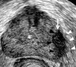
Identify this image.
Prostatic cancer
What is brachytherapy?
Surgical implantation of radioactive seeds in those with non-aggressive in situ prostate cancer
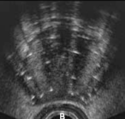
Identify this image.
Brachytherapy
What are corpus cavernosa?
Two main erectile structures of penis
What is the corpus spongiosum?
Singular penile structure that consists of urethra and urethral arteries
Identify this image.
Corpus spongiosum
Corpus cavernosum
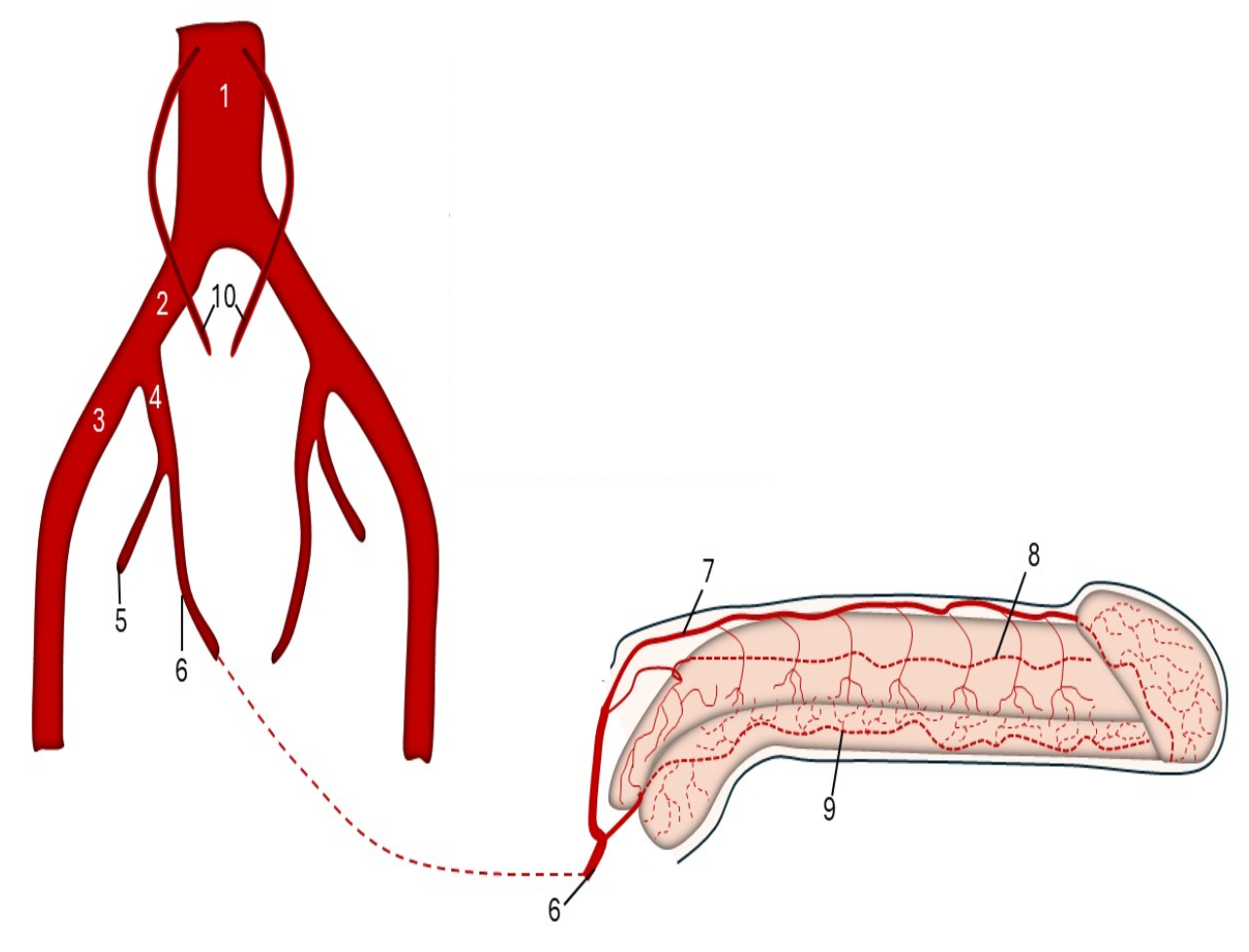
Identify this image.
Aorta
Common iliac artery
External iliac artery
Internal iliac artery
Gluteal artery
Internal pudendal artery
Dorsal artery
Cavernosal artery
Bulbourethral artery
Gonadal arteries
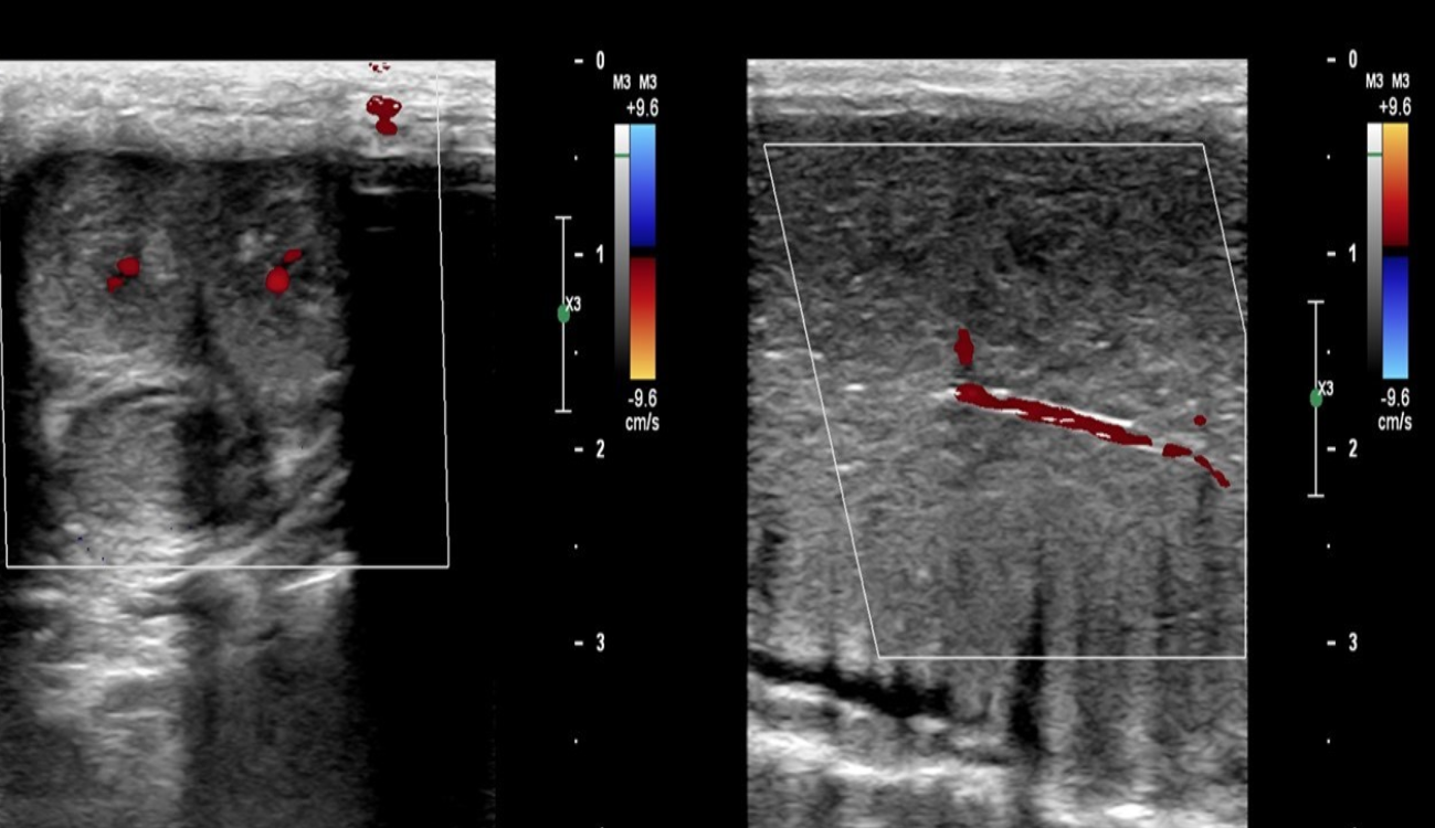
Identify this image.
Cavernosal arteries
What is erectile dysfunction?
Inability to achieve a full and persistent erection due to vascular insufficiency
What is priapism?
Unwanted painful erection that lasts more than four hours
What is Peyronie disease?
Development of scar tissue and plaque involving tunica albuginea that causes curvature of affected side of penis during erection
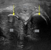
Identify this image.
Thin tunica albuginea
Plaque formation seen with Peyronie disease
Identify this image.
Peyronie disease
What is the most common cancer to affect the penis?
Squamous cell carcinoma
Where is penis cancer most commonly located?
Glans penis or foreskin of uncircumcised men
What is the most common portion of the penis affected by trauma?
Corpus cavernosum
What are the values for penile brachial index (PBI)?
≥ 0.75: Normal
0.65-0.75: Marginal reduction
< 0.65: Abnormal
What is the normal sonographic appearance for a penile Doppler?
Cavernosal artery PSV > 30 cm/sec
Cavernosal artery EDV < 1 cm/sec
Cavernosal artery diameter increases by at least 75%
Cavernosal artery RI > 0.99
Clinical response to Papaverine injection is greater than 90 degrees