5B
 Call Kai
 Learn
Learn Practice Test
 Spaced Repetition
 Match
Match Flashcards
 Knowt Play
1/76

There's no tags or description
Looks like no tags are added yet.
Name | Mastery | Learn | Test | Matching | Spaced | Call with Kai |
|---|
No analytics yet
Send a link to your students to track their progress
77 Terms
Prokaryote genome features
No nuclear membrane; genome located in cytoplasm (nucleoid); small genome; circular chromosome; no histones; no introns; RNA not processed; transcription and translation occur together because there is no nucleus
Eukaryote genome features
Nuclear membrane present (double lipid bilayer); large genome; linear chromosomes; histones and chromatin present; introns in most genes; RNA processed; transcription occurs in nucleus and translation in cytoplasm
Location of prokaryote genome
Nucleoid region within cytoplasm
Location of eukaryote genome
Stored in chromosomes inside nucleus
Prokaryote chromosome
Circular DNA chromosome
DNA storage in prokaryotes
E.coli chromosome is folded and supercoiled so genome fits into small cell and remains usable
Why prokaryote DNA folds
Allows circular chromosome to fit into small cell and remain functional
Human somatic cell chromosomes
46 chromosomes total
Human chromosome composition
22 pairs autosomes + 1 pair sex chromosomes
Female karyotype
46, XX
Male karyotype
46, XY
Diploid
Cells with two copies of each chromosome; 2n = 46 in humans
Haploid
Cells with one copy of each chromosome; n = 23 in humans
Human gametes
Egg and sperm cells; haploid
Human haploid genome
3.2 × 10⁹ nucleotides packaged into 23 chromosomes
The "packaged into 23 chromosomes" part tells you those 3.2 billion nucleotides aren't one continuous string; they're divided up among 23 separate chromosomes.
Important chromosome fact
One chromosome = ONE molecule of double-stranded DNA
DNA length problem
Haploid genome DNA length ≈ 1.02 m; diploid cell ≈ 2.04 m but nucleus only ~10 μm so DNA requires packaging
Unwound chromosome
Single chromosome not duplicated or condensed
Chromosome duplication
After DNA replication one chromosome contains two sister chromatids
Homologous chromosomes
Pair of chromosomes inherited from different parents with same genes in same locations
Chromosome telomeres
Protective ends of chromosome
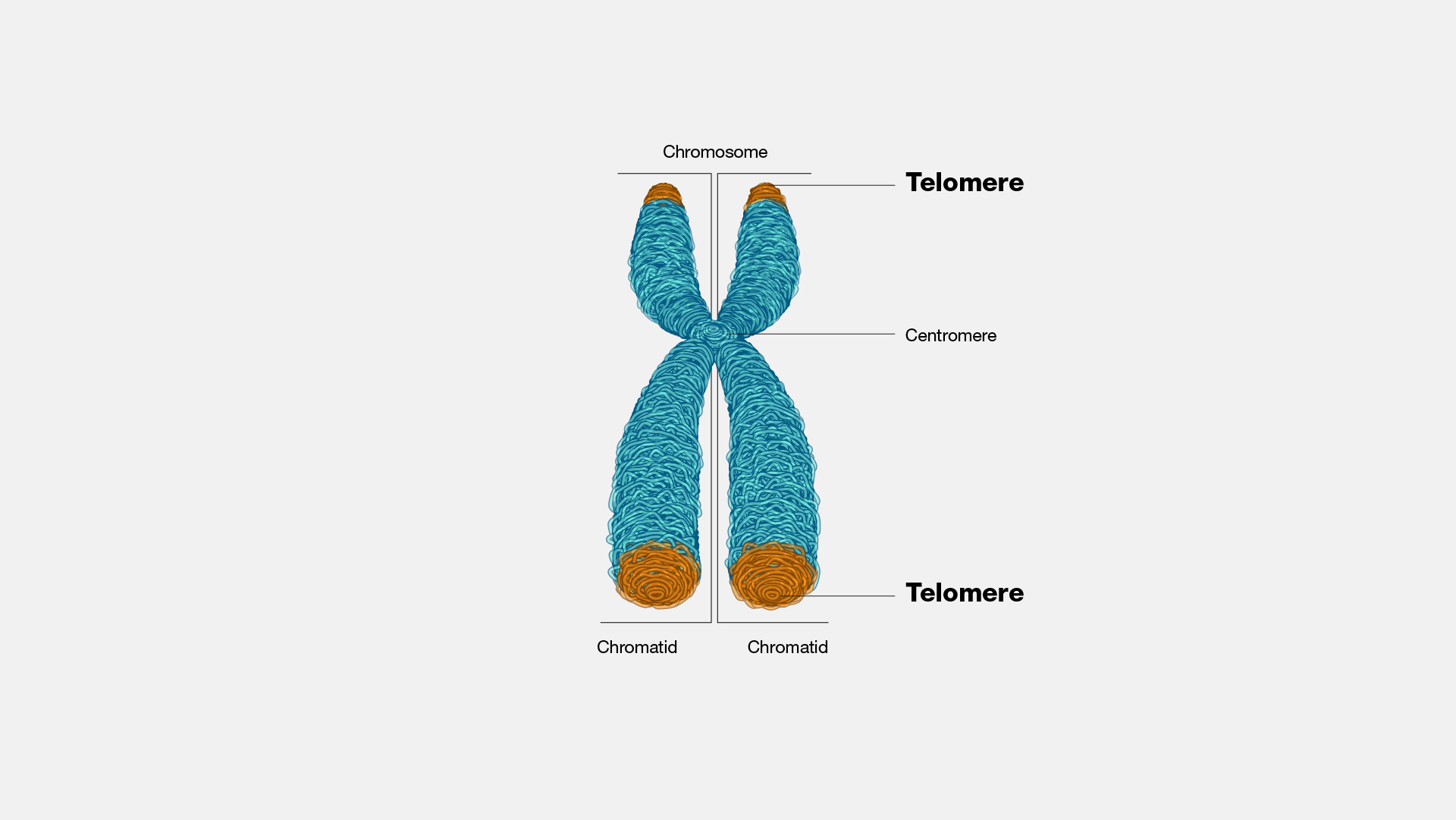
Centromere
Region joining sister chromatids and containing centromeric proteins
Chromosome p arm
Short arm (“petit”)
Chromosome q arm
Long arm (“q” = next letter after p)
Metaphase chromosome
Fully condensed duplicated chromosome seen during metaphase
Stages of eukaryotic DNA packaging
Double helix → nucleosome/chromatin → 30 nm fibre → looped domains → metaphase chromosome
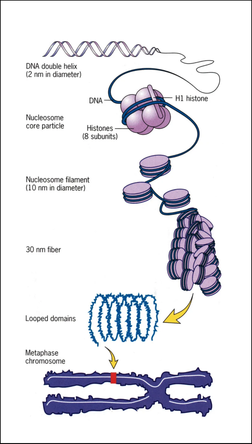
1st level DNA packaging
Double-stranded DNA helix (2 nm)
2nd level DNA packaging
Nucleosomes / chromatin (11 nm)
The second level of DNA packaging is the nucleosome — the "beads on a string" arrangement that gives chromatin its 11 nm width
3rd level DNA packaging
30 nm fibre
the 11 nm beads-on-a-string fiber coils up on itself into a thicker, more compact thread about 30 nanometres across.
it folds and coils so that neighbouring nucleosomes pack tightly against each other, roughly tripling the width from 11 nm to 30 nm.
4th level DNA packaging
Looped domains (~300 nm)
The 30 nm fiber doesn't just keep coiling uniformly; instead it gets organized into a series of big loops,
The bases of these loops attach to a central protein framework called the nuclear scaffold (or chromosome scaffold).
5th level DNA packaging
Further coiling to form metaphase chromosome (~700 nm per chromatid)
The looped domains from level four (the 300 nm fiber) coil and fold even more tightly on themselves, packing the looped fiber into the dense, compact rod-shaped structure of a chromosome.
only occurs in meiosis/mitosis
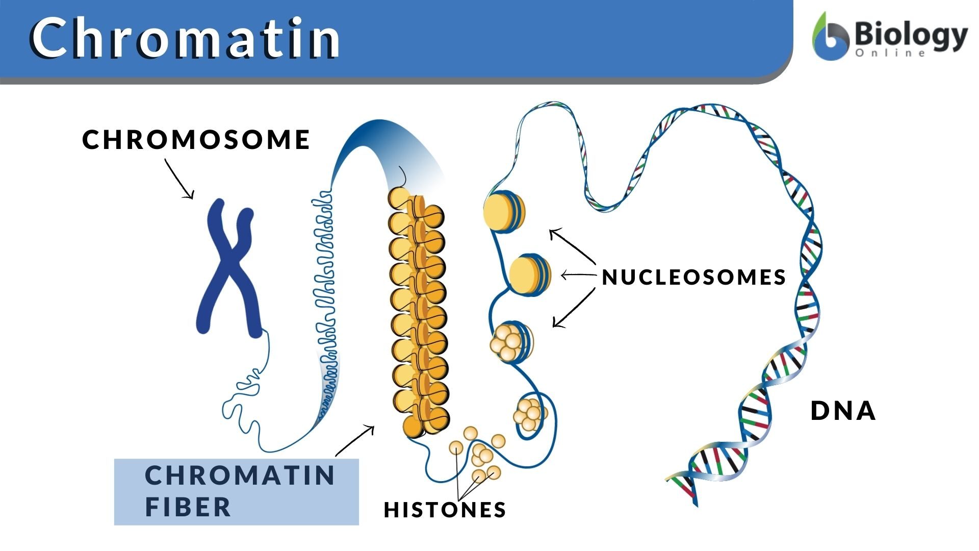
Chromatin
DNA + histone proteins
DNA charge
DNA carries net negative charge due to phosphate groups
Reason DNA is negatively charged
Phosphate groups ionise at cellular pH giving negative charge
Histone charge
Histones are positively charged due to many basic amino acids
Histone amino acids
Arginine and lysine contribute positive charge
DNA-histone interaction
Negative phosphate groups of DNA interact ionically with positively charged histones
Nature of DNA-histone interaction
Electrostatic, reversible, forms without damaging DNA or histones
Nucleosome
DNA wrapped around histone octamer
Histone octamer composition
2 × H2A + 2 × H2B + 2 × H3 + 2 × H4
Histones in nucleosome
H2A, H2B, H3, H4 (2 copies each)
Histone conservation
Histones are highly conserved proteins with little variation between species
Core nucleosome DNA
145 bp wrapped around histone core
Linker DNA
DNA segment between nucleosomes
Linker DNA length
8–114 bp
Chromatin appearance
“Beads on a string” = nucleosomes joined by linker DNA
10 nm fibre
Nucleosome chain / beads-on-string chromatin
Histone H1 function
Binds nucleosome and adjoining linker DNA
Role of histone H1
Links nucleosomes into 30 nm fibre
The four core histones (H2A, H2B, H3, H4) form the octamer that DNA wraps around to make the nucleosome bead itself — that's level 2. H1 is not part of that octamer.
the core histones build the nucleosome; H1 is the separate linker histone that stabilizes the nucleosome and drives the packing of the 11 nm fiber into the 30 nm fiber.
30 nm fibre
More compact chromatin formed by nucleosome packing with H1
Looped domains
30 nm fibres looped and linked by DNA-binding proteins
300 nm fibres
Further compaction level formed by looped domains
700 nm chromatid
Highly condensed metaphase chromatid
Proteins linking loops
DNA-binding proteins connect looped domains
The proteins that link the loops together are the non-histone scaffold proteins — condensin and topoisomerase II,
Interphase chromosomes
More extended and less condensed than metaphase chromosomes
Replication and transcription effects
Usually only slightly disrupt beads-on-string chromatin
Fully condensed chromosomes
Only metaphase chromosomes are fully condensed
Chromatin composition
DNA + histones
Other nuclear components
Nonhistone proteins + RNA + nonchromatin nuclear constituents
Examples of nonchromatin nuclear proteins
Transcription factors, telomere-binding proteins, methyl-CpG binding proteins, centromere proteins, repressors, promoters, DNA replication enzymes, transcription enzymes, DNA repair proteins
Human genome protein coding
Only ~1.5% of genome codes for proteins
Regulatory DNA
~20% of genome
Repetitive DNA
~50% of genome
Importance of repeats
They drive genome evolution in three ways:
Rearrange the genome — recombination between similar repeats causes deletions, duplications, and translocations
Create new genes — duplicated copies can mutate and evolve novel functions
Modify existing genes — repeating gene inserts into or near another gene can disrupt, alter, or regulate it
Human non-protein coding DNA
Humans have highest proportion; important for regulation
Humans have one of the highest proportions of non-coding DNA, and this correlates with biological complexity.
Regulating gene expression — promoters, enhancers, silencers, and regulatory non-coding RNAs (miRNA, lncRNA)
Maintaining chromosome structure — telomeres and centromeres
Human genome project
Started 1990; draft genome announced 2000; published 2001; completed 2003
Genome size facts
Human genome ≈ 3164.7 million bases
Average gene size
~3000 bases
Largest known gene
Dystrophin ≈ 2.4 million bases
Human genetic similarity
99.9% of nucleotide bases identical between people
SNPs
~1.4 million estimated single-base variation sites
Unknown gene function
~50% of discovered genes still have unknown functions
Packaging DNA exam focus
Differences between prokaryote/eukaryote genomes; chromatin structure; nucleosomes; histones; DNA-histone interaction; packaging hierarchy; chromatin fibres; loops; nuclear proteins
euchromatin and heterochromatin
Euchromatin is loosely packed DNA. It is highly accessible, contains actively expressed genes, and is involved in transcription.
Heterochromatin is densely condensed DNA. It is tightly packed, making it generally inaccessible to the proteins that drive gene expression, leaving it genetically and transcriptionally inactive
How does DNA interact with histone proteins to form chromatin?
DNA wraps around histone proteins to form the basic units of chromatin. The negatively charged phosphate backbone of DNA is attracted to the positively charged (basic) amino acids — lysine and arginine — of the histones. About 147 base pairs of DNA wrap roughly 1.65 turns around a core of eight histones (the octamer: two each of H2A, H2B, H3, and H4), forming a nucleosome. Repeated nucleosomes connected by linker DNA give chromatin its "beads on a string" appearance.
nuceleosome
A nucleosome is the basic structural unit of chromatin: a segment of DNA (~147 bp) wrapped about 1.65 turns around a core of eight histone proteins (a histone octamer of two each of H2A, H2B, H3, H4). Histone H1, the linker histone, binds outside the core where DNA enters and exits, stabilizing the structure. Nucleosomes joined by linker DNA produce the 11 nm "beads on a string" fiber.
How is human genomic DNA further compacted into a chromosome?
Through successive levels of coiling and folding:
The 11 nm nucleosome fiber coils (helped by histone H1) into a thicker 30 nm fiber
The 30 nm fiber folds into looped domains (~300 nm) anchored to a non-histone protein scaffold (condensin, topoisomerase II)
These loops coil and condense further into the fully compacted metaphase chromosome (~1400 nm)