Lecture 3 -- Structures and functions of Distal Hindlimb
1/34
There's no tags or description
Looks like no tags are added yet.
Name | Mastery | Learn | Test | Matching | Spaced | Call with Kai |
|---|
No analytics yet
Send a link to your students to track their progress
35 Terms
What are the components of the stifle joint?
Femur + tibia + Patella
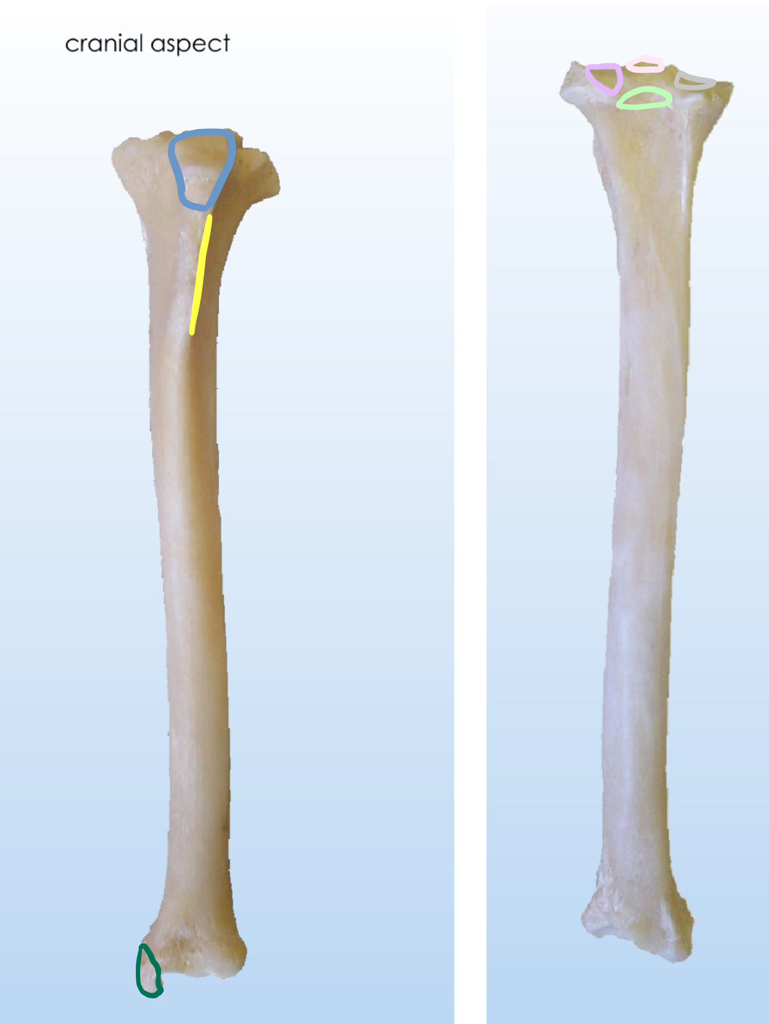
What bone is this? Describe the bony structure of this bone.
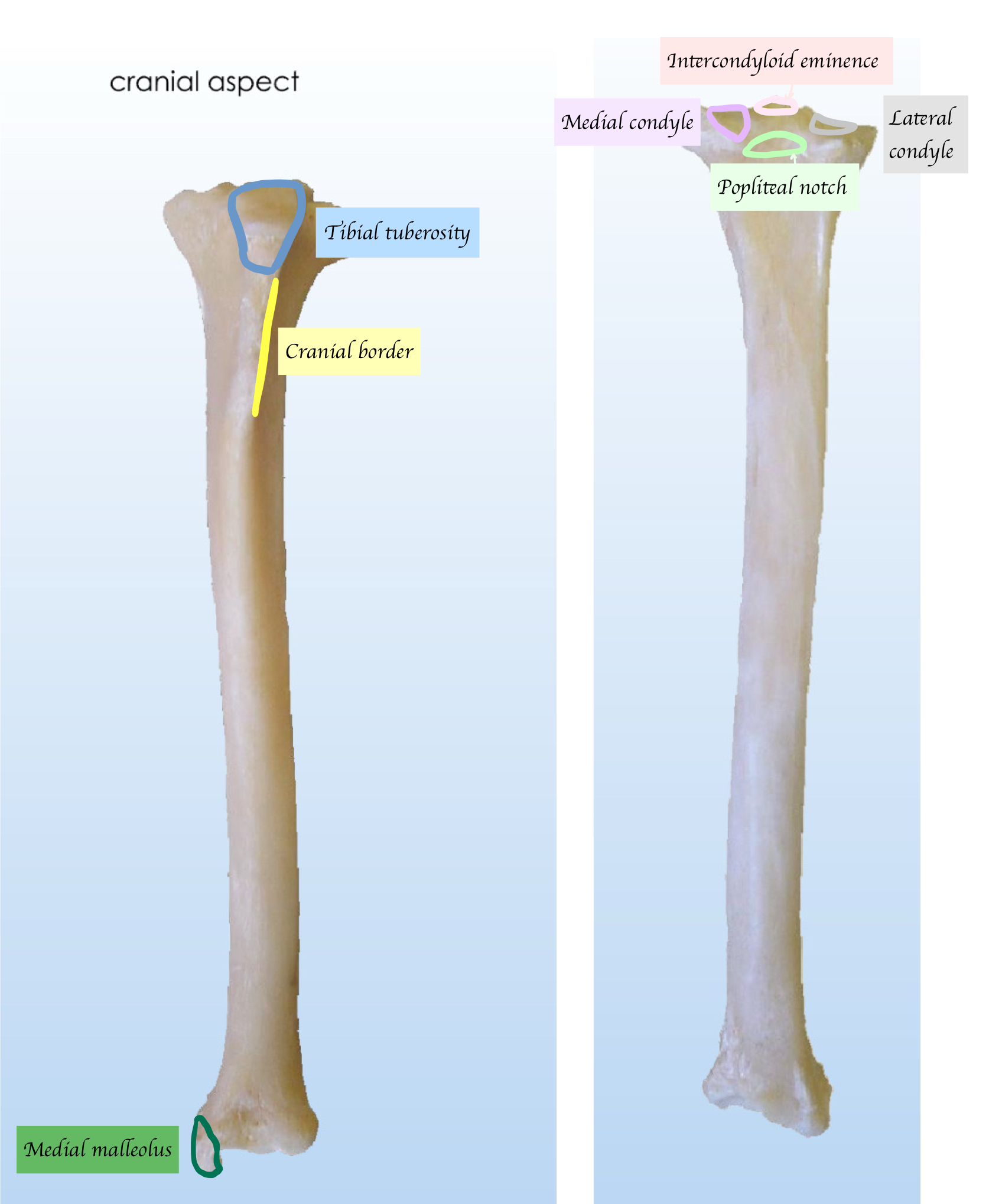
What structures make up the femora-tibial components of the stifle joint?
Femoral condyles, Tibial condyles
Intercondylar fossa of femur, Intercondylar eminence of tibia (Non-articular)
What is the function of the non-articular component of stifle joint
Allow ligament attachment
What are the C-shaped cartilage in stifle joint?
Meniscus
What are the functions of meniscus?
Stabilise joint
Cushioning
Proprioception (Contain nerve ending → Painful if damaged)
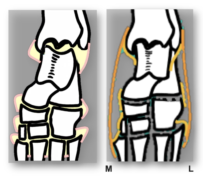
To which part of the tibia does the meniscus attach?
Tibial condyles
List out all three meniscal ligaments.
Menisco-tibial ligament
→ Hold meniscus - intercondylar eminence of tibiaTransverse ligament
→ Hold between the cranial aspects of menisci
Menisco-femoral ligaments
→ Hold lateral meniscus to intercondylar fossa of femur
Apart from meniscal ligaments, what other ligaments support the stifle joint?
Collateral ligaments (Medial and Lateral)
→ Medial epicondyle femur - tibia
→ Lateral epicondyle femur - fibula + tibia
Cranial cruciate ligaments
→ Divide into craniomedial + caudolateral band
Medial femoral condyle + Lateral femoral condyle - Intercondylar eminence of tibia
Caudal cruciate ligaments
→ intercondylar fossa of femur - Intercondylar eminence of tibia
What is the function of the Cruciate ligaments?
Maintain femur on menisci
Resist rotation
What happens if cranial cruciate rupture?
Joint instability
Positive cranial draw sign → Excessive forward (cranial) movement of tibia relative to femur
Which cruciate ligament is most prone to damage?
Cranial cruciate ligament
Because cranial cruciate ligaments is shorter than caudal cruciate ligaments
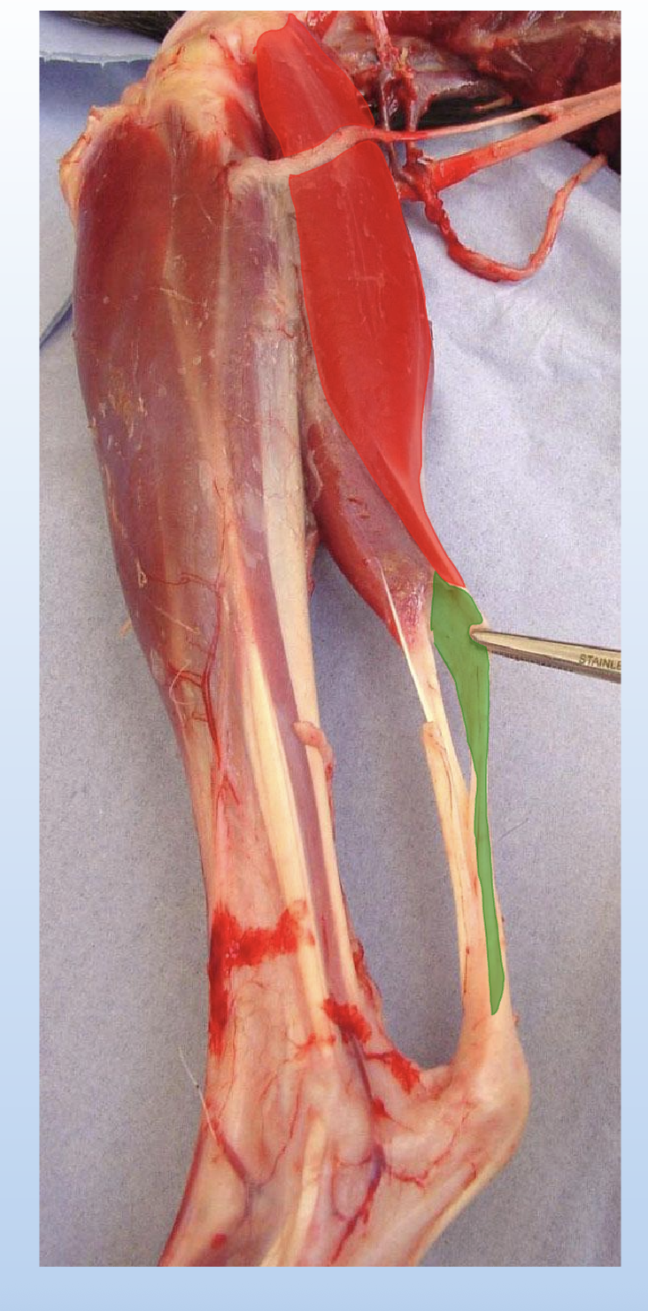
Which muscle does the patellar ligament allow to exert force?
Patellar ligament allows the insertion of quadriceps (& sartorius) on tibial tuberosity
Where is the patella held? How does the patella move within the stifle joint?
Patella is held in trochlear groove → No lateral movement is allowed
When patella pulled proximally → Extension;
When patella pulled distally → Flexion
What supports the patella and holds it in place within the trochlear groove?
Medial and lateral trochlear ridges
Lateral & medial femoro-patellar ligaments (Orange)
Fascia / retinaculum (Light yellow) → Joint capsule reinforcement within the retinaculum
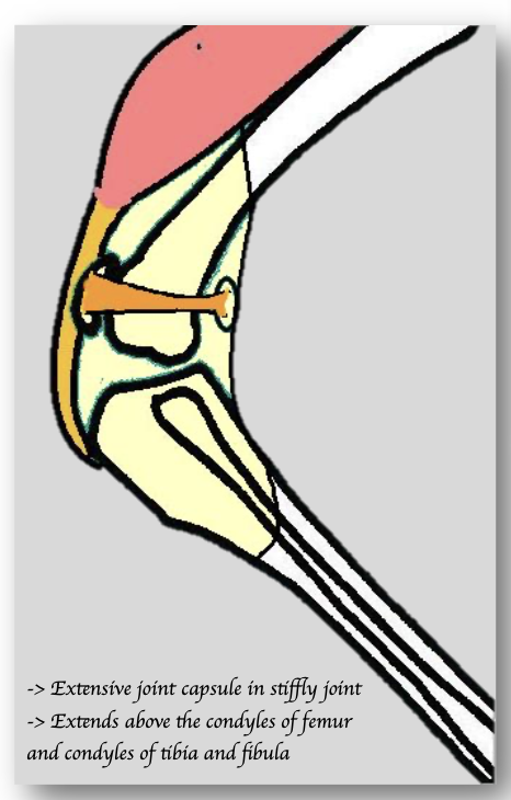
What are the 3 Synovial joint compartments in the stifle joint?
Femoro-patellar
Medial femoro-tibial
Lateral femoro-tibia
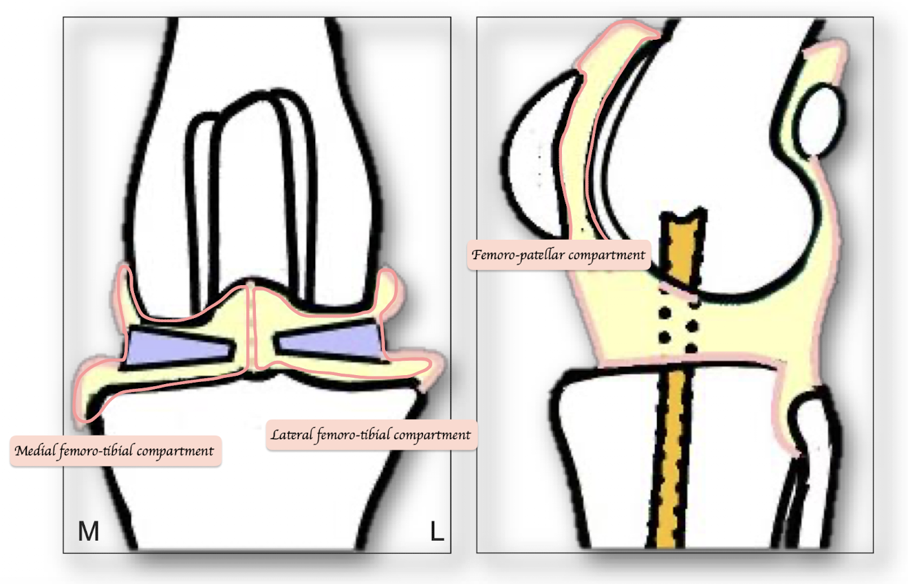
What structures make up the femoro-patellar components of the stifle joint?
Medial and lateral trochlear ridges
Medial and lateral femoro-patella ligament
Retinaculum
What are the special features of a cat’s stifle joint in radiography?
Pointed patella compared to dog
Medial labella often not mineralised (Still present! Just cannot be seen in X ray)
Lateral fabella + politeal sesamoid is visible
Name the stifle extensors
Sartorius (2 heads), Quadriceps muscle
How many head does sartorius have? What are the origin and insertion points of the sartorius? Which nerves innervate sartorius muscle?
2 heads
Cranial part
O: Crest of ilium
I: FemurCaudal part
O: Ventral iliac spine of ilium
I: Tibial tuberosity (Join the patellar ligament together with rectus femoris)
Nerve: Femoral nerve
How many head does quadriceps have? What are the origin and insertion points of all heads of quadriceps? Which nerves innervate quadriceps?
Quadriceps has four heads:
Innervated by femoral nerve
Rectus femoris
O: Ventral ilium
I: Tibial tuberosity via patellar ligament
Vastus lateralis
O: Lateral Femur
I: Tibial tuberosity via patellar ligament
Vastus medialis
O: Medial Femur
I: Tibial tuberosity via patellar ligament
Vastus intermedius
O: Cranial femur
I: Tibial tuberosity via patellar ligament
What are the stifle flexors muscles?
Biceps femoris, Semitendinosus, Semimembranosus, Gastrocnemius
Where are the stifle flexors located in relation to the stifle joint?
Caudal to stifle joint
What are the origin and insertion points of Gastrocnemius? Which nerves innervate Gastrocnemius?
O: Caudal aspect of femur
I: Calcaneus via the common calcanean tendon
N: Tibial nerve (Branch of sciatic nerve)
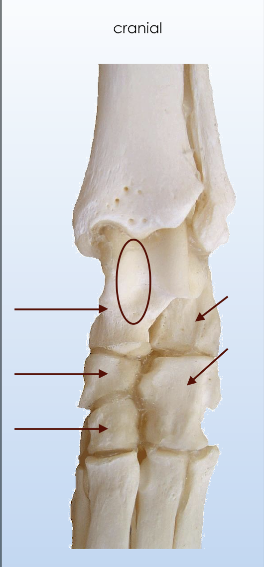
How many rows are the tarsal bones arranged in, and how are they distributed?
3 rows
Proximal row:
Talus + Calcaneus
Middle row:
Central + IV
Distal row:
I, II, III and IV
Which tarsal bone is the largest?
Calcaneus
Name the key structure of tarsus
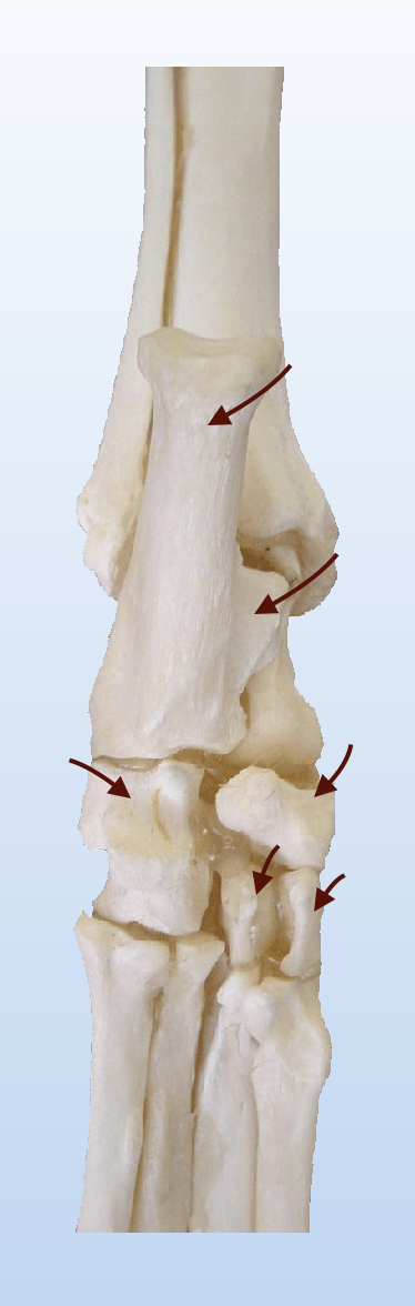

Tuber calcanei = Calcanea tuberosity
What are the function of sustentaculum tali of calcaneus?
Projects medially → Allow the passage of DDFT
What is the centre of ossification of tarsus?
Other than calcaneus (2 centre of ossification), other tarsal bone have one centre of ossification
List out all the tarsus joint
Tarso-crural joint = Tarso-tarsal joint
Proximal inter-tarsal joint
Distal inter-tarsal joint
Tarso-metatarsal joint
Which tarsal joint has the most movement?
Tarso-crural joint
What is the Proximal intertarsal joint?
Talus & calcaneus – Central & IV
What is special about the trochlea of the talus bone?
Trochlea of talus bone is not vertical = Not straight → When there is flexion, the joint and the hindlimb automatically move laterally instead of in a straight line → Avoid overreaching or hitting the forelimb especially when they are doing long strides e.g. running
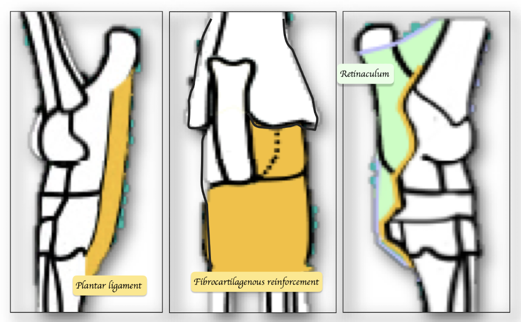
What supports the tarsus joint?
Collateral ligaments
Calcaneus - metatarsal bone
Plantar ligament
Fibrocartilagenous of joint capsule
Retinaculum (On top of the plantar ligament and fibrocartilagenous)
On top of the palmar ligament and fibrocartilagenous
What types of collateral ligaments are there in the tarsus joint?
Long collateral ligaments (medial and lateral):
Medial: from the tibia to the 2nd metatarsal
Lateral: from the fibula to the 5th metatarsal
Short (intertarsal) collateral ligaments:
Between adjacent tarsal (bridge) bones