Peritoneal Fluid and Synovial
1/50
There's no tags or description
Looks like no tags are added yet.
Name | Mastery | Learn | Test | Matching | Spaced | Call with Kai |
|---|
No analytics yet
Send a link to your students to track their progress
51 Terms
ascites
Peritoneal Fluid
Is the accumulation of fluid between the peritoneal membranes
ascitic fluid
Peritoneal Fluid
is used to refer to the fluid instead of peritoneal fluid
cirrhosis
Peritoneal Fluid
is the frequent cause of ascitic transudates
Exudative fluids
Peritoneal Fluid
In peritoneal it leads to;
Bacterial Infection/Peritonitis (intestinal perforation or ruptured appendix)
Malignancy
peritoneal lavage
Peritoneal Fluid
Is a sensitive test to detect intraabdominal bleeding in blunt trauma cases
>100,000/mL
Peritoneal Fluid
RBC count can be used along with radiographic procedures to aid in determining the need for surgery
RBC count ________are indicative of blunt trauma injuries
Eosinophil count can be used to detect allergic reactions to the equipment used in peritoneal dialysis
serum ascites albumin gradient
Peritoneal Fluid
Is recommended over the Fluid:Serum Total Protein and LDH Ratio to detect hepatic transudates
SAAG = serum albumin – fluid albumin
Fluid and serum albumin levels are measured concurrently
>_1.1
Peritoneal Fluid
For SAAG, a difference/gradient of ____ suggest a hepatic transudate
SAAG = serum albumin – fluid albumin
>500
Peritoneal Fluid
WBC count of <500 cells/uL is normal
WBC count of _______ cells/uL indicates bacterial peritonitis and cirrhosis
Absolute Neutrophil Count
Peritoneal Fluid
To differentiate between bacterial peritonitis and cirrhosis, perform an?
>250
Peritoneal Fluid
To differentiate between bacterial peritonitis and cirrhosis, perform an Absolute Neutrophil Count
An absolute neutrophil count of ______ cells/uL or >50% of the total WBC count indicates infection
Note: Lymphocytes are the predominant cells in tuberculosis (Same with Pleural Fluid)
green
Significance of Peritoneal Fluid Testing
Bile, gallbladder, pancreatic disorders
alkaline phosphatase
Significance of Peritoneal Fluid Testing
Increased in gastrointestinal perforation
BUN/ creatinine
Significance of Peritoneal Fluid Testing
Ruptured or punctured bladder
lipophages
Peritoneal Fluid Cells
Macrophages containing fat droplets
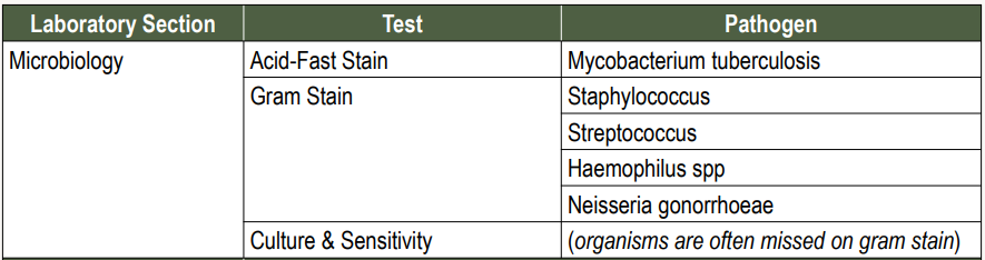
Budding yeast
Ascitic Fluid Cells: Peritoneal Fluid
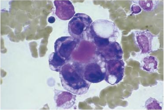
ovarian carcinoma
Ascitic Fluid Cells: Peritoneal Fluid
Showing community borders (Cytoplasmic molding) , nuclear irregularity and hyperchromatic nucleoli
adenocarcinoma
Ascitic Fluid Cells: Peritoneal Fluid
Prostate showing cytoplasmic vacuoles, community borders, and hyperchromatic nucleoli
colon carcinoma
Ascitic Fluid Cells: Peritoneal Fluid
Containing mucin vacuoles and nuclear irregularities
psammoma bodies
Ascitic Fluid Cells: Peritoneal Fluid\
exhibiting concentric striations
cells that have calcium deposits
synovial fluid
Often referred to as “Joint Fluid”
A viscous liquid found in the cavities of the movable joints (diarthroses)
An ultrafiltrate of plasma, most of its constituents have concentrations similar to plasma values
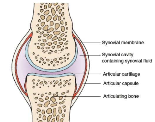
hyaluronic acid
Synovial Fluid
The synovial membrane contains specialized cells called synoviocytes, which secrete a mucopolysaccharide containing ___________, which polymerizes and contributes to the viscosity of synovial fluid
arthritis
Synovial Fluid
is the collective term for any damage to the articular membranes that produces pain and stiffness in the joints
Arthrocentesis
Synovial Fluid
Is the medical procedure done to collect synovial fluid via needle aspiration
heparin
Synovial Fluid
Normal synovial fluid does not clot, but a diseased joint may contain fibrinogen and will clot, therefore collection is often done with a syringe that has been moistened with ?
gram stain and culture
Required Tube Types for Synovial Fluid Tests
Sterile heparinized or SPS
cell counts
Required Tube Types for Synovial Fluid Tests
Heparin or liquid EDTA
Note: Since Synovial is viscous, don’t use regular diluting fluids like glacial acetic acid, but you need to use NSS 0.9% to dilute
glucose analysis
Required Tube Types for Synovial Fluid Tests
Sodium Fluoride
1 hour
Synovial Fluid: Technical Tip
To prevent falsely decreased values caused by glycolysis, specimens should be analyzed within _____ or preserved with Sodium Fluoride
<3.5 mL
Normal Synovial Fluid Values
Volume?

4-6 cm
Normal Synovial Fluid Values
its Viscosity is able to form a string _____ long (2.3 Inches)
If very viscous; pre -treat with
1 drop of 0.05 % hyaluronidase in phosphate buffer per mL of fluid and incubate at 37*C for 5 min
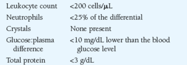
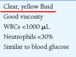
noninflammatory
Classifications and Pathologic Significance of Join disorders (Synovial)
Degenerative joint disorders
Osteoarthritis
infammatory
Classifications and Pathologic Significance of Join disorders (Synovial)
septic
Classifications and Pathologic Significance of Join disorders (Synovial)
Microbial infection
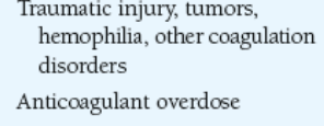
hemorrhagic
Classifications and Pathologic Significance of Join disorders (Synovial)
noninflammatory
Laboratory Findings in Joint Disorders(Synovial)
Note: You will notice, all other than this have a poor viscosity.
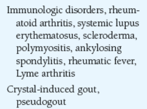
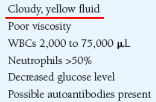
inflammatory : immunologic origin
Laboratory Findings in Joint Disorders
Poorly viscous, because diseases affects the production and polymerization of hyaluronic acid
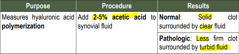
ropes/mucin clot test
Test for Hyaluronic Acid
Note: Not routinely performed because all forms of arthritis decrease viscosity and little diagnostic information is obtained
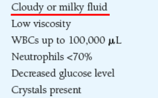
inflammatory : crystal induced origin
Laboratory Findings in Joint Disorders
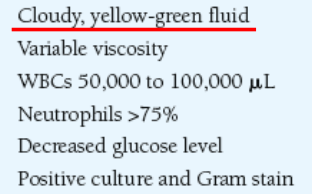
septic
Laboratory Findings in Joint Disorders
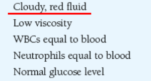
hemorrhagic
Laboratory Findings in Joint Disorders
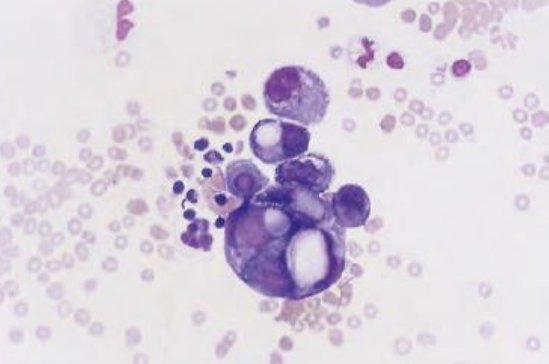
macrophages, synovial lining cell
Cells and Inclusions Seen in Synovial Fluid
Normal significance
Reiter cell
Cells and Inclusions Seen in Synovial Fluid
Reactive arthritis (Infection in another part of the body)
rice bodies
Cells and Inclusions Seen in Synovial Fluid
Tuberculosis
Septic and RH
fat droplets
Cells and Inclusions Seen in Synovial Fluid
Traumatic injury
Chronic Inflammation
hemosiderin
Cells and Inclusions Seen in Synovial Fluid
Pigmented villonodular synovitis
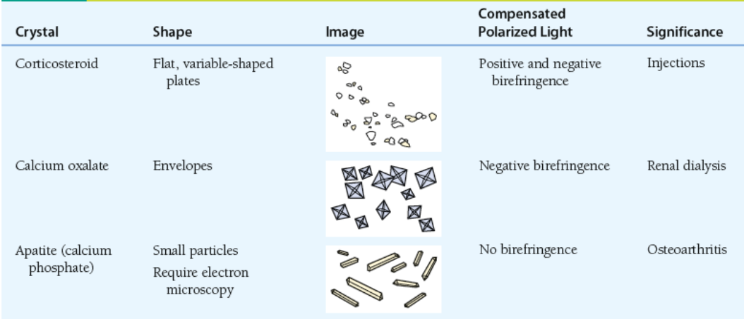
Monosodium urate and calcium pyrophosphate
Characteristics of Synovial Fluid Crystals
Commonly seen crystals in synovial fluid
monosodium urate
Characteristics of Synovial Fluid Crystals
Usually seen in cytoplasm
Shape: Needles
Compensated Polarized light: (-) Bifringence
Significance: Gout
Attached photo are just other types lang
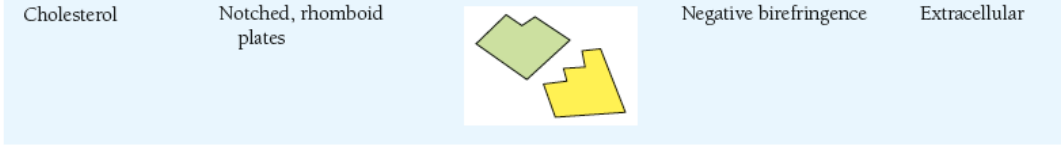
calcium pyrophosphate
Characteristics of Synovial Fluid Crystals
Usually seen in vacuoles
Shape: Rhomboid square, rods
Compensated Polarized light: (+) Bifringence
Significance: Pseudogout (e.g athletes)
Attached photo are just other types lang
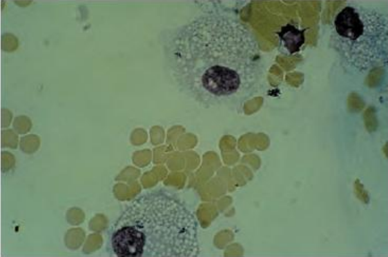
lyme disease
Laboratory Section : Synovial Fluid
SEROLOGY
Most test are performed on serum, synovial fluid is used as a confirmatory measure for cases that difficult to diagnose
Arthritis is a frequent complication of ________, therefore demonstrating antibodies against Borrelia burgdorferi can confirm the cause.
Attached Photo is for Microbiology

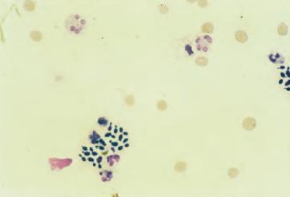
glucose
Laboratory Section : Synovial Fluid
CHEMISTRY
Most frequently tested is _________ (8 hours fasting), because markedly decreased ________levels indicate inflammatory (group II) or septic (group III) disorders
Attached Photo is for Microbiology