Lecture 24: Mycobacterium species
1/23
There's no tags or description
Looks like no tags are added yet.
Name | Mastery | Learn | Test | Matching | Spaced | Call with Kai |
|---|
No analytics yet
Send a link to your students to track their progress
24 Terms
Salient features of Mycobacterium species
Aerobic, non-spore forming, non-motile, rods
Cytochemically Gram-positive
Acid-fast (ZN-positive) due to high lipid and mycolic acid in their cell walls
Mycobacterium species multiply
intracellularly and cause chronic, granulomatous infections
Major diseases caused by Mycobacterium species include
tuberculosis (TB), Johne’s disease and feline leprosy
M. aviumcomplex main hosts are
most avian species except psittacines
Mycobacterium are ___ to adverse environmental conditions due to their lipid-rich walls
resistant
Mycobacterium are identified by
Acid-fast staining (Ziehl-Neelsen)
Mycobactin is required for growth for
M. avium subsp. paratuberculosis
Recently highly reliable and fast molecular techniques such as PCR and 16s rRNAgene sequencing have been developed to identify
Mycobacterium
Johne’s disease is caused by
M. avium subsp. paratuberculosis (MAP)
Johne’s disease or Paratuberculosis is a worldwide -
chronic, contagious granulomatous enteritis of mainly ruminants
Johne’s disease is characterized in cattle by
persistent diarrhea, progressive weight loss, debilitation, eventually death
With Johne’s disease, M. avium subsp. paratuberculosis is excreted in
large numbers in feces of infected animals and in lower numbers in their colostrum and milk
How is Johne’s infection spread
fecal-oral route
Introduction of Johne’s disease into a non-infected herd is usually through
herd expansion or replacement purchases via subclinically infected carriers
After ingestion and uptake in the Peyer’s patches of the lower small intestine, the organism of Johne’s disease infects
macrophages in the GI tract and associated lymph nodes
In most cases, the organisms (Johne’s disease) multiply and eventually provoke a
chronic granulomatous enteritis that interferes with nutrient uptake and processing, leading to the cachexia typical of advanced infections
Clinical findings of Johne’s disease
Infected animals can appear healthy for months to years
In cattle, weight loss and diarrhea
ventral and intermandibular edema (due to a protein-losing enteropathy)
Clinical findings of Johne’s disease in sheep and goats and other ruminants
maybe diarrhea
milk yield may drop
as many as 50% of animals may be infected subclinically with associated production losses
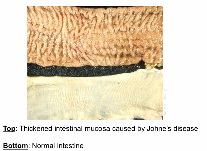
Johne’s disease causes lesions which include
Thickened and corrugated intestine with enlarged and edematous neighboring lymph nodes
Lesions caused by Johne’s disease cause
Thickened and corrugated intestine with enlarged and edematous neighboring lymph nodes
Intestinal lesions from Johne’s disease may be mild but typically,
distal small-intestinal wall is diffusely thickened
Diagnosis of Johne’s disease
Organism based test - culture, PCR
Antibody based test - ELISA, AGID
Is there a treatment for Johne’s disease?
No satisfactory treatment
How can we prevent Johne’s disease?
Prevent new infection
• keep young away from adult animals
• colostrum from paraTB-free animals only
Remove the source of MAP from the farm