post mortem change
1/25
There's no tags or description
Looks like no tags are added yet.
Name | Mastery | Learn | Test | Matching | Spaced | Call with Kai |
|---|
No analytics yet
Send a link to your students to track their progress
26 Terms
3 types of change
due to disease- lesions
occur just prior to or at the time of death- agonal change
those that occur after death and before the cadaver is examined- post mortem change
post mortem change
algor mortis- cooling
rigor mortis- rigidity
post mortem clotting
livor mortis- hypostatic congestion
post mortem staining
autolysis
putrefication
algor mortis
cooling of the cadaver to the temp of the environment
practical importance in meat production- reduces post mortem bacterial growth, affects tenderness of meat for consumption
may aid in estimating time of death
rigor mortis
what is it
what does it lead to
when does it begin
what is affected first and last
stiffening of skeletal muscle after death
leads to immobilisation of joints
begins 1-6hr after death
smaller muscles affected first and those in excessive use before death affected last
skeletal muscle post mortem
immediately after death the cell tries to maintain ATP production by ADP to ATP through the phosphagen system and the glycogen/lactic acid ssytem
reduced atp availability reduces ca sequesteration in sr and build up of ca in cytosol
myosin heads cant be released from binding sites in actin
only released when there is sufficient enzymatic digestion of the contractile proteins to remove cross bridges
when you forcibly break rigor you are mechanically overcoming the cross bridges
rigor mortis- variation in
time of onset
intensity
between species- poultry 1-2hrs, pork 4-6 and beef/lamb 7-15
what is rigor mortis influenced by
pre mortem glycogen reserves- low levels quick onset
ph of muscles at time of death- low ph quick onset
env temp- warm is quick
algor and rigor morits
• Warm and flaccid – Dead less than 3 hours
• Warm and stiff – Dead 3-8 hours
• Cold and stiff – Dead from 8-36 hours
• Cold and flaccid – Dead more than 36 hours
Body held at 10-20ºC
timing of rigor mortis
what does it occur rapidly in
when might it not develop
what is this called
occurs rapidly in exccited or stressed- as total energy supply is less and critical level of atp needed to delay it is soon reached
may not develop in emaciated animals
cadaveric spasm
in emaciated
normally l ventricle stiffens leaving it empty of blood
in extreme emaciation rigor mortis wont occur and blood will remain in both chambers of the blood
post mortem clotting
2 layers
colours
how to distinguish between post mortem clotting and thrombus
-separates into an upper plasma layer and a lower red blood layer
upper is translucent and lower is intense red
no damage to the inner surface of the vessel and the clot can be easily removed
thrombus is firmly attached to the underlying vascular endothelium, has a white interior and is layered.
hypostatic congestion- livor mortis
when does it begin
when can it become fixed
where is it best appreciated
hypostatic congestion discolouring the skin of man and pigs
gravitational pooling of blood which occurs post mortem
begins an hour after death and clotted blood can become fixed in place 12-24 hours so movement will not influence distribution
may be apparent in animals with little hair cover
best appreciated in the dependent lung and kidneys in animals
post mortem imbibition of haemoglobin
haemoglobin pigment diffuses out of erythryocytes and through the walls of small vessels
in cases where there has been a haemolytic crisis, staining of tissues occurs early after death
most common obscurer of lesions in post mortem examination
post mortem imbibition of bile pigment
cholebillirubin readily diffuses out of the gall bladder
very common finding which occurs within hours of death
stains tissues adjacent to gall bladder yellow/green
pseudomelanosis
what is it caused by
caused by iron sulphide (hydrogen sulfide produced by bacteria reacts with iron from haemoglobin)
staining
haemolytic staining- red
biliary imbibition- yellow
pseudomelanosis- green
autolysis
tissue breakdown due to anoxia and cell death
in a hypoxic or anoxic cell ATP levels fall and are depleted
cellular membrane damage which destory selective permeability that retains proteins and electrolytes within the cell and restricts na, ca and water entry from extracellular spance
involves no inflammatory responses
process is enhanced by higher temps and failure to cool body after death
putrefication
what is it and hwat happens
what is produced and examples
what other process can occur as as result
dead tissue is invaded by anaerobic saprophytic bacteria
proteins, fats and carbs in the body substances are attacked by enzymes produced by these putrefactive bacteria
producesgas and colour change
foul smelling substances, hydrogen sulphide , indole/skatole (degredation product of tryptophan)
pseudomelanosis
gaseous distention of the alimentary tract
bacterial fermentation producing gas continues after death
may be surface pallor of adjacent organs due to pressure of this distension squeexing blood out of this adjacent tissue
abdominal viscera may be pale in comparison to thoracic viscera or the musculature of hind lumbs
gas rises- distended loops may be pale and lower loops hypostatically congested
if gaseous distention is excessive, distended intestine may tear
autolysis and putrefaction
autolysis plus putrefaction equal decomposition
decomposition occurs faster at high temps
is faster in air compared to water
deep burial is equivalent to storage at 4 degrees
post mortem damage by insects
bluebottles and flies
maggots
eggs are laid on the lips, nostrils or genitals of the fallen carcase
secrete digestive fluid that soften tissue
creophilus maxillosus feed on larvae- imply longer
mummification
occurs in dry conditions
dryness inhibit bacterial groth
skin and tissue become hard and leathery
occurs in foetuses when hte skin is thick enough to resist autolysis and fluids are resorbed
why freeze a carcass
to preserve and halt decomposition i immediate pm not possible
body outside in cold env, ay have diet from hypothermia
poach fish/ deer out of season then freeze and thaw in legal
to hide evidence
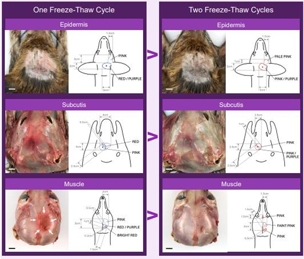
freeze thaw artefact
damage occurs to tissues during freezing due to fluid movement between intra and extra cellular spaces, due to ice crystal formation
alters the gross and histological appearance of tissues which may mimic or obscure lesions
effects of multiple freeze thaw cycles