1.7. Kidney, urinary tract and endocrine organs
1/78
Earn XP
Description and Tags
Niere, ableitende Harnwege und endokrine Organe
Name | Mastery | Learn | Test | Matching | Spaced | Call with Kai |
|---|
No analytics yet
Send a link to your students to track their progress
79 Terms
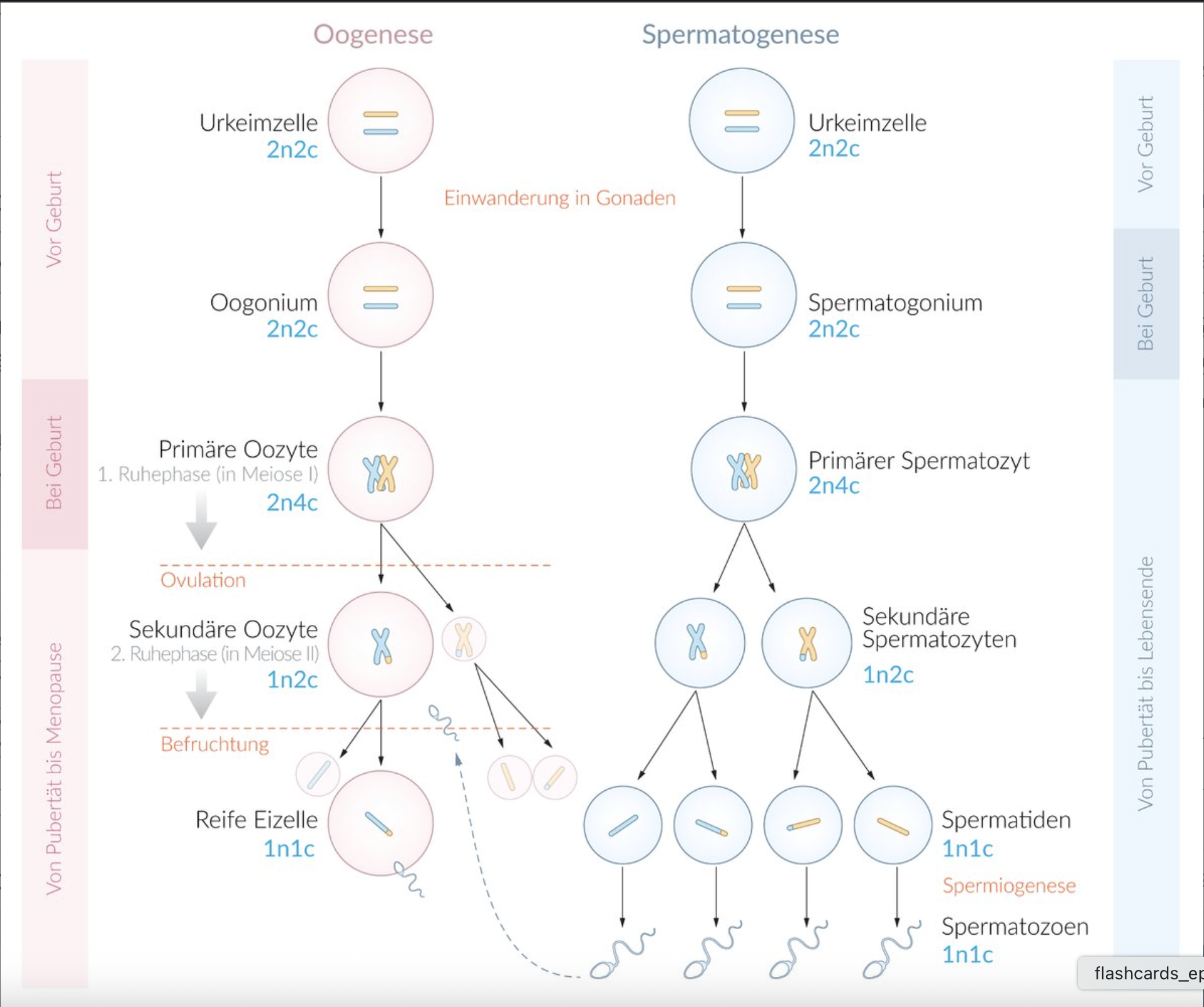
1. Already during the embryonic period, the formation of oocytes begins, which later develop into fertilizable egg cells (oogenesis). Initially, there is a proliferation phase, which is completed before birth. Meiosis also begins before birth, during which primary oocytes are formed. However, this process is interrupted for about 12–50 years.
Which statement characterizes the primary oocyte at birth, where n = number of chromosome sets and C = number of chromatids?
The primary oocyte remains, until shortly before fertilization, in the so-called resting phase 1, which genetically corresponds to prophase 1 of meiosis.
A) 2n, 1C
B) 2n, 2C
C) 2n, 4C
D) 1n, 1C
E) 1n, 2C
C) 2n, 4C
2. Developmental errors can lead to the death of the embryo or fetus, or to malformations.
Which process during development is referred to as gastrulation?
A. The subdivision of the blastocyst into embryoblast and trophoblast
B. The formation of the 3 germ layers
C. The formation of the primitive gut (gut tube)
D. The rotation of the stomach
E. The differentiation processes in the ventral and dorsal mesogastrium
B. The formation of the 3 germ layers
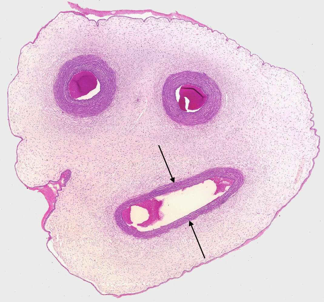
3. The image shows a cross-section of the umbilical cord.
In which of the following structures in an adult human can a remnant of the structure marked by arrows be found?
A. Ligamentum teres hepatis (Round ligament of the liver)
B. Ligamentum teres uteri (Round ligament of the uterus)
C. Plica umbilicalis lateralis (Lateral umbilical fold)
D. Plica umbilicalis medialis (Medial umbilical fold)
E. Plica umbilicalis mediana (Median umbilical fold)
A. Ligamentum teres hepatis (Round ligament of the liver)
4. In general, the different hormone-producing cell types in endocrine organs show little morphological difference.
Which of the following methods can be used to specifically identify them in histological sections?
A. Using polarization microscopy
B. Using the Feulgen reaction with counterstaining
C. Using Masson's trichrome stain
D. Using immunohistochemistry
E. Using silver staining techniques
D. Using immunohistochemistry
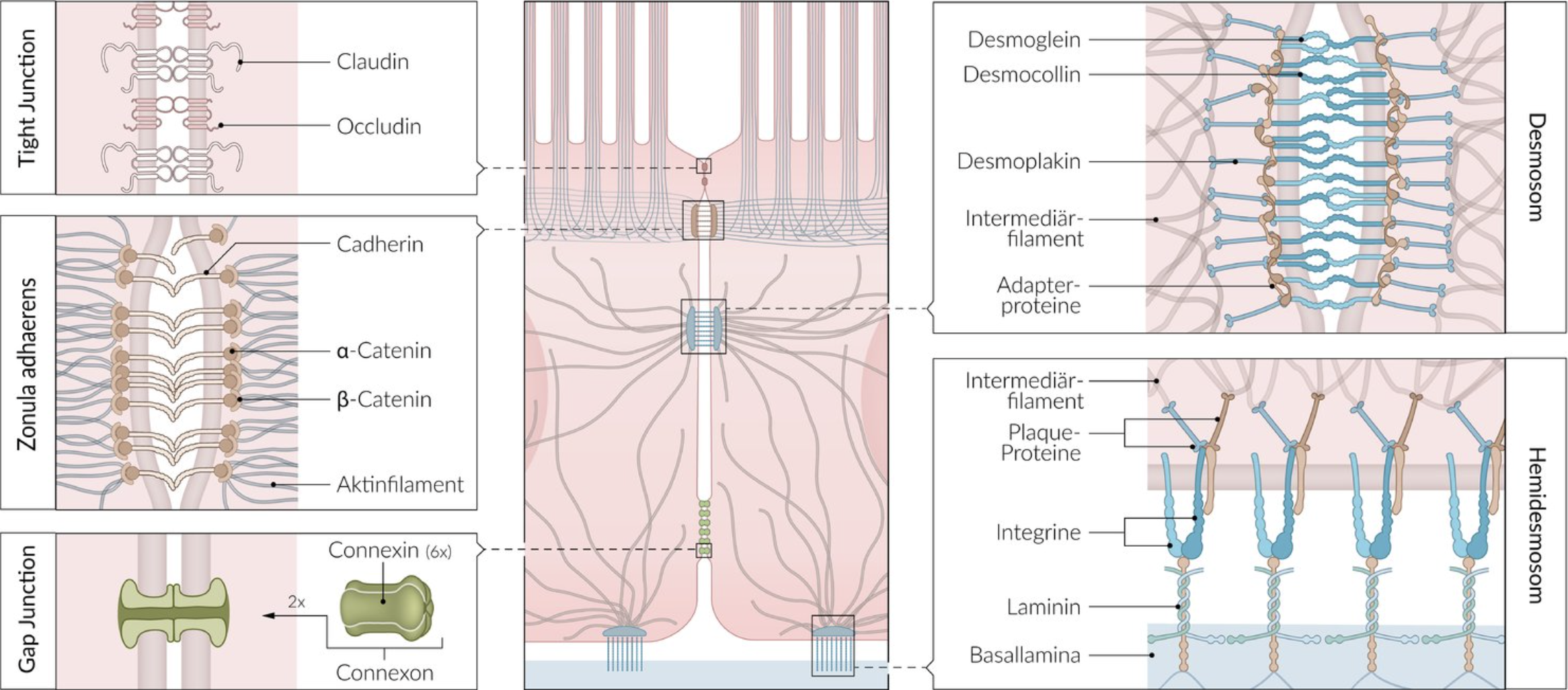
5. Cell junctions are essential for the structure and function of tissue assemblies.
What are the characteristics of hemidesmosomes?
A. They form the so-called crusta of the transitional epithelium.
B. They connect the cells in the stratum spinosum of the stratified keratinized squamous epithelium.
C. They connect endothelial cells to each other in larger lymphatic vessels.
D. They connect enterocytes with goblet cells.
E. They connect epithelial cells with the extracellular matrix.
E. They connect epithelial cells with the extracellular matrix.
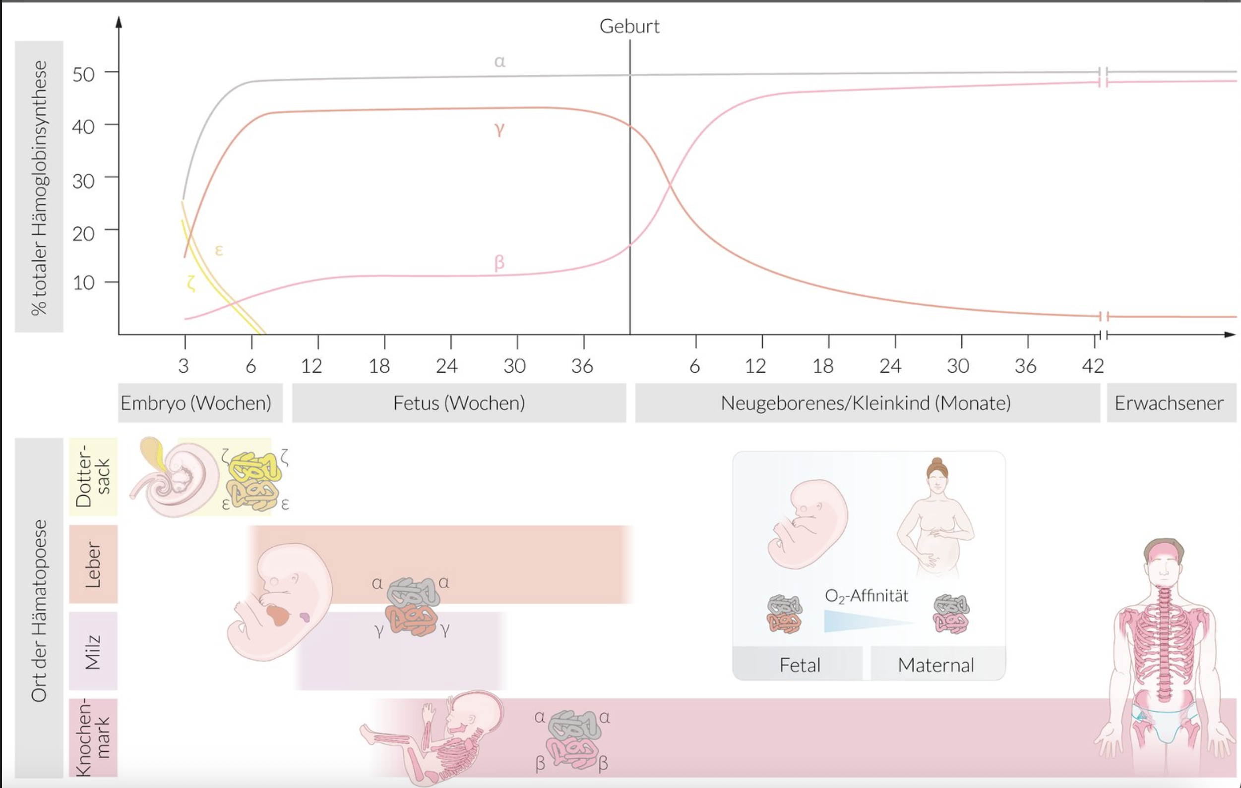
6. The site of blood formation changes multiple times during prenatal development.
The first blood formation during embryonic development takes place in the:
A. Crypts of the palatine tonsil
B. Paracortical zones of the lymph nodes
C. Sinuses of the spleen
D. Wall of the yolk sac
E. Central veins of the liver
D. Wall of the yolk sac
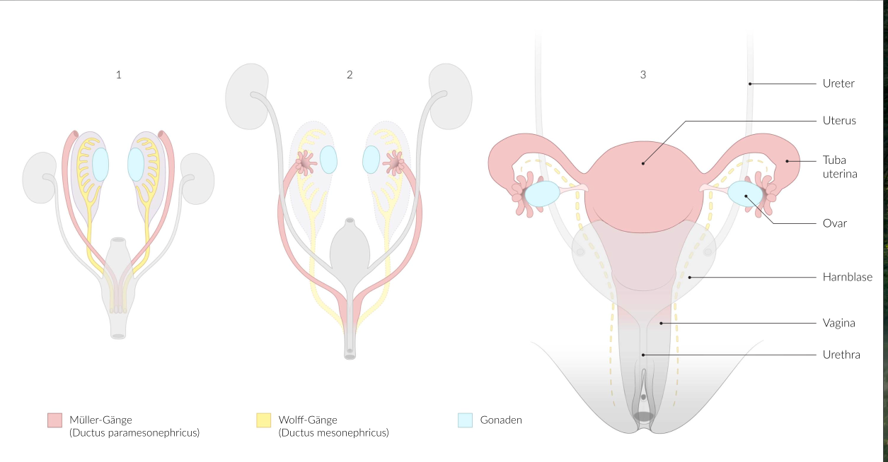
7. The embryonic genital ducts are partly involved in the development of the reproductive organs and the urinary tract. Developmental disturbances can lead to malformations.
What develops from the Müllerian ducts (also known as paramesonephric ducts)?
A. Ductus deferens (vas deferens)
B. Tuba uterina (fallopian tube)
C. Ureter
D. Urethra feminina (female urethra)
E. Urethra masculina (male urethra)
B. Tuba uterina (fallopian tube)
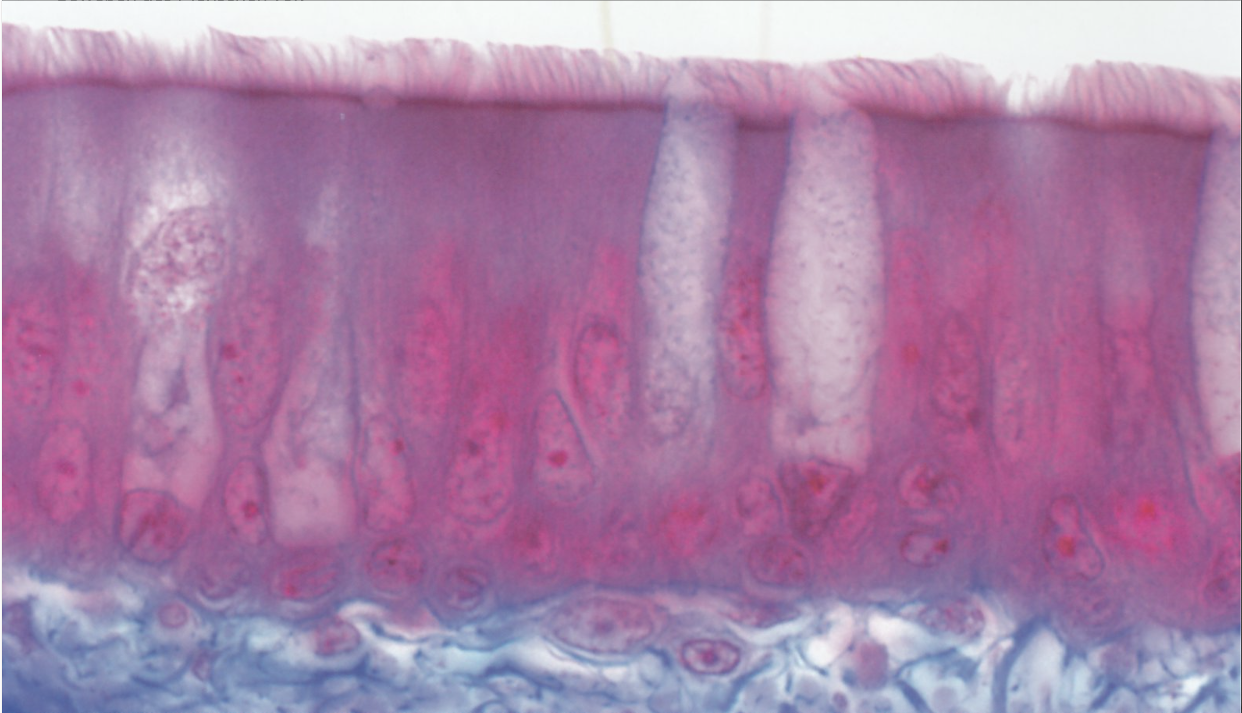
8. Multinucleation (i.e., two or more nuclei per cell) occurs regularly under normal physiological conditions in several human tissues.
Which of the following cells or tissues usually has only one nucleus per cell?
A. Superficial (umbrella) cells of the urothelium
B. Osteoclasts
C. Respiratory epithelium
D. Skeletal muscle
E. Syncytiotrophoblast
C. Respiratory epithelium
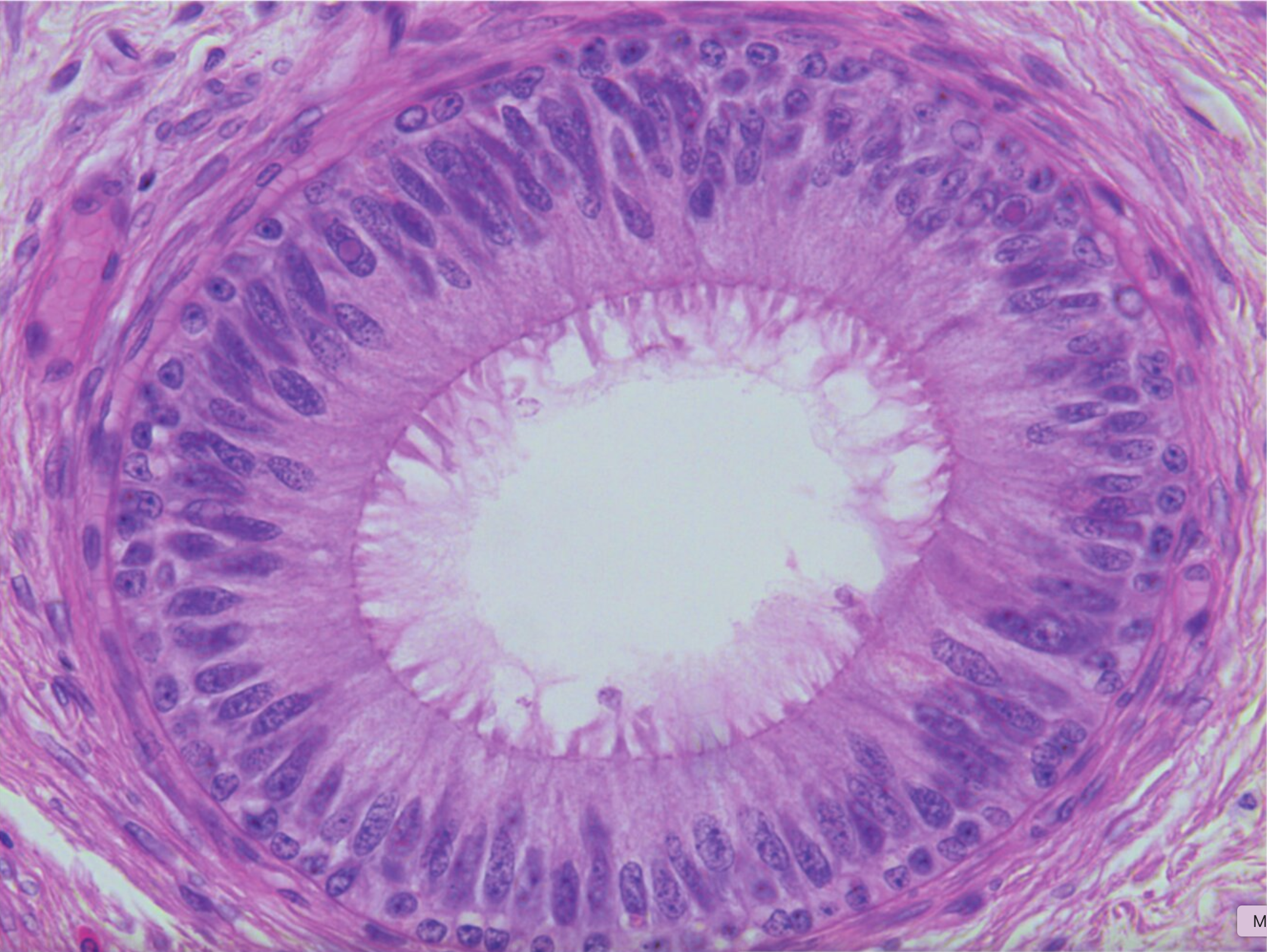
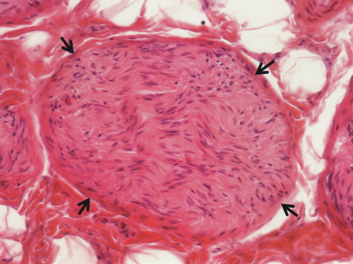
9. The image shows a section of a histological slide of an organ.
Which statement best describes the function or characteristic of this organ?
A. It serves in the absorption of nutrients.
B. It stores sperm cells.
C. It transports egg cells.
D. It transports urine.
E. Its epithelium is involved in mucociliary clearance.
B. It stores sperm cells.
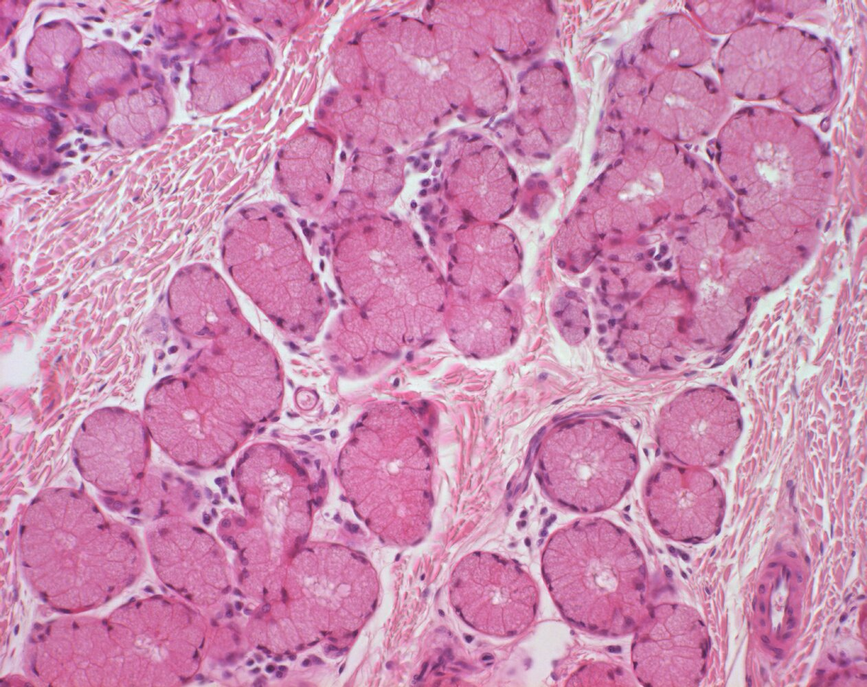
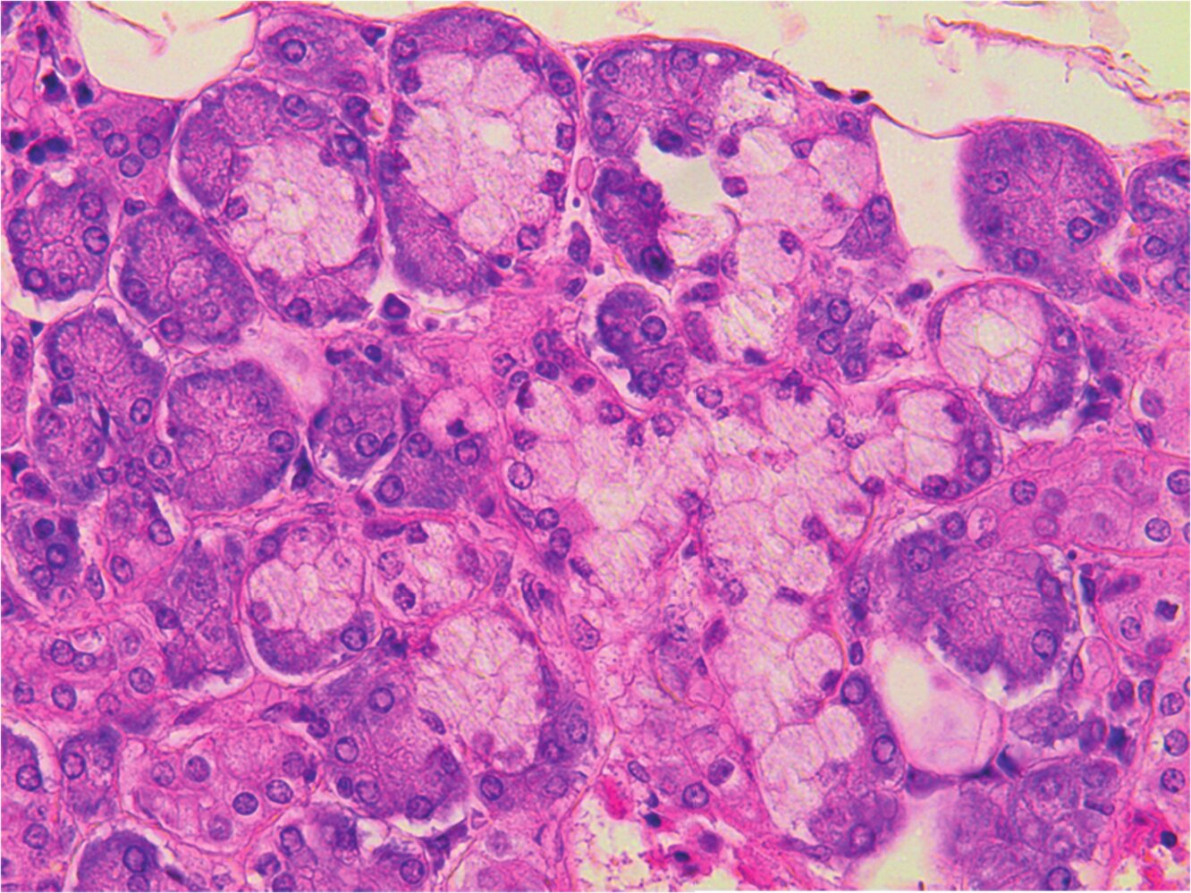
10. The image shows the following structures:
A. Alveoli of the lactating mammary gland
B. Scent (apocrine) glands
C. Mucous glands
D. Sweat glands
E. Serous glands
C. Mucous glands
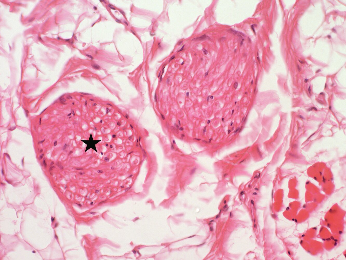
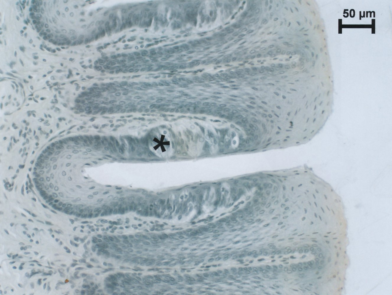
11. The structure marked with a star in the image is:
A. A blood vessel with red blood cells
B. A bundle of smooth muscle cells
C. A peripheral nerve
D. A primary bundle of skeletal muscle fibers
E. A tendon fiber bundle
C. A peripheral nerve
12. According to current studies, glial cells may partly perform neuron-like functions.
Which function is not performed by astrocytes?
A. Antibody production
B. Barrier functions
C. Gliotransmission
D. Metabolic interaction with neurons
E. Production of extracellular matrix
A. Antibody production
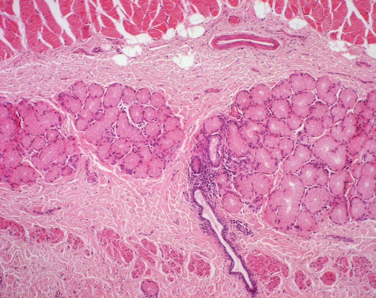
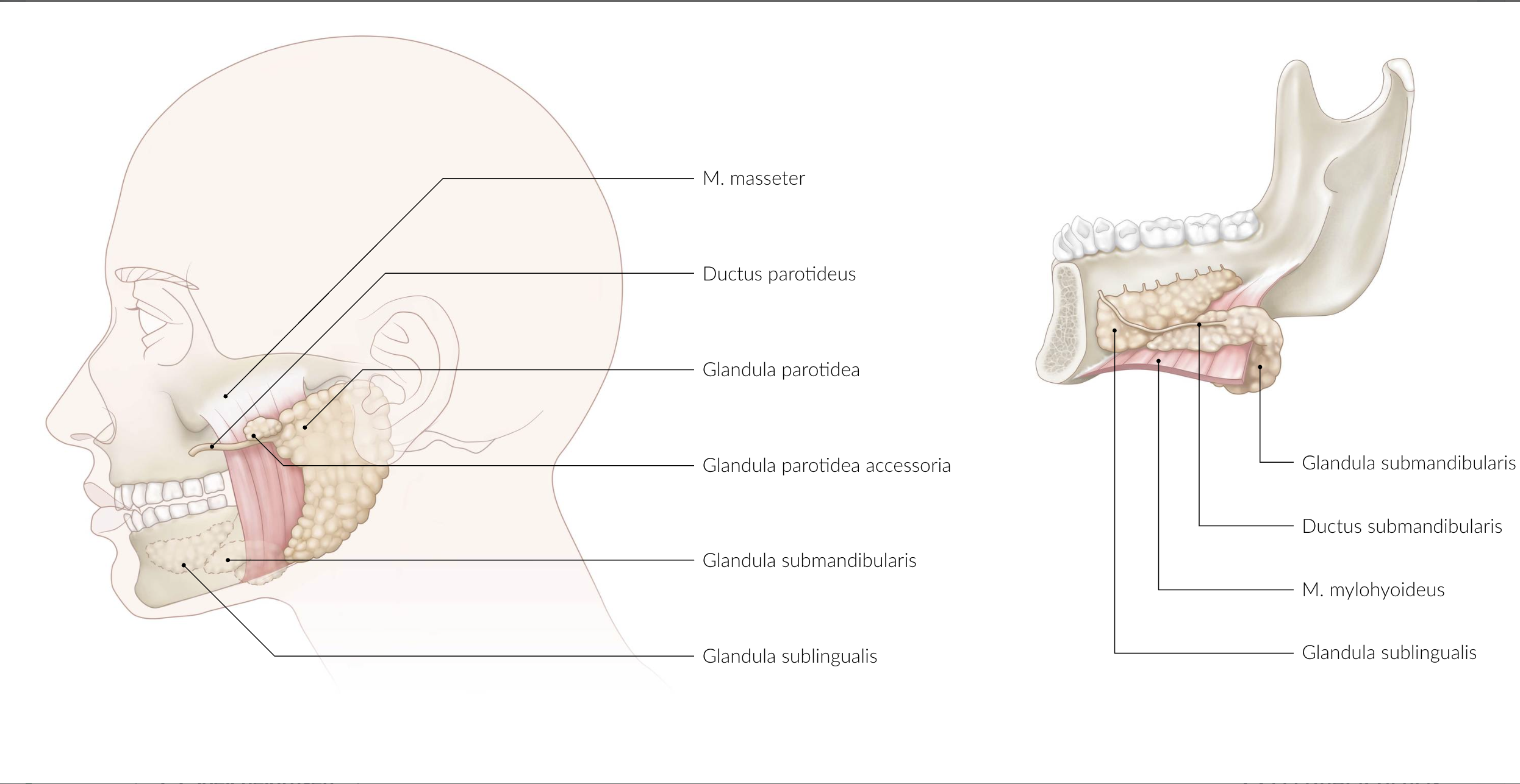
13. The image shows a section of a histological slide of an organ.
Where is the largest portion of this organ located?
A. Posterior to the masseter muscle (M. masseter)
B. Caudal (below) to the mylohyoid muscle (M. mylohyoideus)
C. Between the palatoglossal arch and palatopharyngeal arch (Arcus palatoglossus and Arcus palatopharyngeus)
D. Caudal to the thyroid cartilage (Cartilago thyroidea)
E. Secondarily retroperitoneal between the descending part of the duodenum and the spleen
B. Caudal (below) to the mylohyoid muscle (M. mylohyoideus)
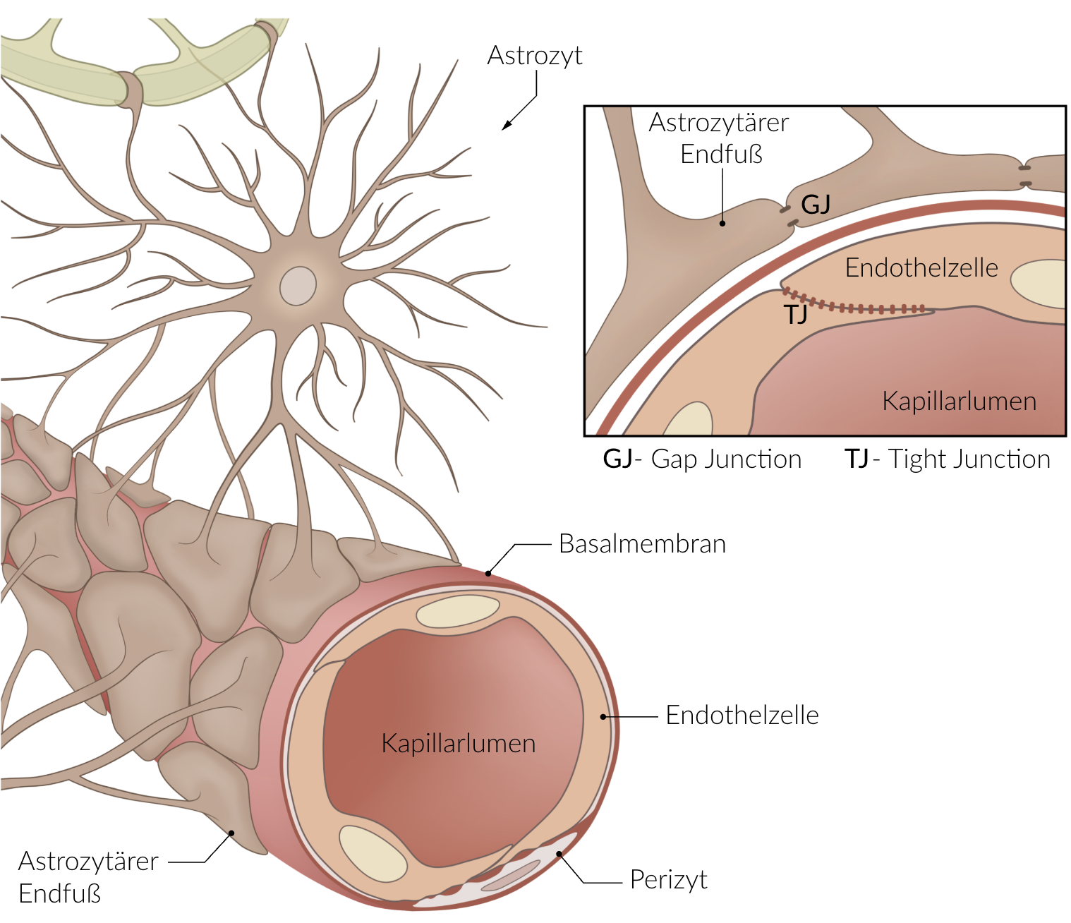
14. In large parts of the brain, there is a barrier between the bloodstream and the brain tissue (the so-called blood-brain barrier), which prevents certain medications from entering the brain.
Which cell types are involved in forming the blood-brain barrier?
A. Astrocytes and the endothelium of capillaries
B. Ependymal cells and microglial cells
C. Smooth muscle cells and the endothelium of arterioles
D. Smooth muscle cells and the endothelium of venules
E. Pericytes and the endothelium of lymphatic vessels
A. Astrocytes and the endothelium of capillaries
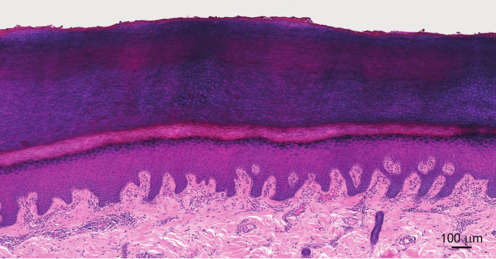
15. The image shows a section from a histological specimen.
Which of the cell types found in this organ can differentiate into cells capable of presenting antigens to both CD4+ and CD8+ T-lymphocytes?
A. Granulocytes
B. Langerhans cells
C. Melanocytes
D. Merkel cells
E. Plasma cells
B. Langerhans cells
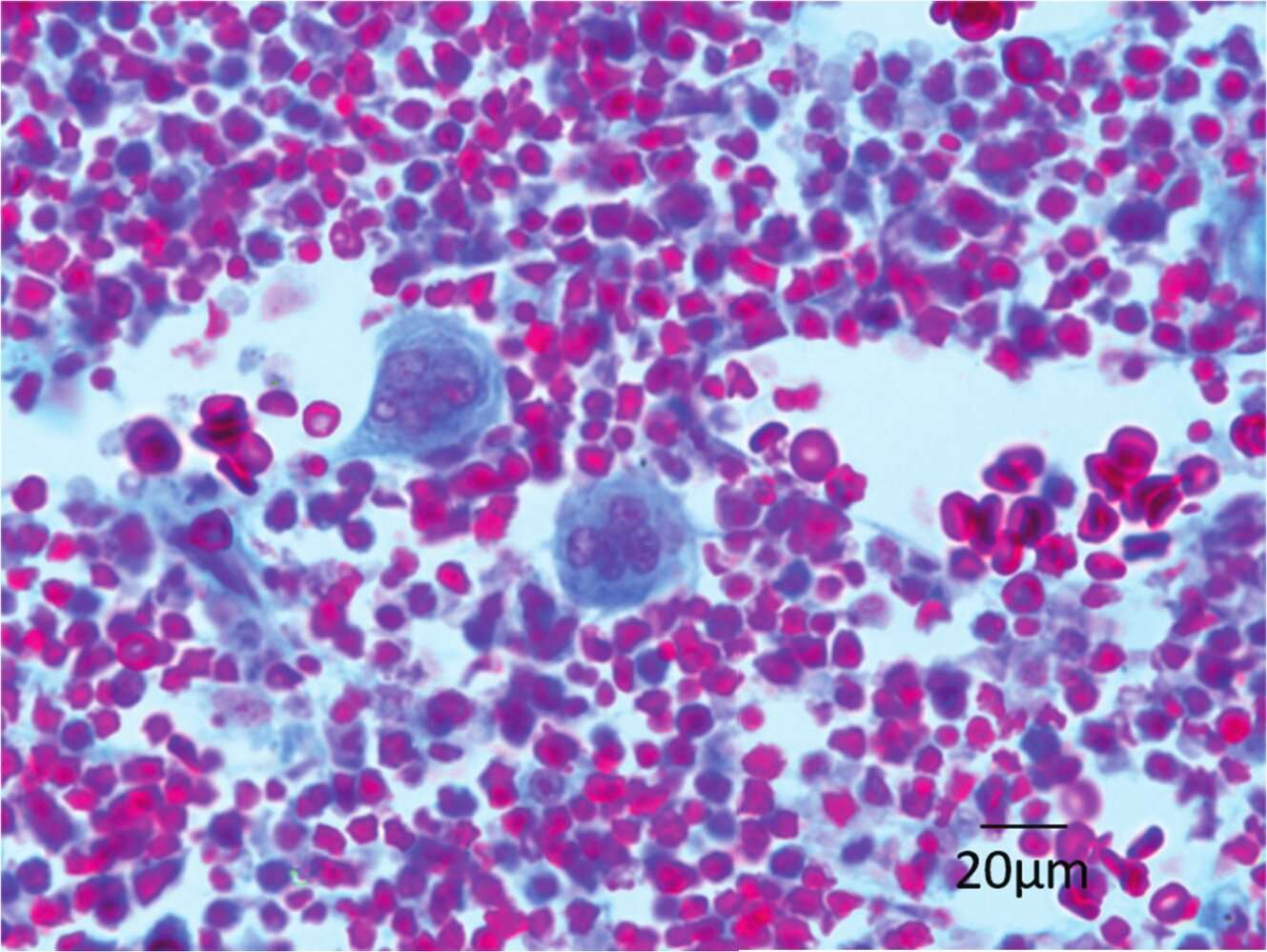
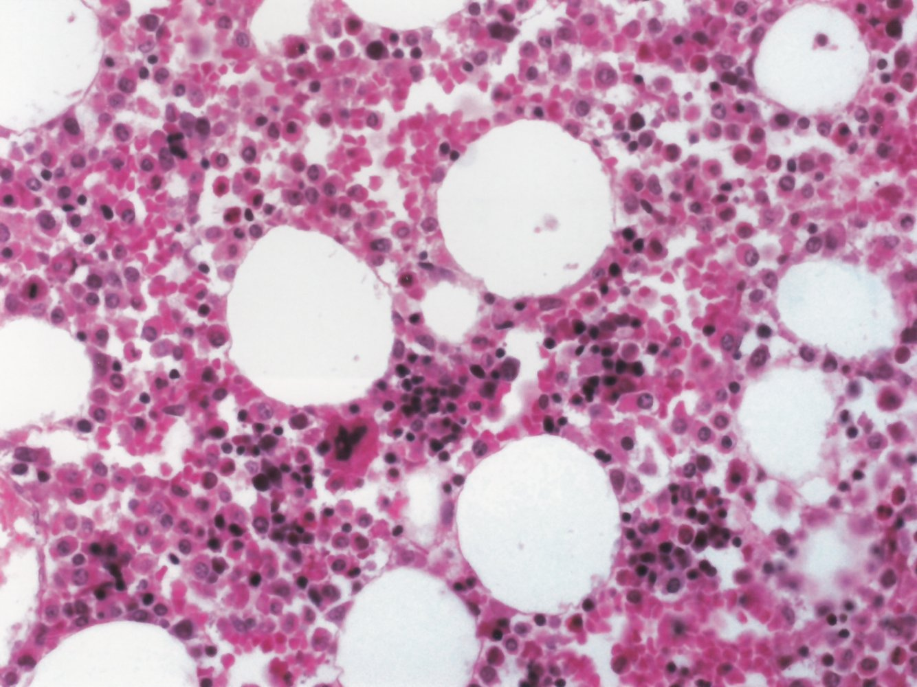
16. Organs that serve immune defense are classified into primary and secondary lymphatic organs.
From which lymphatic organ was the histological specimen shown in the image obtained?
A. Bone marrow
B. Lymph node
C. Spleen
D. Thymus
E. Palatine tonsil (Tonsilla palatina)
A. Bone marrow
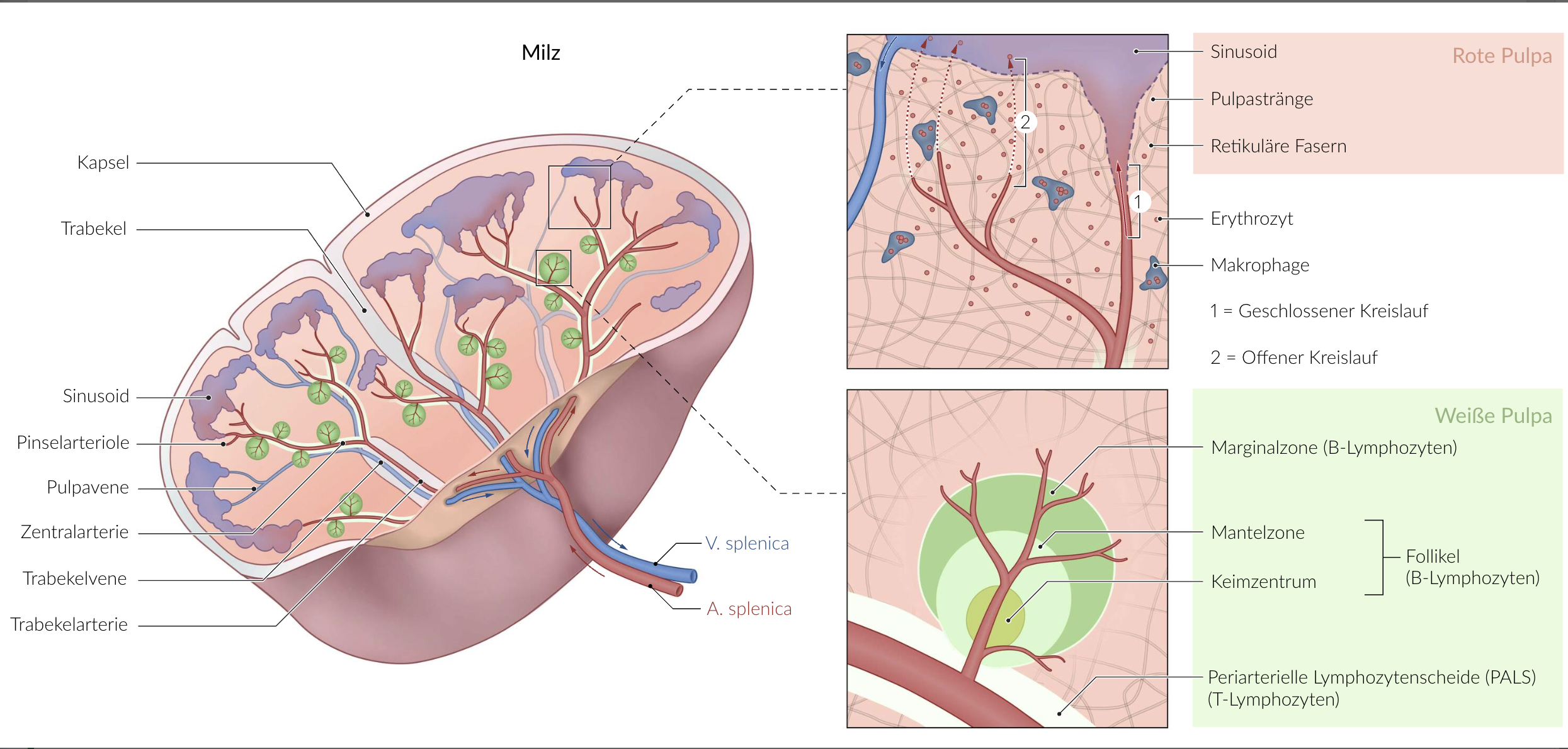
17. The parenchyma of the spleen is divided into white pulp and red pulp.
Which of the following structures is not part of the histological structure of the white pulp?
A. Germinal center of the follicle
B. Mantle zone
C. Marginal zone
D. Periarteriolar lymphoid sheath (PALS)
E. Sinusoids
E. Sinusoids
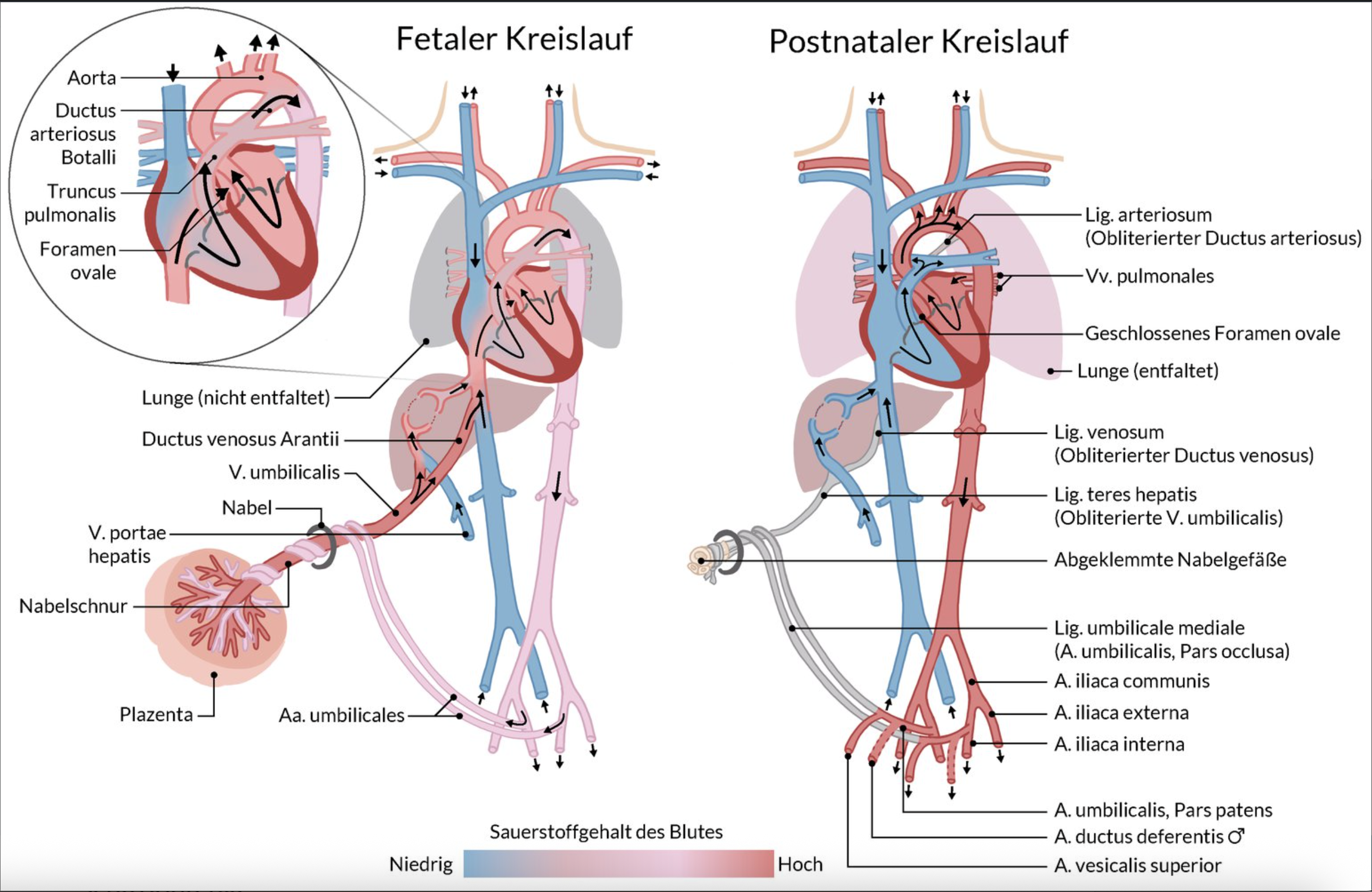
18. In the fetal circulation, there are shortcut connections (shunts) between vessels or heart chambers that close after birth.
What does the ductus arteriosus connect?
A. Umbilical artery and internal iliac artery (A. umbilicalis and A. iliaca interna)
B. Umbilical artery and umbilical vein (A. umbilicalis and V. umbilicalis)
C. Right atrium and left atrium (Atrium dextrum and Atrium sinistrum)
D. Pulmonary trunk and aorta (Truncus pulmonalis and Aorta)
E. Right ventricle and left ventricle (Ventriculus dexter and Ventriculus sinister)
D. Pulmonary trunk and aorta (Truncus pulmonalis and Aorta)
19. In developmental disorders of desmal ossification (bone formation directly from connective tissue), not only the clavicle but also certain skull bones formed through this process can be affected.
Which skull bone or skull bone part is particularly involved?
A. Malleus (hammer – middle ear bone)
B. Maxilla (upper jaw)
C. Os ethmoidale (ethmoid bone)
D. Os hyoideum (hyoid bone)
E. Pars petrosa ossis temporalis (petrous part of the temporal bone)
B. Maxilla (upper jaw)
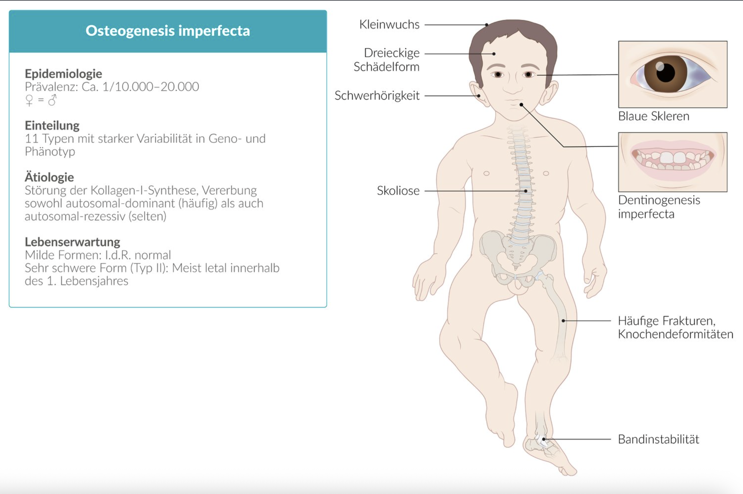
20. A 2-year-old boy is diagnosed with osteogenesis imperfecta, a condition characterized by a tendency for bone fractures. This is typically caused by a congenital disorder affecting the production of a specific type of collagen — the main component of the collagen fibrils found in bone.
Which type of collagen is involved?
A. Collagen type I
B. Collagen type II
C. Collagen type IV
D. Collagen type VII
E. Collagen type IX
A. Collagen type I
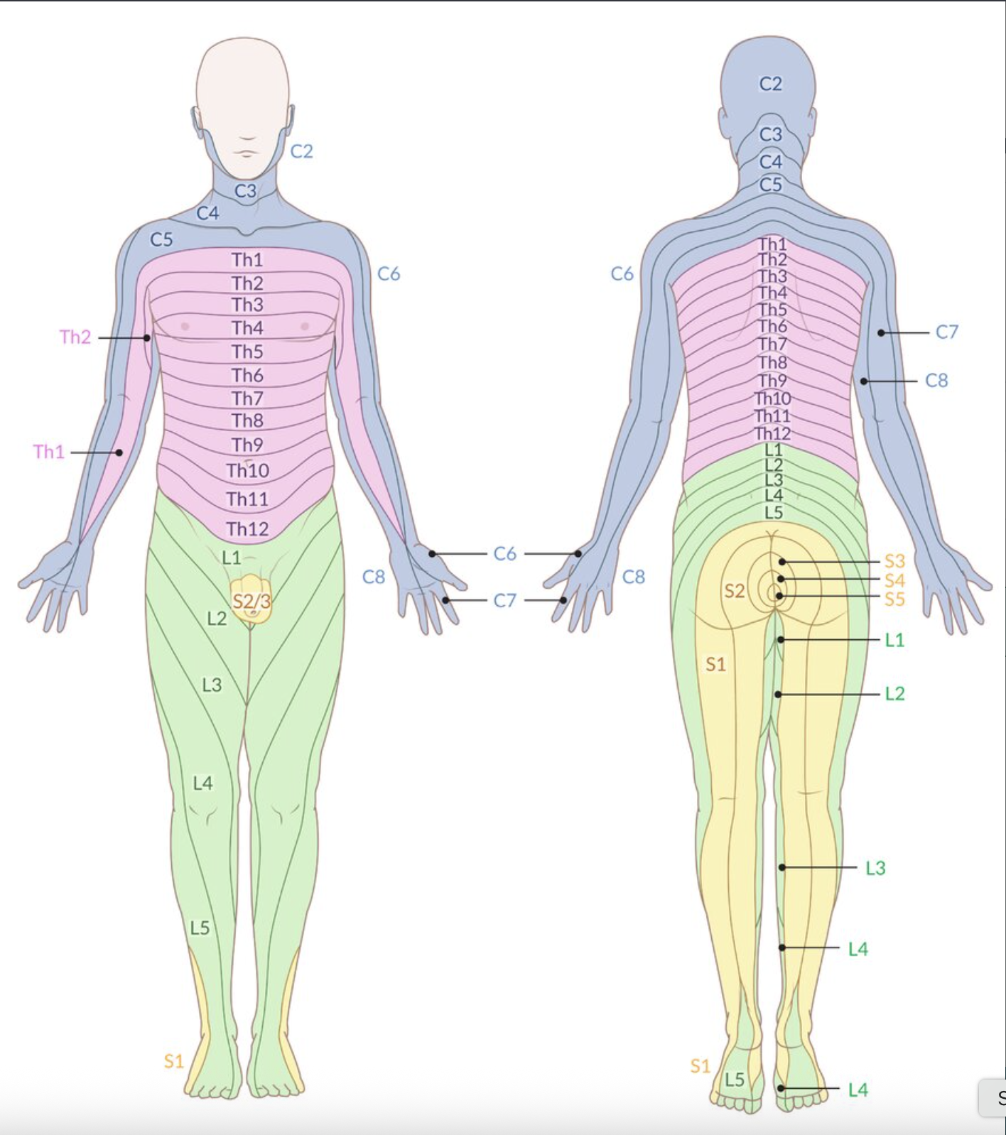
21. The level of a herniated disc can often be inferred from the patient’s described symptoms. Herniated discs can mechanically irritate spinal nerves, leading to pain in the corresponding sensory area.
In which of the following areas is a patient most likely to experience pain if a herniated disc compresses the L5 nerve root?
A. Inner thigh (Oberschenkelinnenseite)
B. Front of the thigh (Oberschenkelvorderseite)
C. Back of the thigh (Oberschenkelrückseite)
D. Front of the lower leg (Unterschenkelvorderseite)
E. Back of the lower leg (Unterschenkelrückseite)
D. Front of the lower leg (Unterschenkelvorderseite)
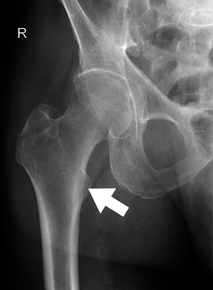
22. The X-ray image (anterior-posterior view) of a right hip joint included in the figure is marked with an arrow pointing to an anatomical structure.
Which hip joint movement is the muscle attaching to this structure primarily responsible for?
A. Flexion
B. Extension
C. Internal rotation
D. External rotation
E. Abduction
A. Flexion
23. After healing from a pelvic fracture, a patient is left with impaired movement of the right leg.
Which of the following symptom combinations most likely indicates damage to the obturator nerve (N. obturatorius) as a result of the fracture?
A. Impairment of active abduction at the hip joint and extension at the knee joint
B. Abduction and internal rotation at the hip joint
C. Adduction at the hip joint and internal rotation at the knee joint
D. Extension at the hip joint and external rotation at the knee joint
E. Flexion at the hip joint and extension at the knee joint
C. Adduction at the hip joint and internal rotation at the knee joint
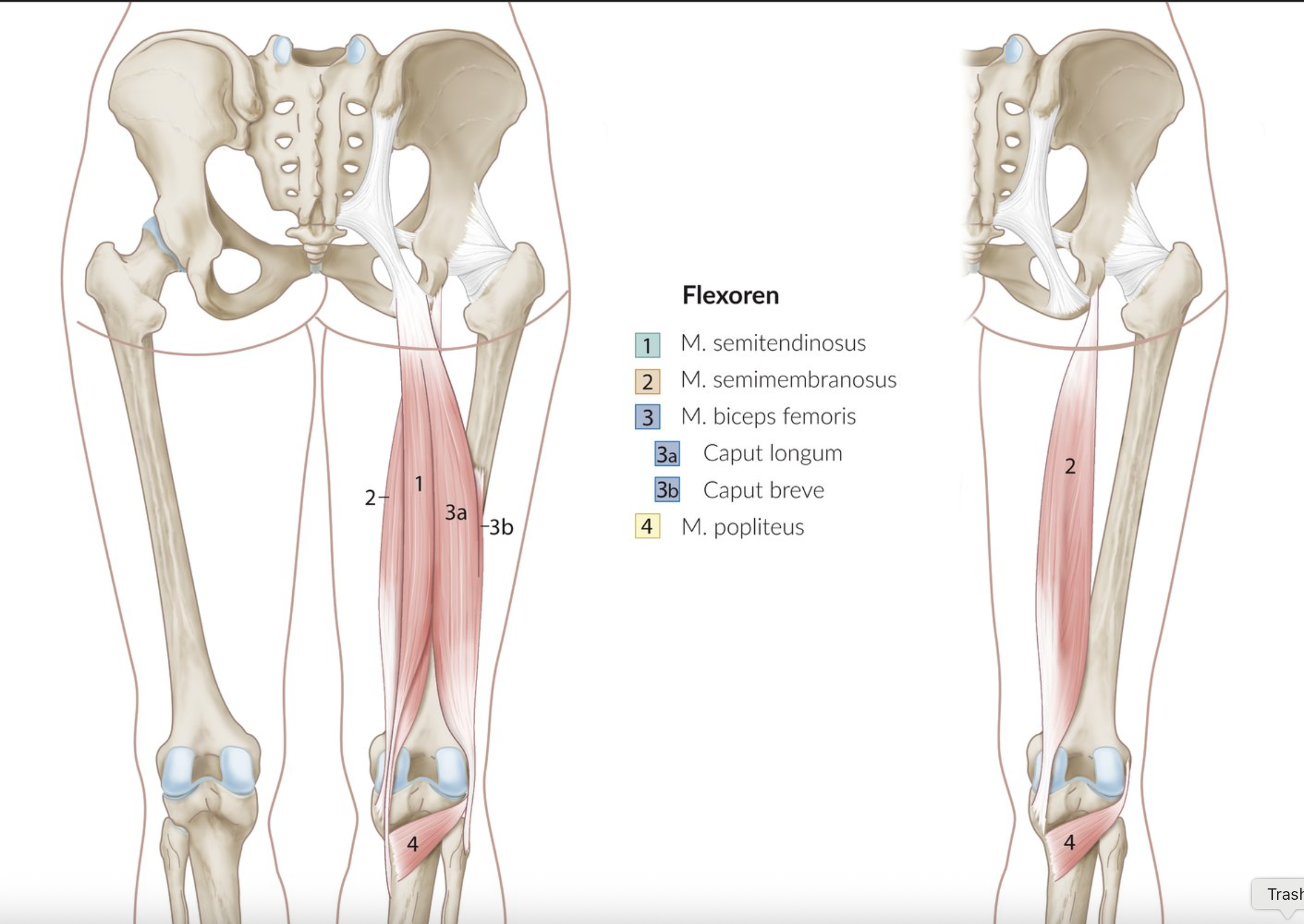
24. The knee joint is a compound joint that allows different movements depending on its position.
Which of the following muscles is involved in both flexion and external rotation of the knee joint?
A. Biceps femoris muscle
B. Gracilis muscle
C. Sartorius muscle
D. Semimembranosus muscle
E. Semitendinosus muscle
A. Biceps femoris muscle
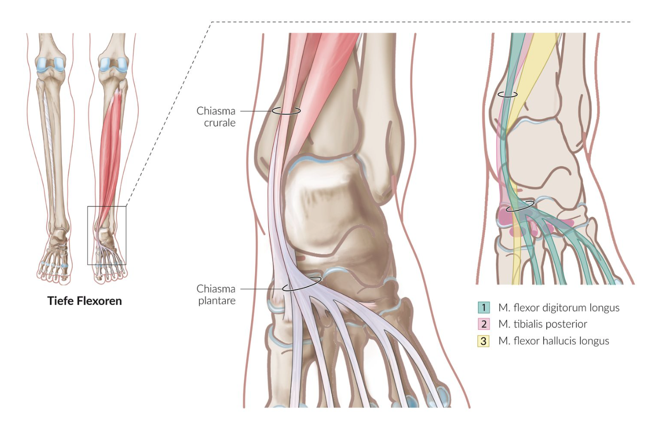
25. In the lower leg and foot region, muscles cross over each other at the crural chiasm (Chiasma crurale) and the plantar chiasm (Chiasma plantare).
At the crural chiasm (Chiasma crurale), which muscles cross over each other?
A. Fibularis longus muscle and lateral head of the gastrocnemius muscle
B. Fibularis longus muscle and fibularis brevis muscle
C. Flexor digitorum longus muscle and flexor hallucis longus muscle
D. Flexor digitorum longus muscle and tibialis posterior muscle
E. Tibialis posterior muscle and plantaris muscle
D. Flexor digitorum longus muscle and tibialis posterior muscle
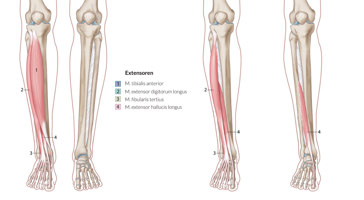
26. Muscle weakness often leads to clinically visible gait abnormalities.
Weakness or failure of which muscle group(s), acting on the upper ankle joint, leads to a steppage gait (also known as high-stepping or stork gait)?
A. Dorsal extensors
B. Plantar flexors
C. Pronators
D. Supinators
E. Pronators and supinators
A. Dorsal extensors
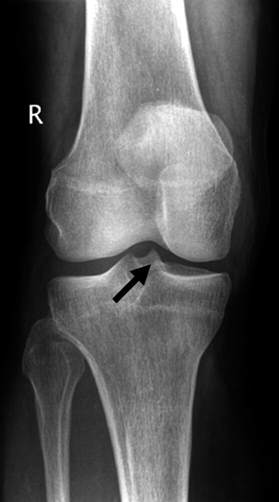
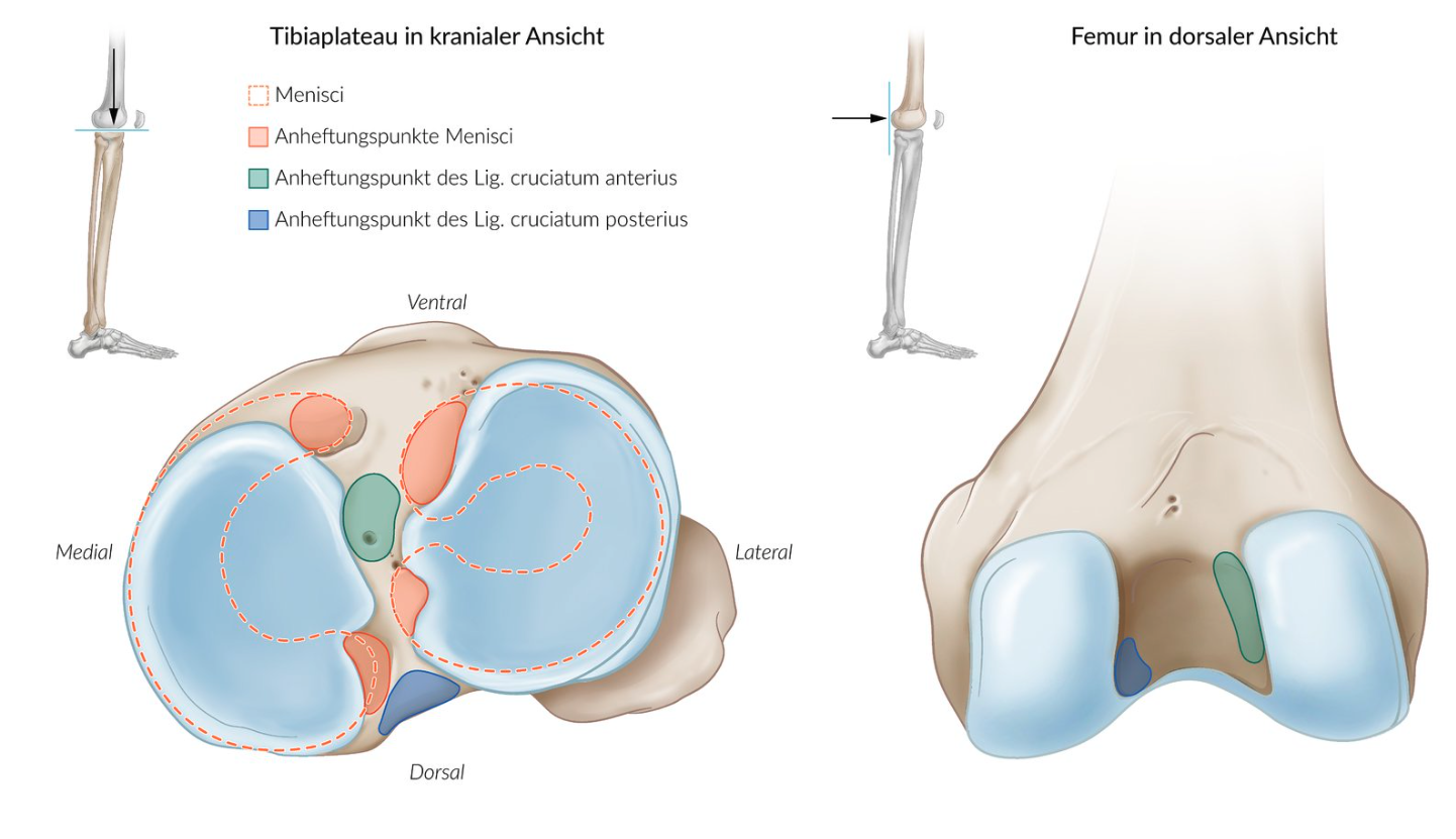
27. The arrow in the X-ray image shown marks the bone attachment, origin, or anchoring point of the:
A. Anterior cruciate ligament (ACL)
B. Patellar ligament
C. Popliteus muscle
D. Soleus muscle
E. Iliotibial tract
A. Anterior cruciate ligament (ACL)
28. Dislocations of the sternoclavicular joint (Articulatio sternoclavicularis) are rare due to strong ligament and muscle support, but dislocation of the sternal end of the clavicle can cause a pneumothorax and lead to life-threatening complications.
Which structure is NOT involved in stabilizing the joint?
A. Costoclavicular ligament
B. Interclavicular ligament
C. Anterior sternoclavicular ligament
D. Anterior scalene muscle
E. Subclavius muscle
D. Anterior scalene muscle
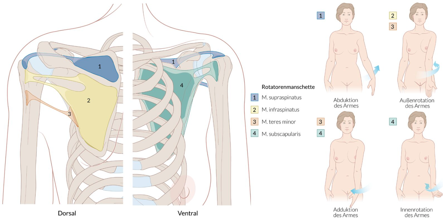
29. Degenerative tears of the rotator cuff can lead to significant loss of function in the affected arm.
Which of the following findings most likely indicates a lesion of the supraspinatus muscle?
A. Weakening of abduction against resistance
B. Weakening of external rotation against resistance
C. Weakening of internal rotation against resistance
D. Weakening of retroversion (extension) against resistance
E. Absence of active hyperadduction in the acromioclavicular joint
A. Weakening of abduction against resistance
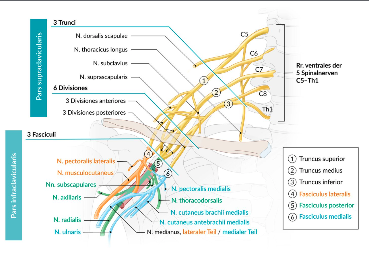
30. The brachial plexus can be understood as a distribution structure for the nerve supply of most parts of the upper limb.
Which nerve belongs to the infraclavicular part (pars infraclavicularis) of this plexus?
A. Axillary nerve
B. Dorsal scapular nerve
C. Phrenic nerve
D. Subclavian nerve
E. Long thoracic nerve
A. Axillary nerve
31. A 35-year-old patient reports to the on-call doctor immediately after surgery due to numbness in the right arm. There are sensory disturbances in the area of the right forearm and back of the hand, with complete loss of sensation over the first dorsal web space. Active extension at the wrist and fingers is no longer possible. However, active extension at the elbow and the triceps reflex are preserved.
Which of the following nerve lesions most likely matches this description?
A. Lesion of the median nerve at the elbow
B. Lesion of the radial nerve in the axilla
C. Lesion of the radial nerve in the upper arm
D. Lesion of the ulnar nerve at the elbow
E. Lesion of the ulnar nerve in the forearm
C. Lesion of the radial nerve in the upper arm
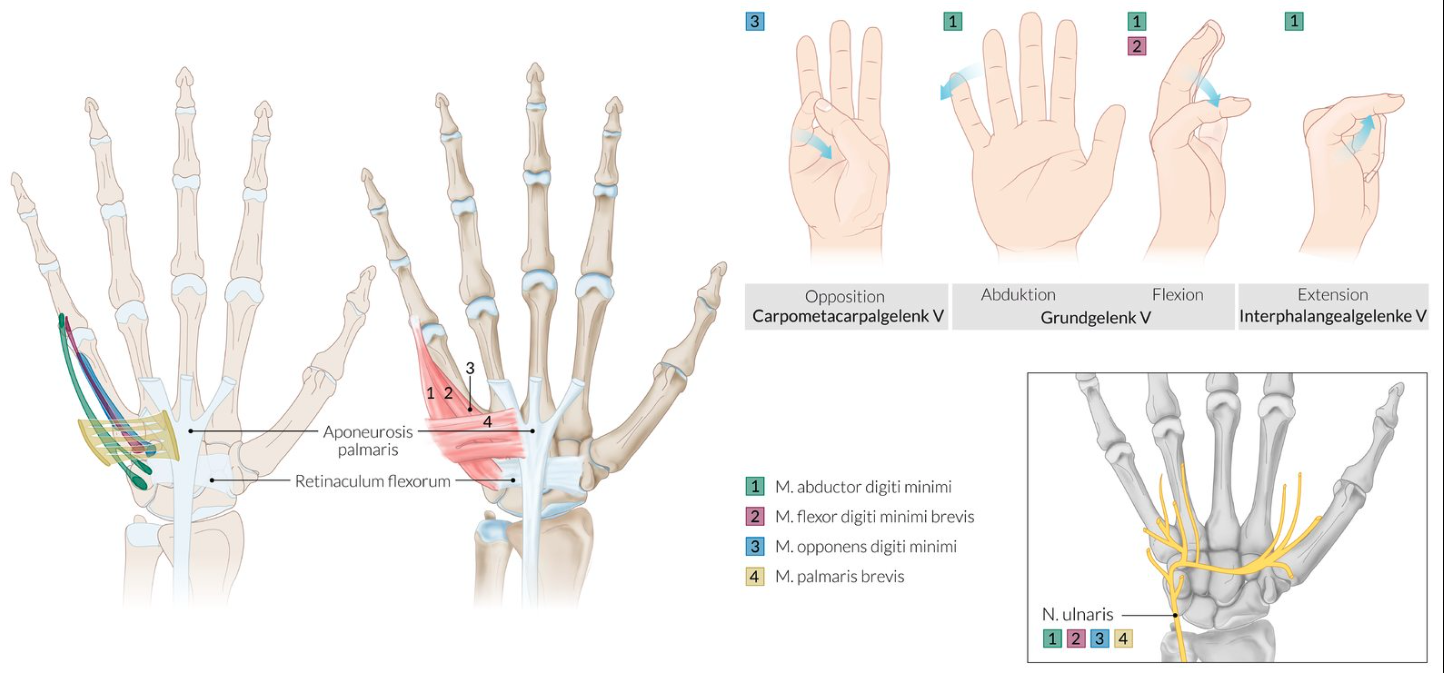
32. In cases of carpal tunnel syndrome with pronounced neurological symptoms, it may be necessary to cut the flexor retinaculum of the hand.
Between which carpal bones does the flexor retinaculum run?
A. Capitate and scaphoid bones
B. Hamate and lunate bones
C. Pisiform and trapezium bones
D. Trapezoid and lunate bones
E. Trapezoid and triquetrum bones
C. Pisiform and trapezium bones
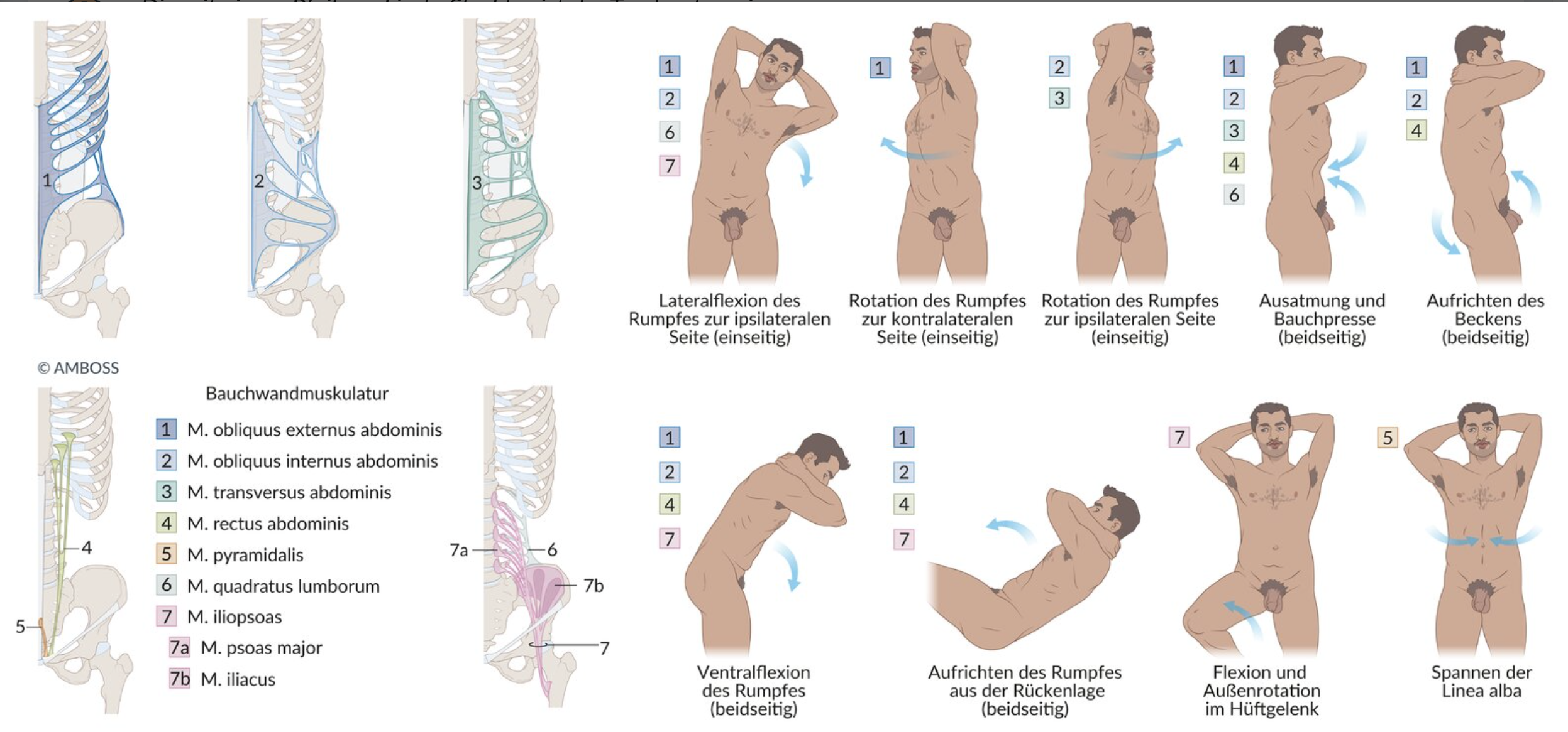
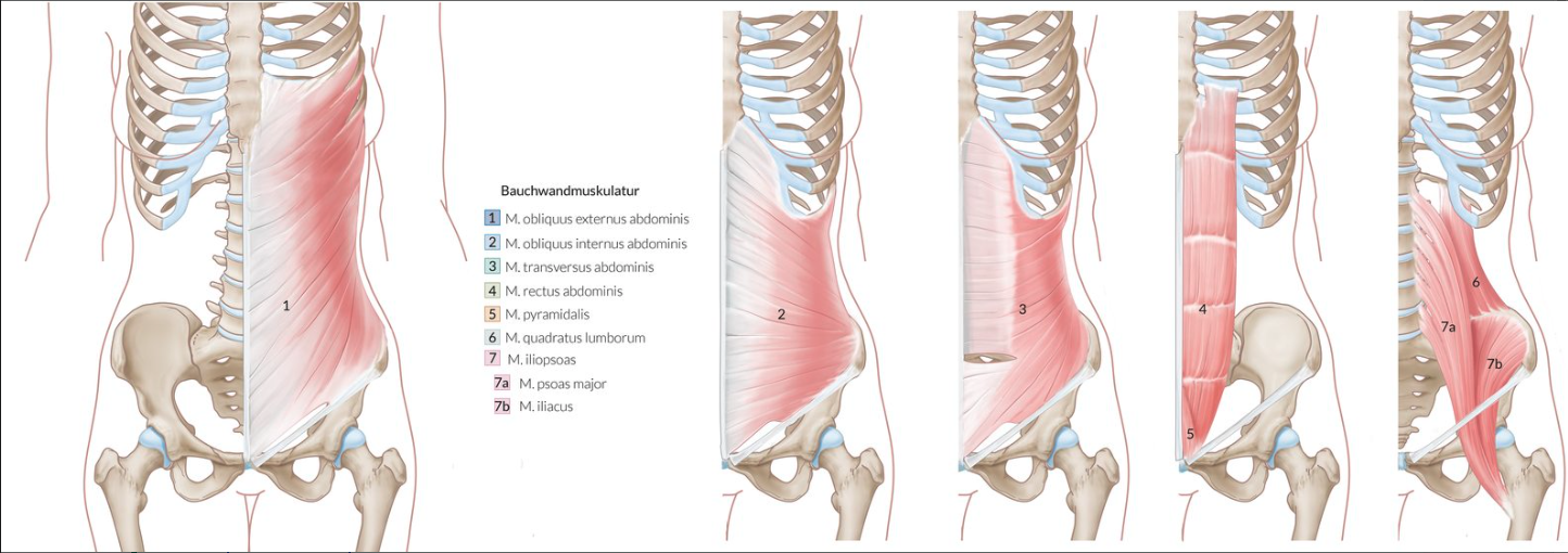
33. The abdominal wall encloses the abdominal cavity and the organs located in the extraperitoneal space of the abdominal cavity (Cavitas abdominalis).
Which of the following muscles is part of the abdominal wall and has its origin, among other places, at the inguinal ligament?
A. External oblique abdominal muscle (M. obliquus externus abdominis)
B. Internal oblique abdominal muscle (M. obliquus internus abdominis)
C. Psoas major muscle
D. Quadratus lumborum muscle
E. Rectus abdominis muscle
B. Internal oblique abdominal muscle (M. obliquus internus abdominis)
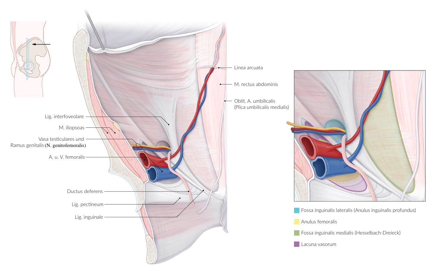
34. An inguinal hernia is the most common type of hernia and primarily affects men. In inguinal hernias, a distinction is made between an internal hernia opening (toward the abdominal cavity) and an external hernia opening (toward the skin/subcutaneous tissue), depending on the location of the hernia defect.
Where is the internal hernia opening located in a direct inguinal hernia (Hernia inguinalis directa)?
A. Lateral inguinal fossa (Fossa inguinalis lateralis)
B. Medial inguinal fossa (Fossa inguinalis medialis)
C. Supravesical fossa (Fossa supravesicalis)
D. Adductor hiatus (Hiatus adductorius)
E. Saphenous opening (Hiatus saphenus)
B. Medial inguinal fossa (Fossa inguinalis medialis)
35. The thoracolumbar fascia serves as an anatomical landmark during retroperitoneal approaches in kidney surgeries.
Which of the following structures is/are least likely to be involved in anchoring the thoracolumbar fascia?
A. Iliac crest (Crista iliaca)
B. Dorsal surface of the sacrum (Facies dorsalis ossis sacri)
C. Costal processes of the lumbar vertebrae (Processus costales)
D. Transverse processes of the thoracic vertebrae (Processus transversi)
E. 12th rib
D. Transverse processes of the thoracic vertebrae (Processus transversi)
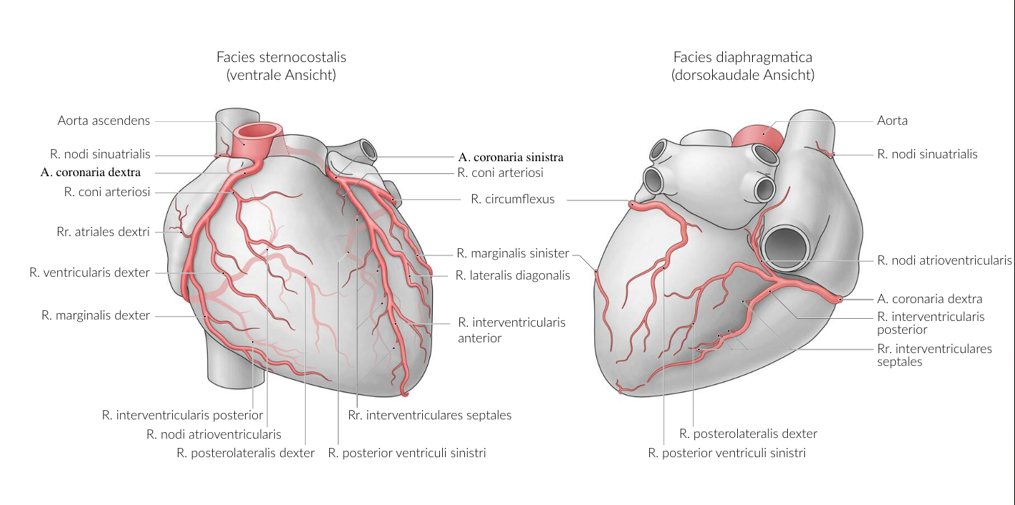
36. In a heart attack, the severity of clinical symptoms depends, among other things, on the type of coronary artery supply.
Which combination of supply type and occlusion location is most likely to result in bradycardic rhythm disturbances, due to the most significant impairment of blood supply to the cardiac conduction system?
A. Proximal occlusion of the right coronary artery (A. coronaria dextra) in a normal supply type
B. Proximal occlusion of the left coronary artery (A. coronaria sinistra) in a right-dominant supply type
C. Proximal occlusion of the anterior interventricular branch (R. interventricularis anterior) in a normal supply type
D. Occlusion of the circumflex branch (R. circumflexus) on the diaphragmatic surface in a normal supply type
E. Occlusion of the circumflex branch (R. circumflexus) on the diaphragmatic surface in a left-dominant supply type
A. Proximal occlusion of the right coronary artery (A. coronaria dextra) in a normal supply type
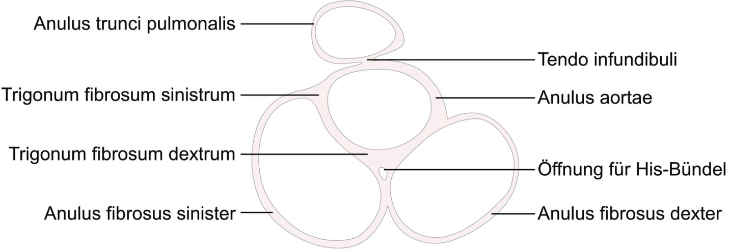
37. Except for the passage of the bundle of His, the cardiac skeleton electrically separates the atria from the ventricles. The bundle of His lies within a triangle that is bordered by three anatomical structures.
Which are these structures?
A. Aortic valve – Mitral valve – Coronary sinus
B. Aortic valve – Mitral valve – Tricuspid valve
C. Aortic valve – Pulmonary valve – Coronary sinus
D. Aortic valve – Pulmonary valve – Tricuspid valve
E. Mitral valve – Pulmonary valve – Tricuspid valve
B. Aortic valve – Mitral valve – Tricuspid valve
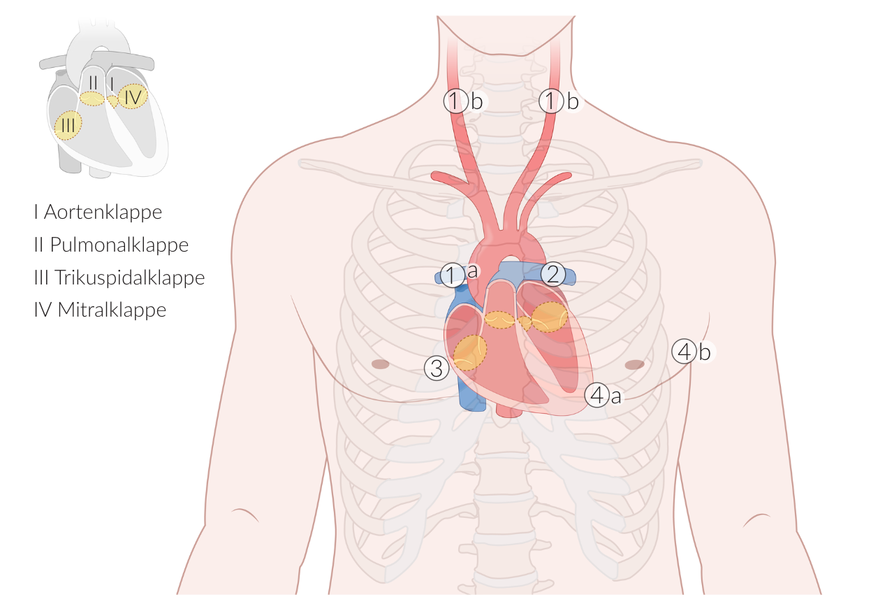
38. Auscultation of the heart valves can provide information about pathological changes. They can be assessed at specific points on the chest wall due to sound conduction.
At which location is the mitral valve particularly well heard?
A. In the 2nd left intercostal space, parasternal
B. In the 5th left intercostal space, midclavicular
C. In the 4th intercostal space, right parasternal
D. At the valvular level of the heart behind the sternum
E. On the dorsal chest wall in the 5th right intercostal space
B. In the 5th left intercostal space, midclavicular
39. During the clinical examination of the size of the spleen and liver, the costal arch (rib margin) is an important anatomical landmark.
How is the costal arch structured?
A. By elastic cartilage of the 8th–10th ribs
B. By fibrocartilage of the 8th–10th ribs
C. By hyaline cartilage of the 8th–10th ribs
D. By fibrocartilage of the 10th–12th ribs
E. By hyaline cartilage of the 10th–12th ribs
C. By hyaline cartilage of the 8th–10th ribs
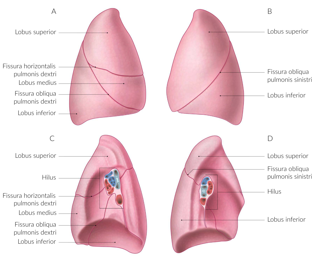
40. In a 57-year-old patient, a bronchial carcinoma (malignant tumor of the bronchi or bronchioles) is to be surgically removed by a lobectomy (removal of a lung lobe). For the most organ-preserving approach, the fissures between the lung lobes are of surgical importance.
The horizontal fissure runs in the anterior section between:
A. Upper lobe and lingula of the left lung
B. Upper and lower lobes of the left lung
C. Lingula and lower lobe of the left lung
D. Upper and middle lobes of the right lung
E. Middle and lower lobes of the right lung
D. Upper and middle lobes of the right lung
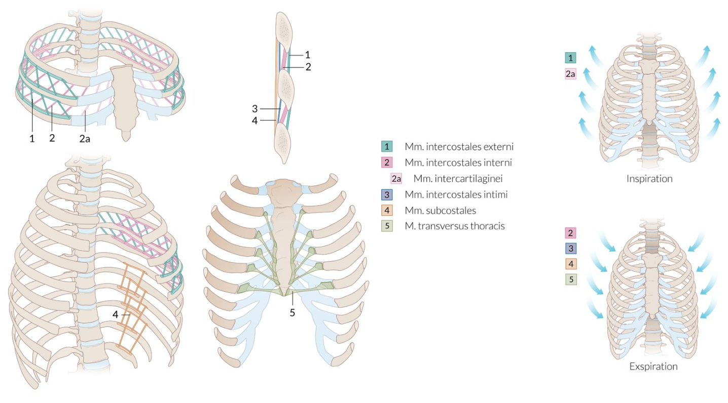
41. A variety of muscles can become more involved in breathing, especially when there is an increased need for oxygen.
Which of the following muscles involved in breathing act(s) in expiration (exhalation)?
A. Mm. intercartilaginei
B. Mm. intercostales externi
C. Mm. intercostales interni
D. Mm. scaleni
E. M. serratus posterior superior
C. Mm. intercostales interni
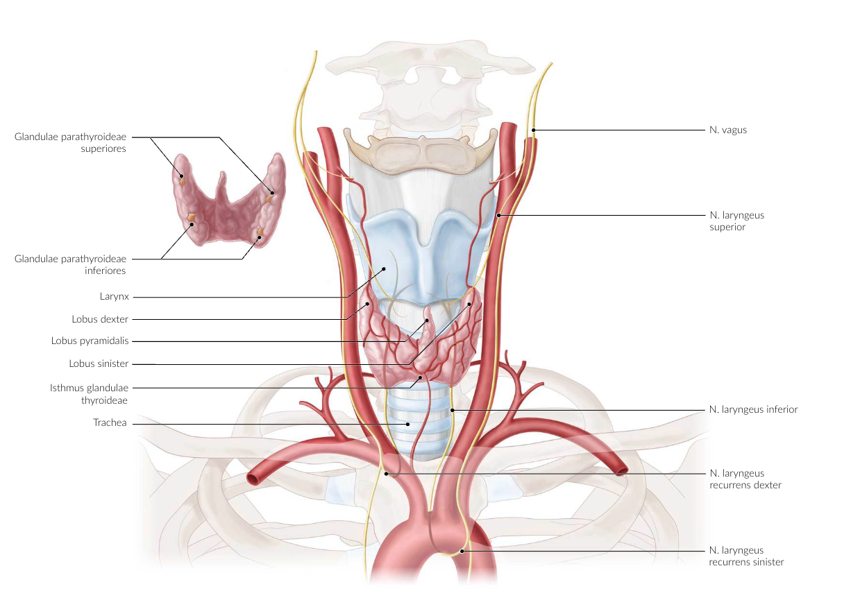
42. An arterial aneurysm is a pathological bulging of the vessel wall. An aneurysm in the area where the aortic arch transitions into the thoracic aorta can irritate a nearby nerve.
Which nerve is most likely to be affected?
A. Right intercostal nerve I (N. intercostalis I dexter)
B. Left recurrent laryngeal nerve (N. laryngeus recurrens sinister)
C. Right phrenic nerve (N. phrenicus dexter)
D. Left greater splanchnic nerve (N. splanchnicus major sinister)
E. Right subclavian nerve (N. subclavius dexter)
B. Left recurrent laryngeal nerve (N. laryngeus recurrens sinister)
43. During surgeries on the esophagus, injuries to the thoracic duct (ductus thoracicus) can occur. These may lead to fluid accumulation in the pleural cavity (pleural effusion), which can cause shortness of breath (dyspnea).
What type of fluid from which body cavity does the effusion primarily contain in this case?
A. Arterial blood from the thoracic cavity
B. Lymph from the abdominal cavity
C. Lymph from the thoracic cavity
D. Venous blood from the upper abdomen
E. Venous blood from the thoracic cavity
B. Lymph from the abdominal cavity
44. In female reproductive anatomy, a distinction is made between external and internal genital organs.
Which structure(s) belong to the internal genital organs?
A. Clitoris
B. Labia majora pudendi
C. Labia minora pudendi
D. Vagina
E. Vestibule of the vagina (Vestibulum vaginae)
D. Vagina
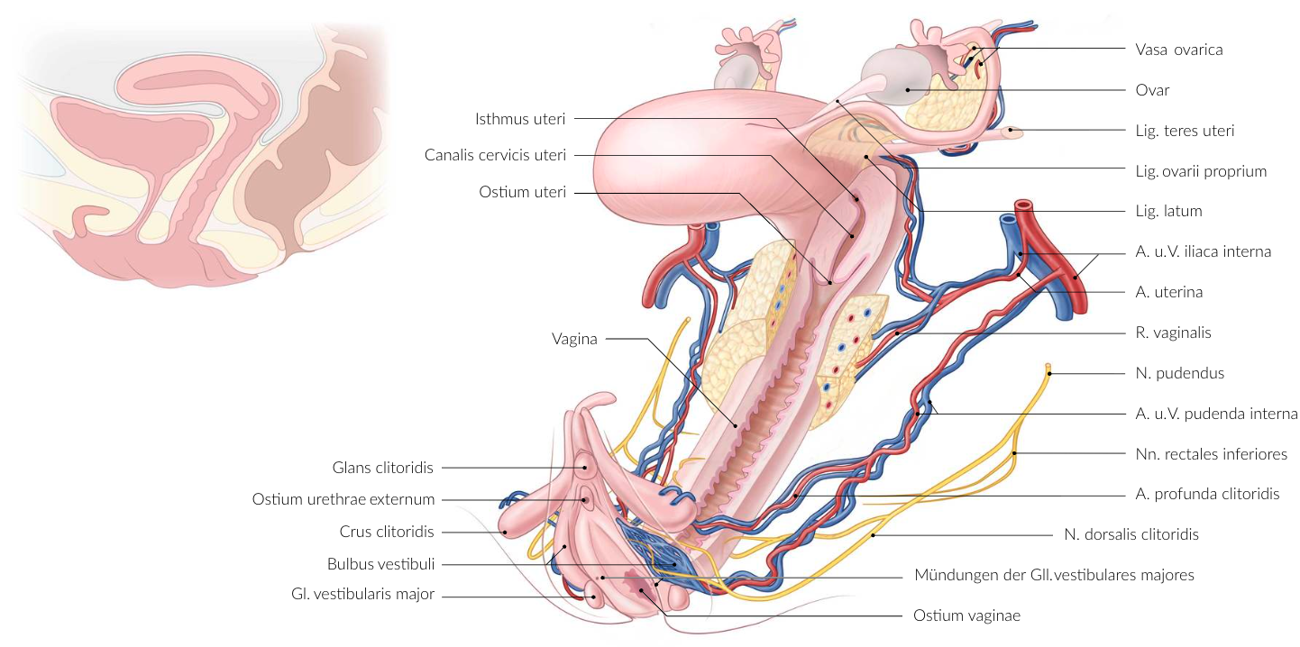
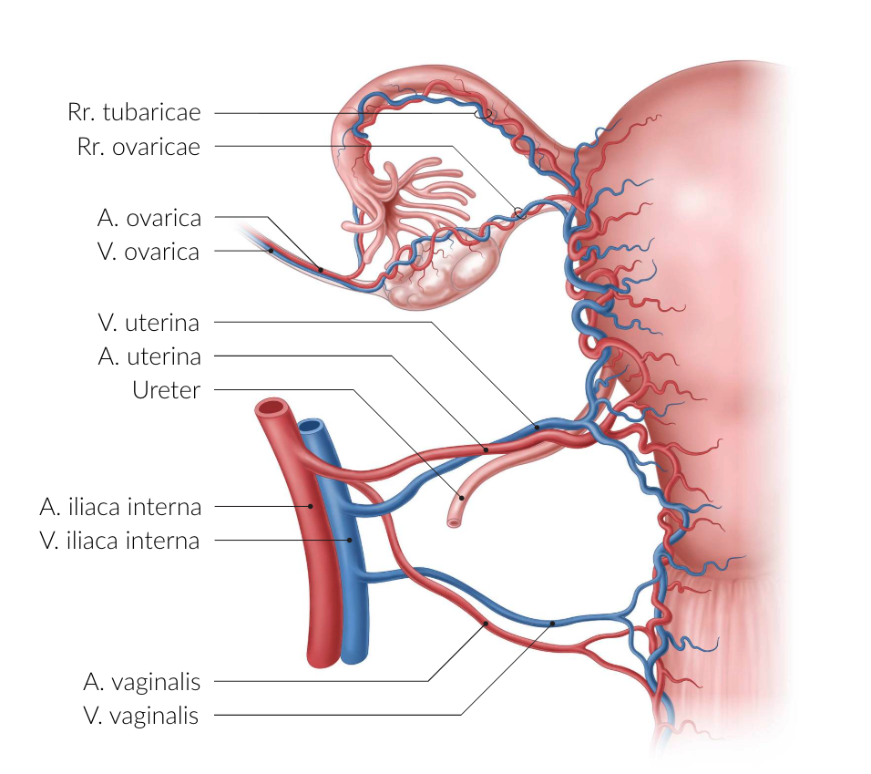
45. The ovary is supplied by two different arteries.
Along which structure does the terminal branch of the artery arising from the uterine artery run?
A. Cardinal ligament (Lig. cardinale)
B. Inguinal ligament (Lig. inguinale)
C. Proper ovarian ligament (Lig. ovarii proprium)
D. Suspensory ligament of the ovary (Lig. suspensorium ovarii)
E. Round ligament of the uterus (Lig. teres uteri)
C. Proper ovarian ligament (Lig. ovarii proprium)
46. As a result of venous congestion in the superior vena cava, inferior vena cava, or common iliac veins, collateral circulation between the superior and inferior vena cava may develop due to various causes.
Through which veins can such a collateral pathway form?
A. Femoral vein – Superficial circumflex iliac vein / Superficial epigastric vein – Thoracoepigastric vein – Axillary vein – Subclavian vein – Brachiocephalic vein
B. Right/Left gastric vein – Esophageal veins – Azygos vein / Accessory hemiazygos vein
C. Inferior mesenteric vein – Testicular/Ovarian vein
D. Superior rectal vein – Middle rectal vein – Inferior rectal vein – Internal iliac vein
E. Paraumbilical veins – Superior epigastric / Inferior epigastric vein – Thoracoepigastric vein – Superficial epigastric vein
A. Femoral vein – Superficial circumflex iliac vein / Superficial epigastric vein – Thoracoepigastric vein – Axillary vein – Subclavian vein – Brachiocephalic vein
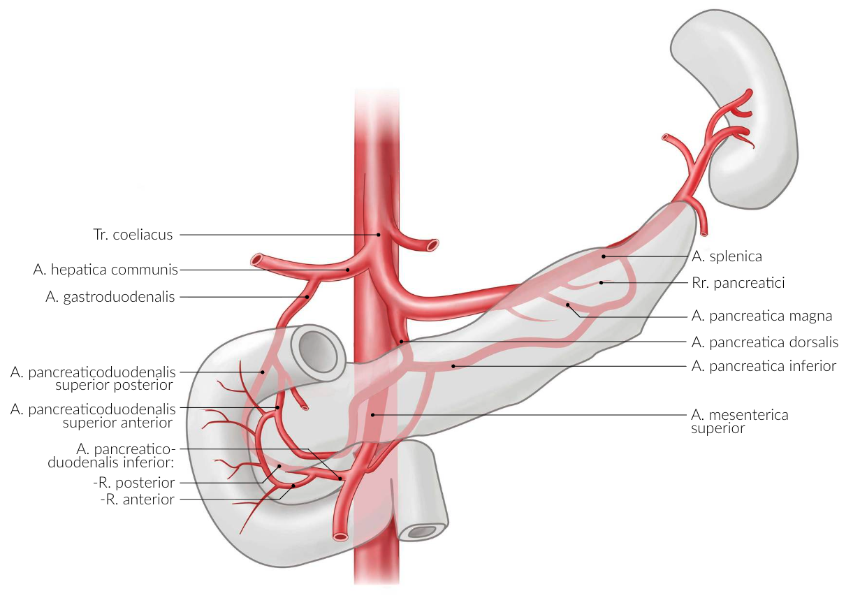
47. Arterial underperfusion and infarctions are relatively rare in the gastrointestinal tract. This is explained by the presence of extensive arterial anastomoses (connections).
Which direct anastomosis exists between the celiac trunk (Truncus coeliacus) and one of the two mesenteric arteries?
A. Middle colic artery – Left colic artery
B. Posterior gastric artery – Inferior pancreatic artery
C. Right gastro-omental artery – Middle colic artery
D. Anterior superior pancreaticoduodenal artery – Anterior inferior pancreaticoduodenal artery
E. Splenic artery – Great pancreatic artery
D. Anterior superior pancreaticoduodenal artery – Anterior inferior pancreaticoduodenal artery
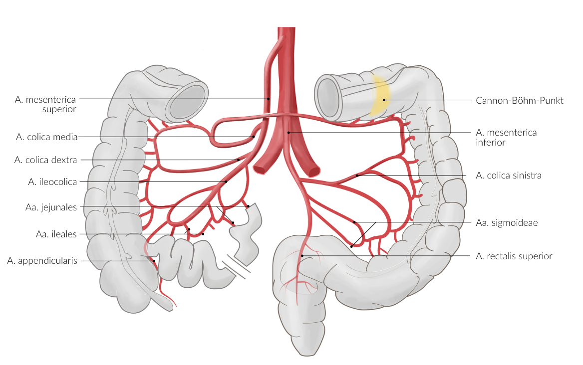
48. Infiltration of the superior mesenteric artery (A. mesenterica superior) by a malignant tumor can lead to inoperability and a correspondingly poor prognosis.
Which organ is not typically supplied by the superior mesenteric artery?
A. Ascending colon
B. Duodenum
C. Ileum
D. Stomach
E. Pancreas
D. Stomach
49. In the fetus, the umbilical artery (A. umbilicalis) carries blood to the placenta. After birth, part of it remains open (pars patens).
In women, which artery arises from the pars patens of the umbilical artery?
A. Artery of the vestibular bulb (A. bulbi vestibuli)
B. Artery of the clitoris (A. clitoridis)
C. Perineal artery (A. perinealis)
D. Urethral artery (A. urethralis)
E. Superior vesical artery (A. vesicalis superior)
E. Superior vesical artery (A. vesicalis superior)
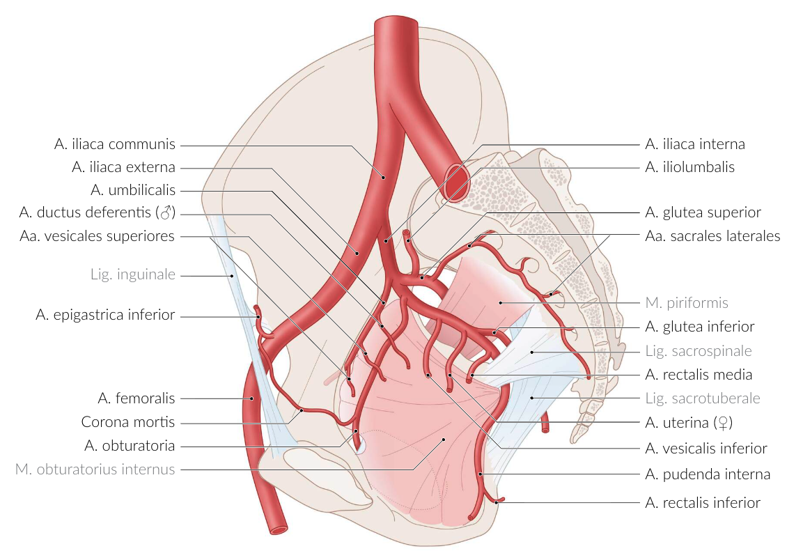
50. During prostate surgery, the supplying blood vessels must be ligated.
Which artery is typically the main supplier of blood to the prostate?
A. Artery of the ductus deferens (A. ductus deferentis)
B. Inferior rectal artery (A. rectalis inferior)
C. Testicular artery (A. testicularis)
D. Inferior vesical artery (A. vesicalis inferior)
E. Superior vesical artery (A. vesicalis superior)
D. Inferior vesical artery (A. vesicalis inferior)
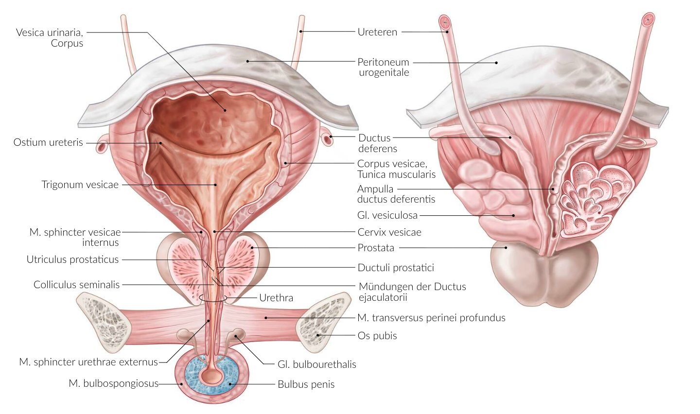
51. When catheterizing the urinary bladder in men, in addition to the external (ostium urethrae externum) and internal urethral openings (ostium urethrae internum), there is another narrowing of the urethra that must be passed.
This narrowing is located:
A. In the navicular fossa (Fossa navicularis)
B. At the transition from the glans to the corpus spongiosum
C. In the area where the ducts of the bulbourethral glands (Glandulae bulbourethrales) enter
D. In the membranous part of the urethra (Pars membranacea)
E. In the prostatic part of the urethra (Pars prostatica)
D. In the membranous part of the urethra (Pars membranacea)
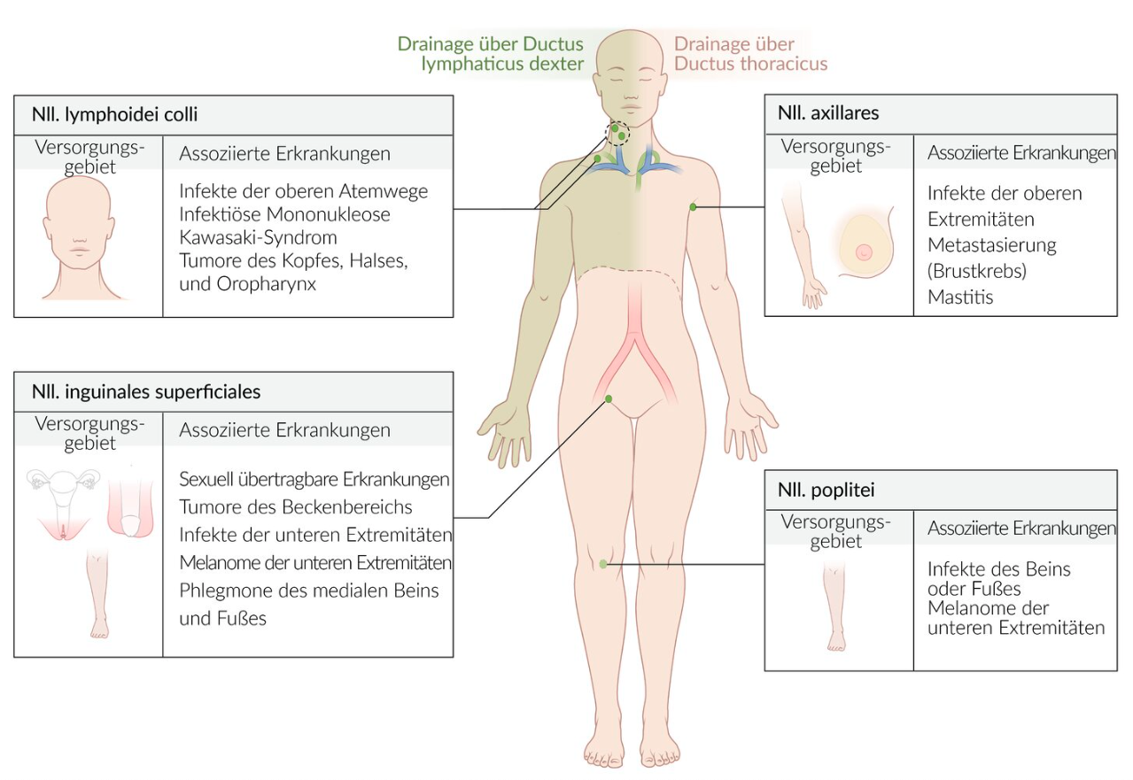
52. Malignant tumors can metastasize through the lymphatic system. A patient has a melanoma (malignant tumor of melanocytes) on the scrotum.
To which of the following lymph nodes is the tumor most likely to metastasize first?
A. External iliac lymph nodes (Nll. iliaci externi)
B. Internal iliac lymph nodes (Nll. iliaci interni)
C. Superficial inguinal lymph nodes (Nll. inguinales superficiales)
D. Lumbar lymph nodes (Nll. lumbales)
E. Popliteal lymph nodes (Nll. poplitei)
C. Superficial inguinal lymph nodes (Nll. inguinales superficiales)
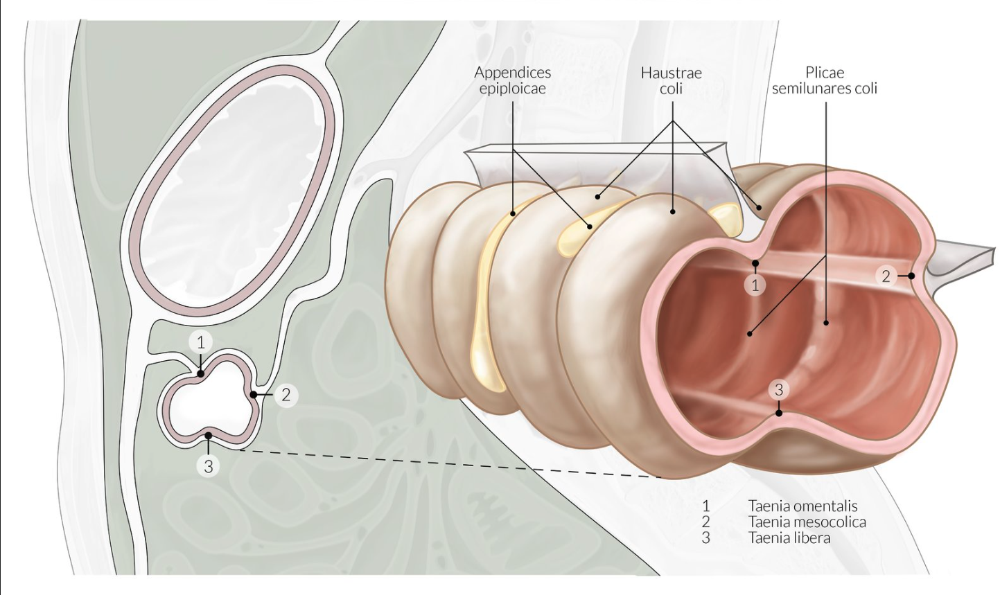
53. During an esophagogastroduodenoscopy, a camera-equipped endoscope is advanced from the mouth into the duodenum and then withdrawn back toward the mouth.
Which of the following physiologically present structures can not be observed during this examination?
A. Pyloric canal (Canalis pyloricus)
B. Angular notch (Incisura angularis)
C. Major duodenal papilla (Papilla duodeni major)
D. Circular folds (Plicae circulares)
E. Taeniae (Tänien)
E. Taeniae (Tänien)
54. Internal hernias can form in the recesses of the peritoneal cavity.
Near which structure are the superior and inferior duodenal recesses most likely located?
A. Superior part of the duodenum (Pars superior duodeni)
B. Superior duodenal flexure (Flexura duodeni superior)
C. Major duodenal papilla (Papilla duodeni major)
D. Inferior duodenal flexure (Flexura duodeni inferior)
E. Duodenojejunal flexure (Flexura duodenojejunalis)
E. Duodenojejunal flexure (Flexura duodenojejunalis)
55. Intraperitoneal sections of the intestine are suspended by mesenteries and have great mobility.
What is the most accurate course of the mesenteric root (Radix mesenterii)?
A. From the greater curvature of the stomach to the junction of the ileum with the cecum
B. From the duodenal bulb (Bulbus duodeni) to the junction of the ileum with the cecum
C. From the duodenojejunal flexure to the junction of the ileum with the cecum
D. From the left colic flexure to the junction of the ileum with the cecum
E. Between the left and right colic flexures
C. From the duodenojejunal flexure to the junction of the ileum with the cecum
56. For assigning clinical symptoms and for topographic classification, the abdominal cavity is divided into upper and lower abdomen.
Which of the following organs/structures belongs to the lower abdomen?
A. Extrahepatic bile ducts
B. Jejunum
C. Liver
D. Spleen
E. Pancreas
B. Jejunum
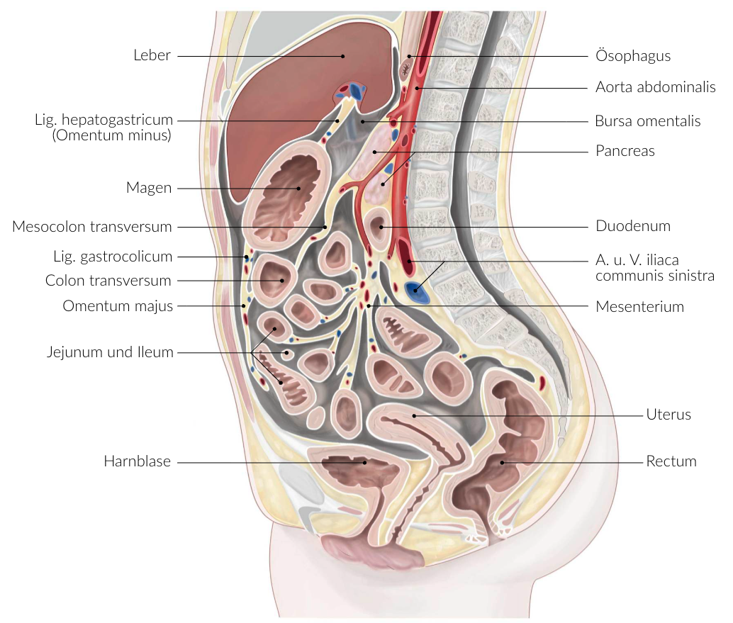
57. To access the pancreas during dissection, the lesser sac (Bursa omentalis) must be opened.
The caudal (lower) boundary of the lesser sac is formed by:
A. Gastrocolic ligament (Lig. gastrocolicum)
B. Gastrosplenic ligament (Lig. gastrosplenicum)
C. Hepatoduodenal ligament (Lig. hepatoduodenale)
D. Transverse mesocolon (Mesocolon transversum)
E. Lesser omentum (Omentum minus)
D. Transverse mesocolon (Mesocolon transversum)
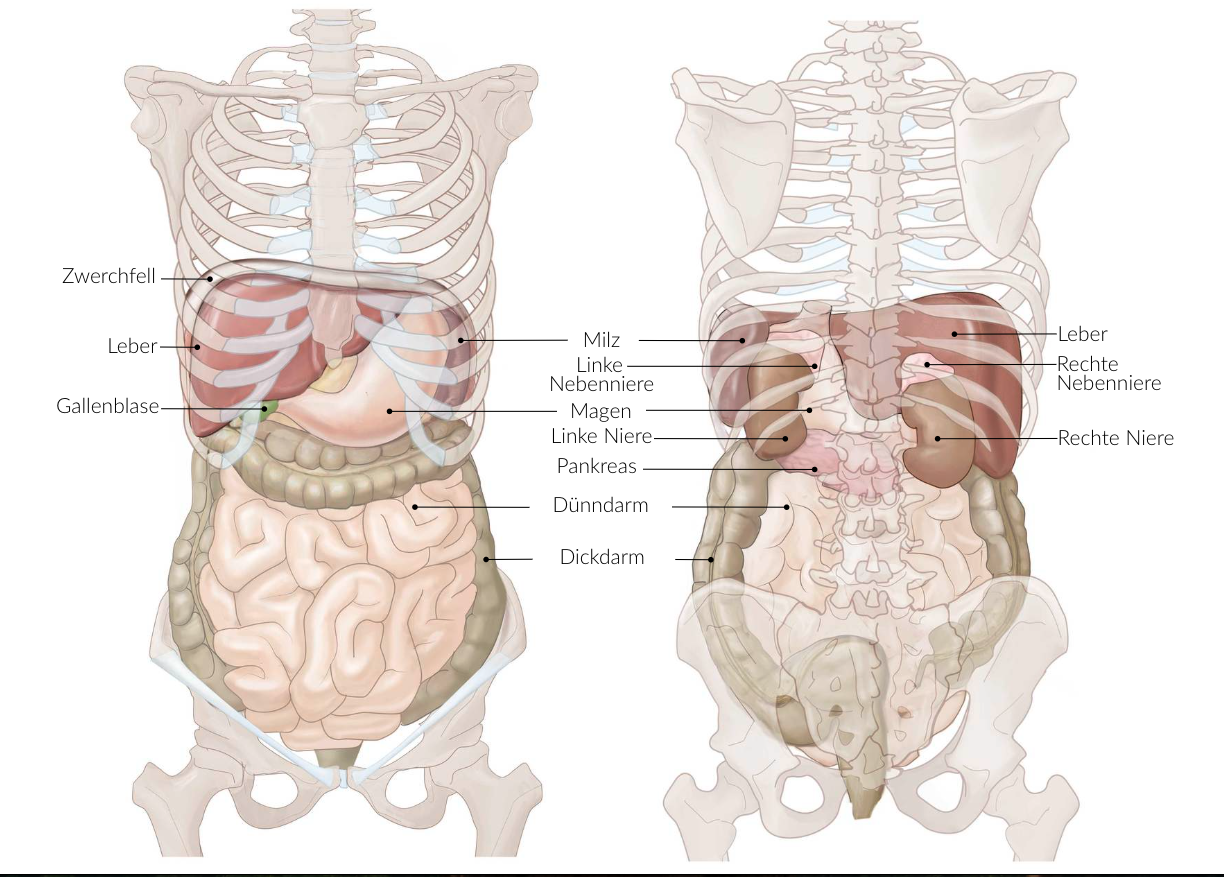
58. The liver is the central metabolic organ in humans. Topographically, large parts of the liver are usually located intraperitoneally in the right upper abdomen.
Before performing a partial resection of the right hepatic lobe (Lobus hepatis dexter), which organ is least likely to be at risk of injury based on its anatomical position — and therefore typically does not require preoperative discussion?
A. Colon
B. Duodenum
C. Pancreas
D. Right adrenal gland
E. Right kidney
C. Pancreas
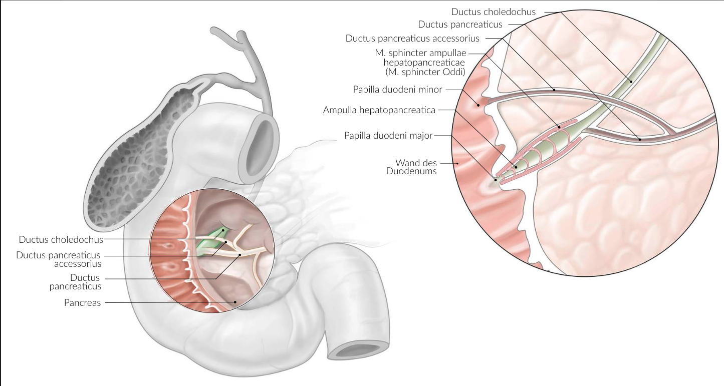
59. A patient has a mechanically caused obstruction of bile flow in the area of the common bile duct (Ductus choledochus).
A mass in which of the following organ regions is most likely to be the cause?
A. Antrum of the stomach (Antrum ventriculi)
B. Head of the pancreas (Caput pancreatis)
C. Right colic flexure (Flexura coli dextra)
D. Left lobe of the liver (Lobus hepatis sinister)
E. Ascending part of the duodenum (Pars ascendens duodeni)
B. Head of the pancreas (Caput pancreatis)
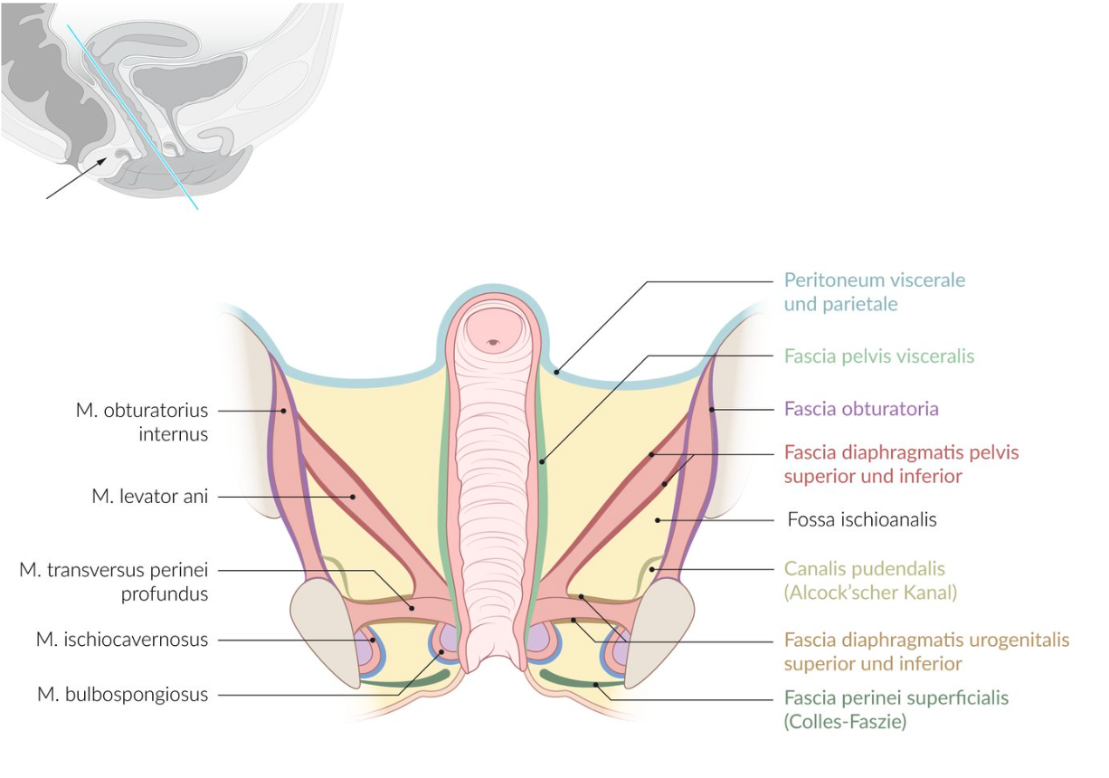
60. The ischioanal fossa is a fat-filled, pyramid-shaped space located on both sides of the anus. It appears largely the same in both sexes and is of significant clinical importance (e.g., abscess formation).
Which of the following structures is not part of the boundaries of the ischioanal fossa?
A. Sacrotuberous ligament (Lig. sacrotuberale)
B. Gluteus maximus muscle (M. gluteus maximus)
C. Levator ani muscle (M. levator ani)
D. External obturator muscle (M. obturatorius externus)
E. External anal sphincter muscle (M. sphincter ani externus)
D. External obturator muscle (M. obturatorius externus)
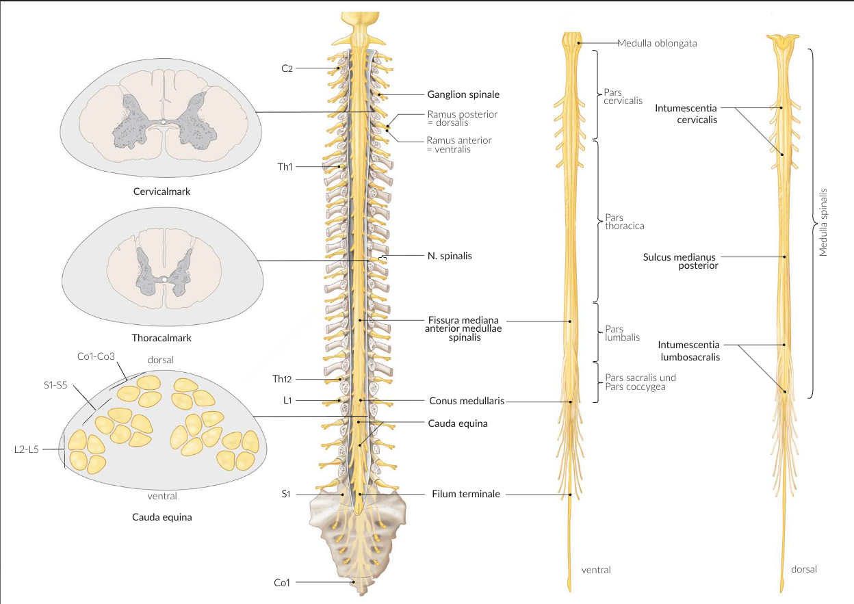
61. During extensive resections of the bony pelvis, the dura mater of the spinal cord must not be injured, as this can lead to cerebrospinal fluid (CSF) fistulas, which may cause severe headaches.
At which vertebral level does the dural sac usually end caudally (at the lower end)?
A. At the level of the 1st lumbar vertebra
B. At the level of the 2nd lumbar vertebra
C. At the level of the 2nd sacral vertebra
D. At the level of the 4th sacral vertebra
E. At the level of the 1st coccygeal vertebra
C. At the level of the 2nd sacral vertebra
62. The pterygopalatine fossa is an important region for the distribution of vessels and nerves at the lateral base of the skull.
To access it from the front (anteriorly), which bony structure must be opened?
A. Greater wing of the sphenoid bone (Ala major ossis sphenoidalis)
B. Lesser wing of the sphenoid bone (Ala minor ossis sphenoidalis)
C. Pterygoid process of the sphenoid bone (Processus pterygoideus ossis sphenoidalis)
D. Perpendicular plate of the palatine bone (Lamina perpendicularis ossis palatini)
E. Maxillary tuberosity (Tuber maxillae)
E. Maxillary tuberosity (Tuber maxillae)
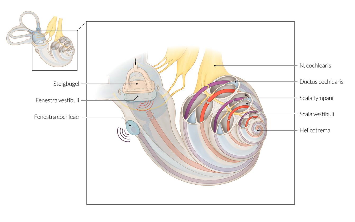
63. In a conductive hearing loss, the transmission of sound waves through the ossicular chain (tiny ear bones) to the perilymph in the cochlea is impaired.
Through which connection are sound waves transmitted from the ossicular chain to the perilymph of the cochlea?
A. Incus at the helicotrema
B. Incus at the oval window
C. Incus at the round window
D. Stapes at the oval window
E. Stapes at the round window
D. Stapes at the oval window
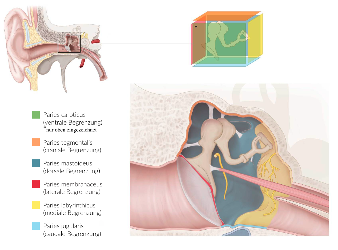
64. Middle ear infections can spread to neighboring structures and cause complications.
Which of the following structures is directly adjacent to the floor of the tympanic cavity (middle ear)?
A. Internal carotid artery (A. carotis interna)
B. Posterior semicircular duct (Ductus semicircularis posterior)
C. Temporal lobe of the brain (Lobus temporalis des Großhirns)
D. Facial nerve (N. facialis)
E. Internal jugular vein (V. jugularis interna)
E. Internal jugular vein (V. jugularis interna)
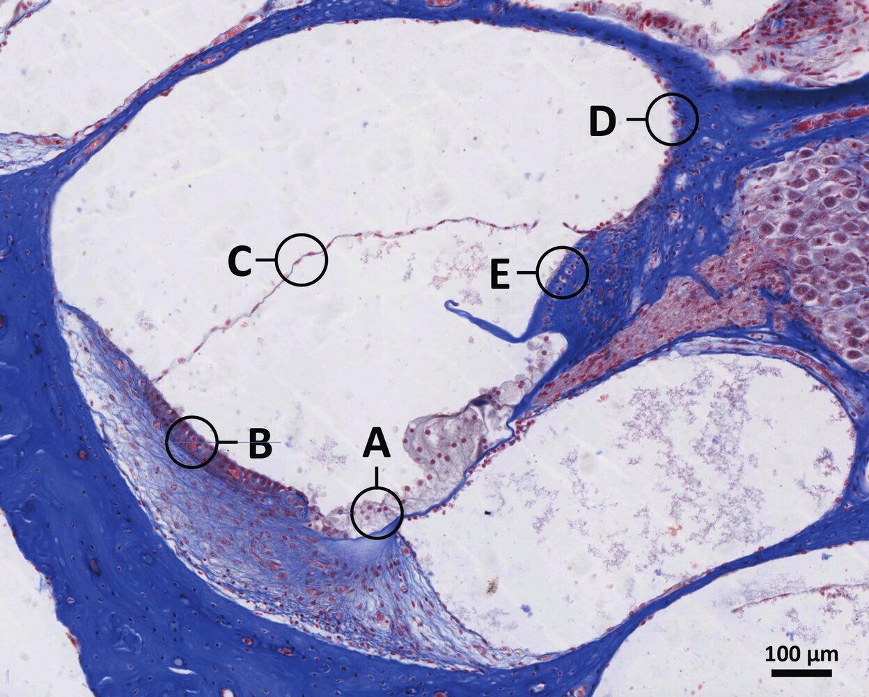
65. Some loop diuretics can cause hearing impairment when used to treat high blood pressure. This side effect occurs due to inhibition of the NKCC1 transporter in cells responsible for the high potassium content of the endolymph in the cochlear duct.
Which letter in the illustration marks these cells?
B
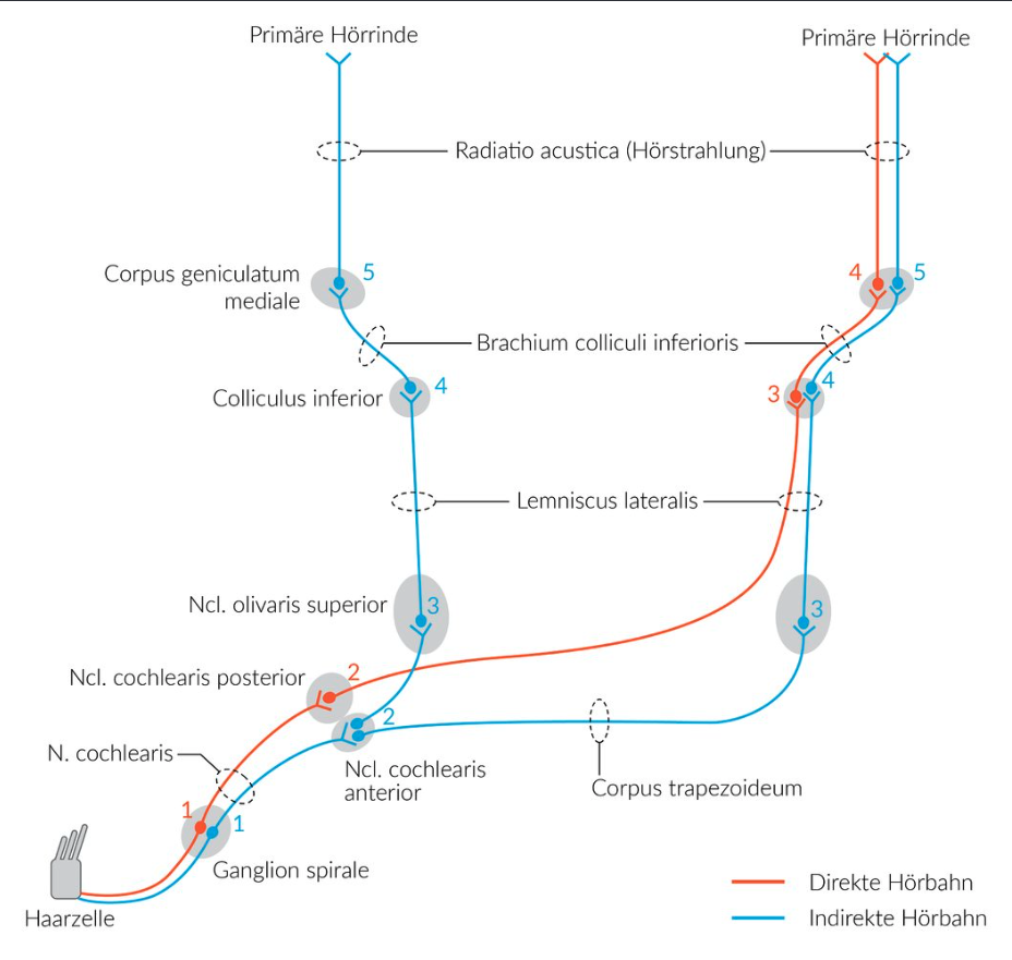
66. In the organ of Corti, sound waves are detected and converted into sequences of action potentials, which are transmitted centrally via neurons of the 8th cranial nerve (vestibulocochlear nerve).
Where do the axons of these neurons mainly terminate?
A. Inferior colliculi (Colliculi inferiores)
B. Trapezoid body (Corpus trapezoideum)
C. Anterior and posterior cochlear nuclei (Nuclei cochleares anterior et posterior)
D. Inferior olivary nucleus (Nucleus olivaris inferior)
E. Superior olivary nucleus (Nucleus olivaris superior)
C. Anterior and posterior cochlear nuclei (Nuclei cochleares anterior et posterior)
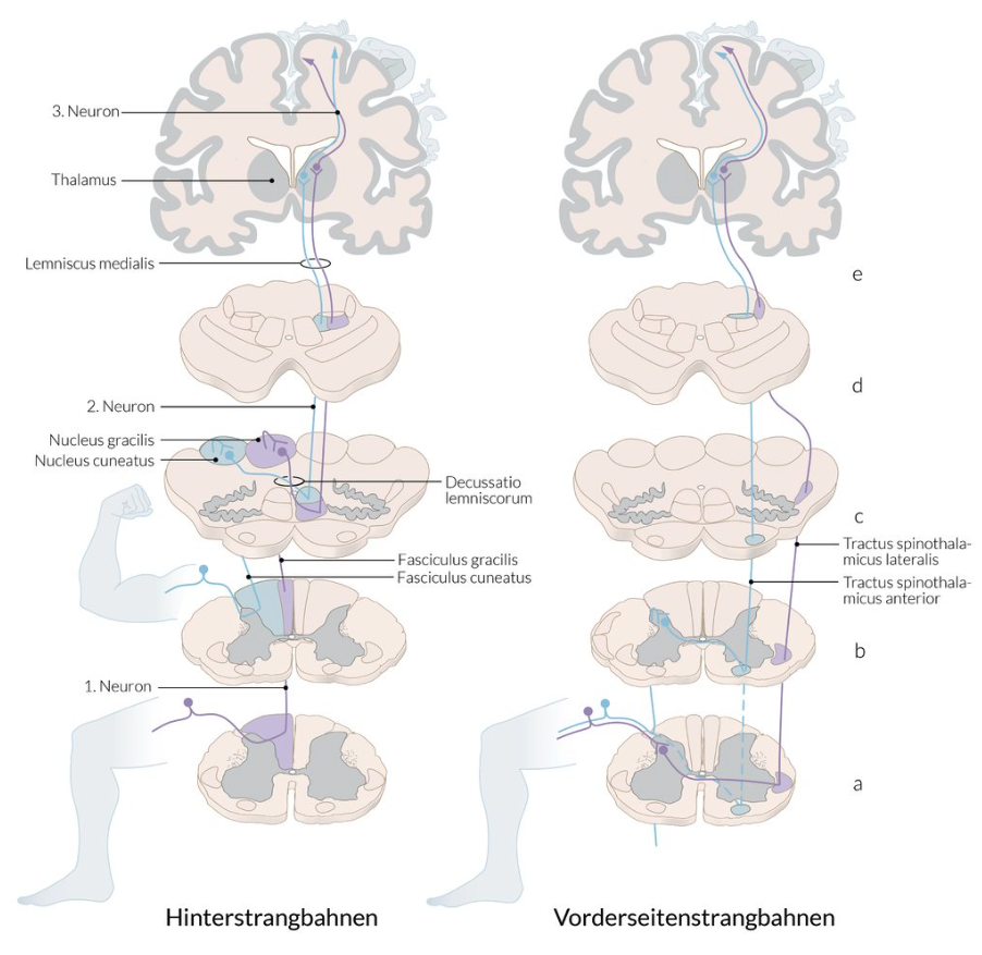
67. The lateral spinothalamic tract is an important ascending pathway from the spinal cord to the brain.
Signals from which of the following receptors are primarily transmitted to the brain via the lateral spinothalamic tract?
A. Free nerve endings
B. Meissner’s corpuscles
C. Muscle spindles
D. Ruffini endings
E. Pacinian corpuscles (Vater-Pacini bodies)
A. Free nerve endings
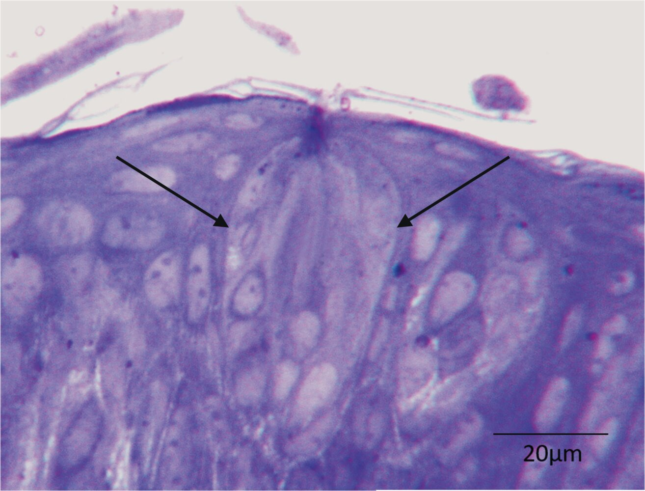
68. Sensory stimuli are detected by various specialized receptors.
Which sensory stimuli are detected by the structure marked with two arrows in the illustration?
A. Acoustic stimuli
B. Taste stimuli
C. Cold stimuli
D. Pain stimuli
E. Vibration stimuli
B. Taste stimuli
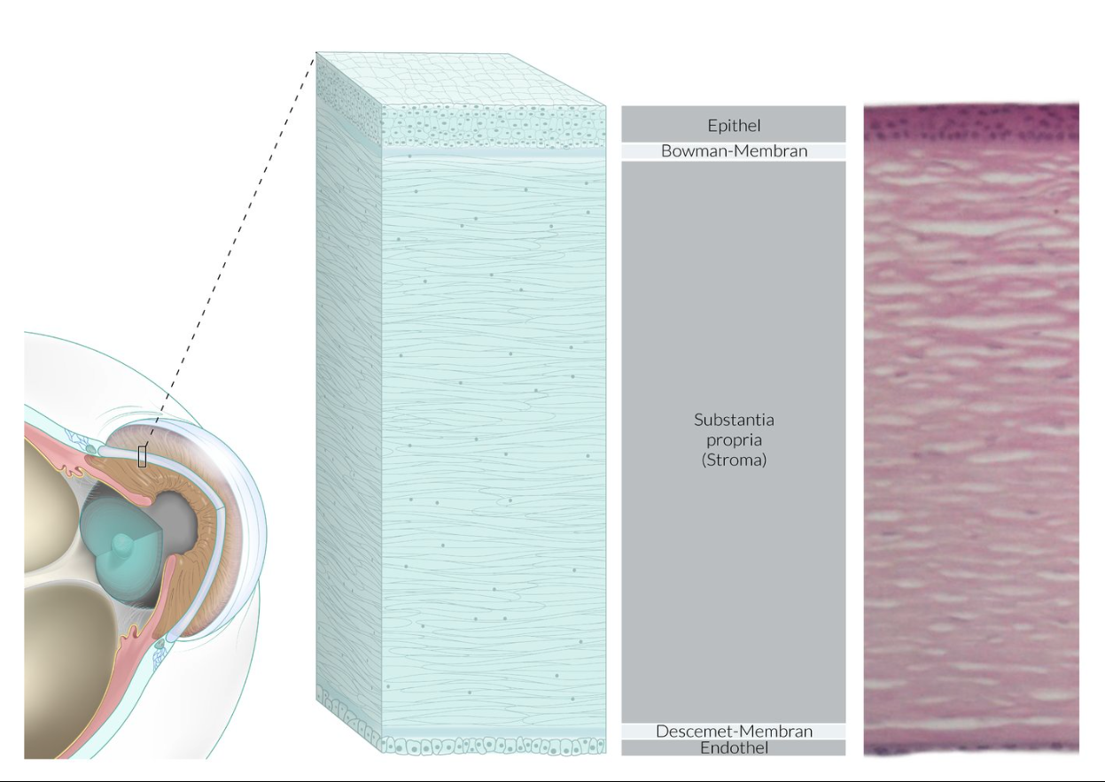
69. Infections of the cornea, such as those caused by herpes zoster viruses, are called keratitis and can damage various corneal layers. As a result, corneal clouding and even blindness may occur.
Which of the listed layers does not belong to the cornea?
A. Bowman’s membrane
B. Descemet’s membrane
C. Anterior epithelium (Epithelium anterius)
D. Posterior epithelium (Epithelium posterius)
E. Reissner’s membrane
E. Reissner’s membrane
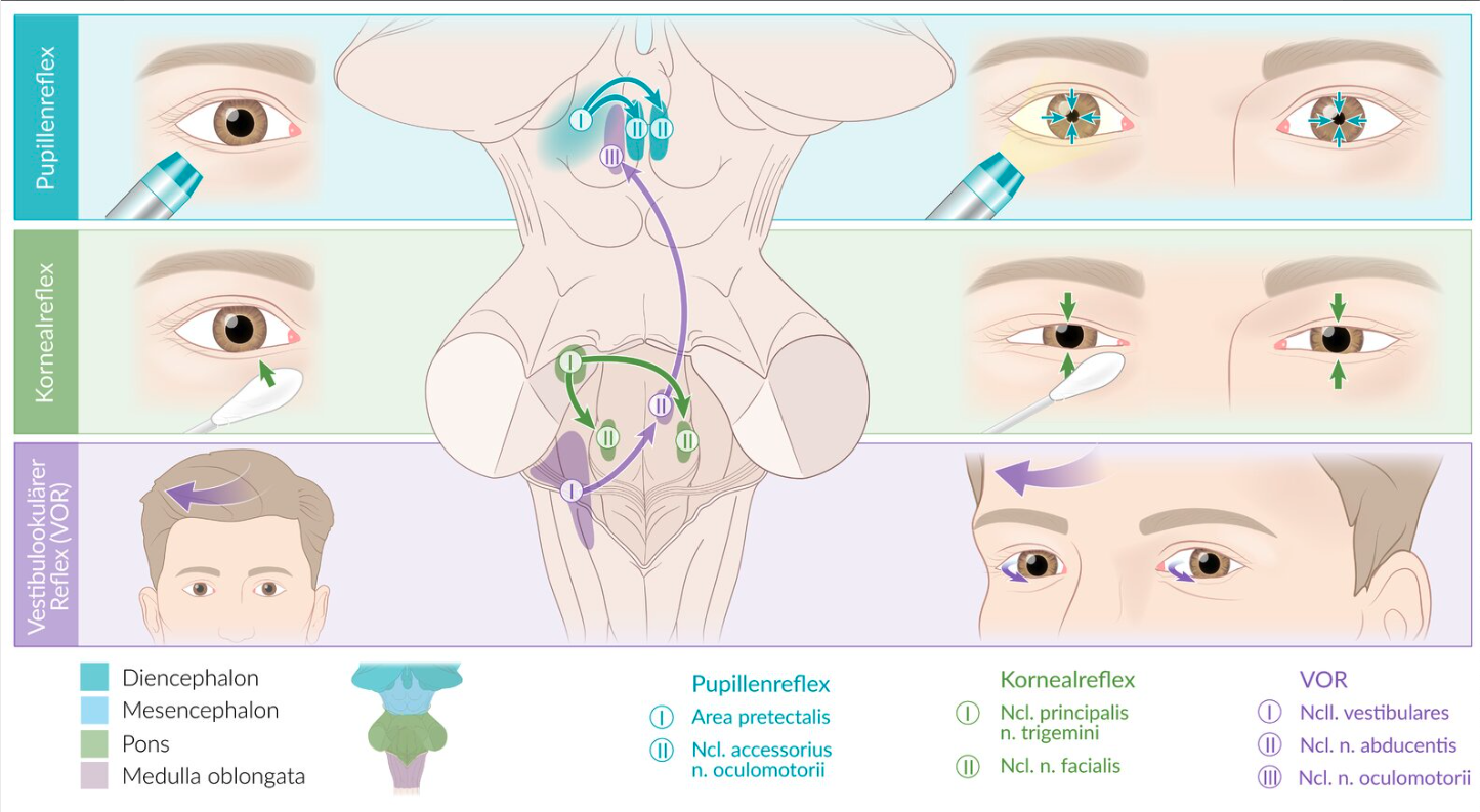
70. The corneal reflex is an important protective mechanism.
In which of the following nerves do the fibers of the efferent limb of the corneal reflex arc run?
A. Facial nerve (N. facialis)
B. Maxillary nerve (N. maxillaris)
C. Ophthalmic nerve (N. ophthalmicus)
D. Greater petrosal nerve (N. petrosus major)
E. Trochlear nerve (N. trochlearis)
A. Facial nerve (N. facialis)
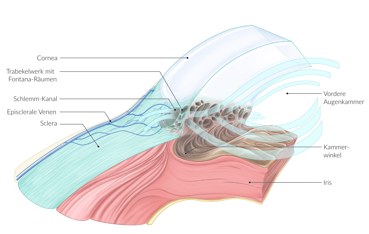
71. Impaired aqueous humor drainage can lead to glaucoma (green star) in the eye.
Through which part of the eye does the majority of aqueous humor normally drain?
A. Iridocorneal angle (Angulus iridocornealis)
B. Cornea
C. Vitreous body (Corpus vitreum)
D. Iris
E. Ciliary processes (Processus ciliares)
A. Iridocorneal angle (Angulus iridocornealis)
72. The cervical portion of the sympathetic trunk plays a central role in the development of Horner’s syndrome.
Which of the following muscles is innervated by axons from ganglia in this part of the sympathetic trunk?
A. Ciliary muscle (M. ciliaris)
B. Levator palpebrae superioris muscle (M. levator palpebrae superioris)
C. Orbicularis oculi muscle (M. orbicularis oculi)
D. Pupillary sphincter muscle (M. sphincter pupillae)
E. Superior tarsal muscle (M. tarsalis superior)
E. Superior tarsal muscle (M. tarsalis superior)
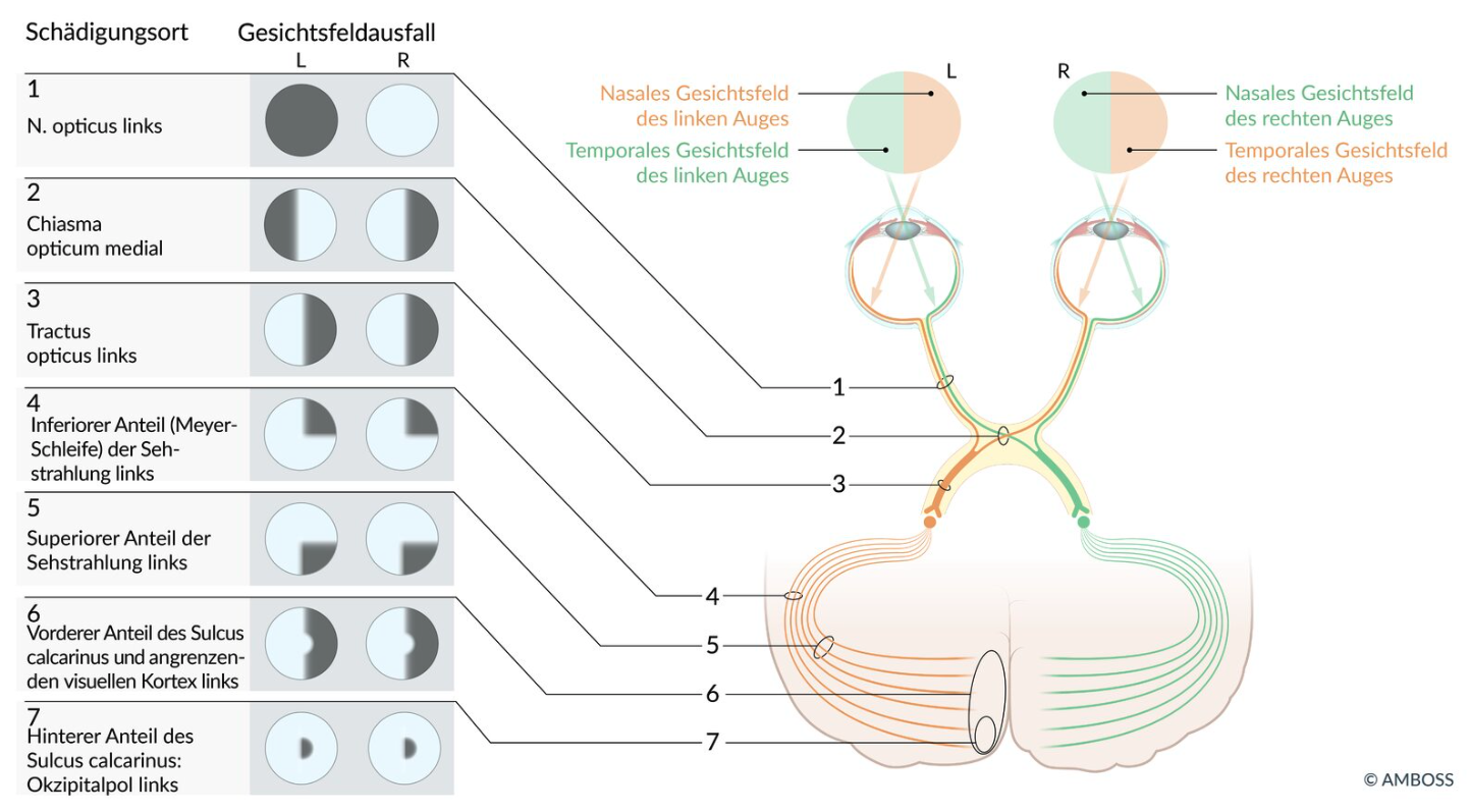
73. A circulation disorder in a brain artery can lead to a stroke, with consequences that depend, among other factors, on the region of the brain supplied.
Which of the following impairments is most likely caused by a cortical lesion due to a circulation disorder in the left posterior cerebral artery (A. cerebri posterior)?
A. Impairment of the right visual field
B. Hearing loss on the right
C. Paralysis of the right hand
D. Paralysis of the right leg
E. Impaired speech production
A. Impairment of the right visual field
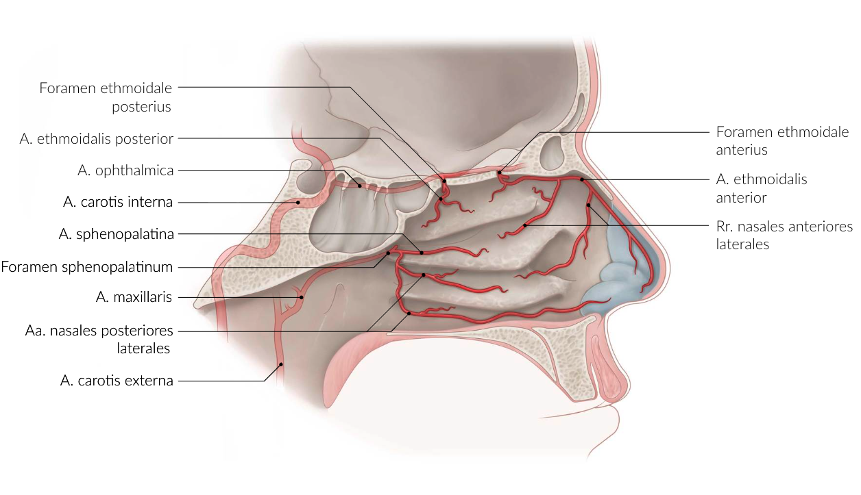
74. Nosebleeds (epistaxis) can occasionally be difficult to control and become life-threatening if arterial in origin. Such severe bleeding can originate from the posterior part of the nasal cavity. If other treatments fail, the affected artery can be therapeutically ligated.
Which artery is most likely involved in such cases?
A. Posterior superior alveolar artery (A. alveolaris superior posterior)
B. Anterior ethmoidal artery (A. ethmoidalis anterior)
C. Nasopalatine artery (A. nasopalatina)
D. Greater palatine artery (A. palatina major)
E. Sphenopalatine artery (A. sphenopalatina)
E. Sphenopalatine artery (A. sphenopalatina)
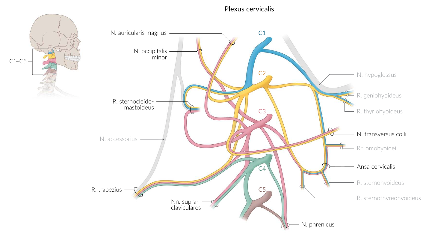
75. In the neck region, there is a nerve loop called the ansa cervicalis (profunda).
Which of the following muscles — or which part of a muscle — is innervated by the ansa cervicalis (profunda)?
A. Anterior belly of the digastric muscle (M. digastricus, Venter anterior)
B. Genioglossus muscle (M. genioglossus)
C. Mylohyoid muscle (M. mylohyoideus)
D. Omohyoid muscle (M. omohyoideus)
E. Stylohyoid muscle (M. stylohyoideus)
D. Omohyoid muscle (M. omohyoideus)
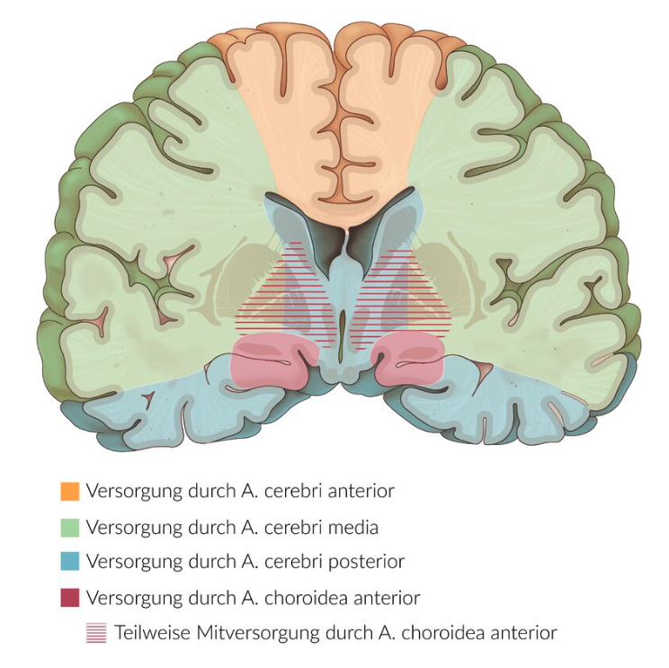
76. Interruption of blood flow in the anterolateral central arteries (lenticulostriate arteries) can lead to infarctions in the region of the basal ganglia.
From which of the following vessels do these arteries originate in most people?
A. Anterior cerebral artery (A. cerebri anterior)
B. Middle cerebral artery (A. cerebri media)
C. Posterior cerebral artery (A. cerebri posterior)
D. Anterior communicating artery (A. communicans anterior)
E. Posterior communicating artery (A. communicans posterior)
B. Middle cerebral artery (A. cerebri media)
77. To identify key brain structures on an MRI scan, understanding their spatial relationships is helpful.
Which of the following brain structures is most closely located to the amygdaloid body (Corpus amygdaloideum)?
A. External capsule (Capsula externa)
B. Mammillary body (Corpus mammillare)
C. Head of the caudate nucleus (Corpus nuclei caudati)
D. Subthalamic nucleus (Nucleus subthalamicus)
E. Foot of the hippocampus (Pes hippocampi)
E. Foot of the hippocampus (Pes hippocampi)
78. The autonomic centers of the brainstem control various functions including the cardiovascular system, respiration, and protective reflexes.
Which of the following structures is involved in the formation of the so-called vomiting center?
A. Area postrema
B. Locus coeruleus
C. Parabrachial nuclei (Ncll. parabrachiales)
D. Pedunculopontine tegmental nucleus (Ncl. tegmentalis pedunculopontinus)
E. Paramedian pontine reticular formation (paramediane pontine Formatio reticularis)
A. Area postrema
79. In central diabetes insipidus, the secretion of ADH (vasopressin) is absent.
In which brain region is ADH synthesized?
A. Pineal body of the epithalamus (Corpus pineale des Epithalamus)
B. Reticular formation nuclei of the midbrain (Kerne der Formatio reticularis des Mesencephalons)
C. Nuclei in the CA1 region of the hippocampus (Kerne in der CA1-Region des Hippocampus)
D. Magnocellular nuclei of the hypothalamus (magnozelluläre Kerne des Hypothalamus)
E. Pontine nuclei of the metencephalon (Nuclei pontis des Metencephalons)
D. Magnocellular nuclei of the hypothalamus (magnozelluläre Kerne des Hypothalamus)