Anat+physiology 2 Midterm practice - Module 1: The Nervous System
 Call Kai
 Learn
Learn Practice Test
 Spaced Repetition
 Match
Match Flashcards
 Knowt Play
1/106

Earn XP
Description and Tags
MAAAAAA mMAAAAAAAAAAAAA MAADGJASJGDSJDSJF
Name | Mastery | Learn | Test | Matching | Spaced | Call with Kai |
|---|
No analytics yet
Send a link to your students to track their progress
107 Terms
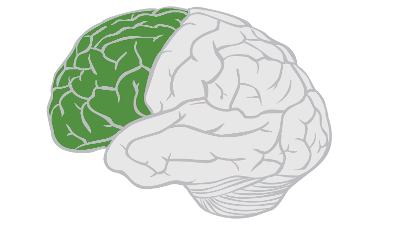
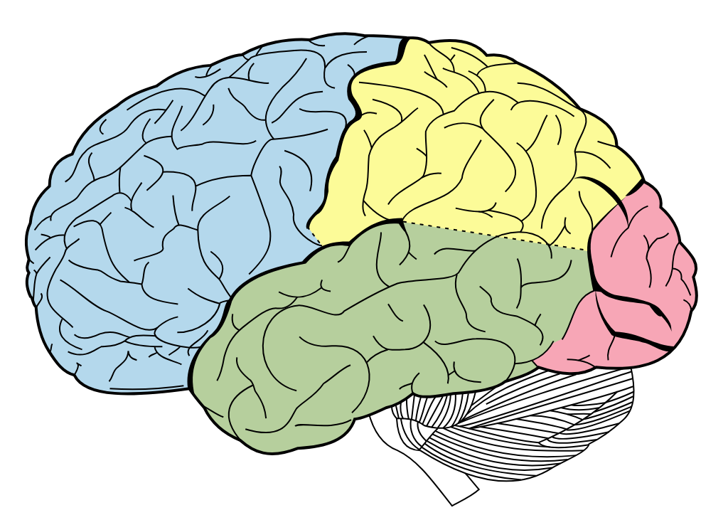
Frontal Lobes
Lobes of the brain that process information involving motor movements, concentration, planning, and problem-solving, as well as the sense of smell and emotions.
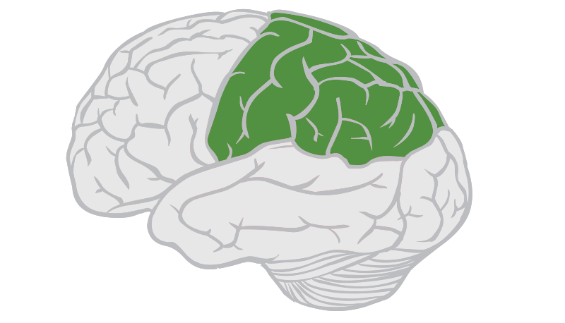
Parietal Lobes
Lobes of the brain that process the sensory information, with the exception of hearing, smell, and vision.
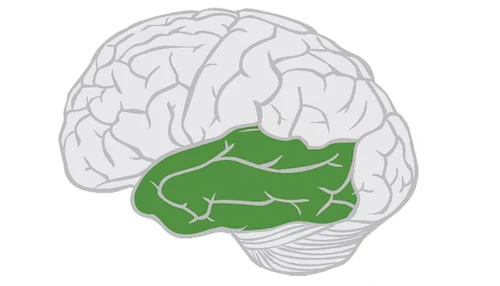
Temporal Lobes
Lobes of the brain that process information related to hearing, smell, and memory, as well as abstract thought and making judgments
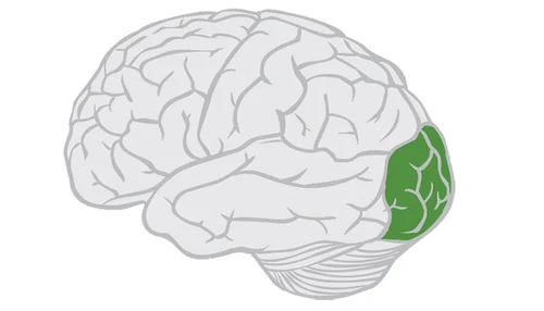
Occipital Lobes
Lobes of the brain that process visual information. Located at the posterior of the brain.
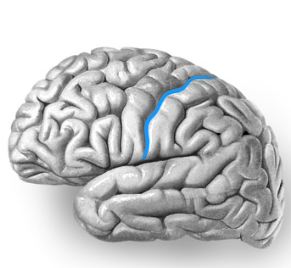
Central sulcus
Groove located midway on the side of the cerebrum that separates the frontal from the parietal lobes
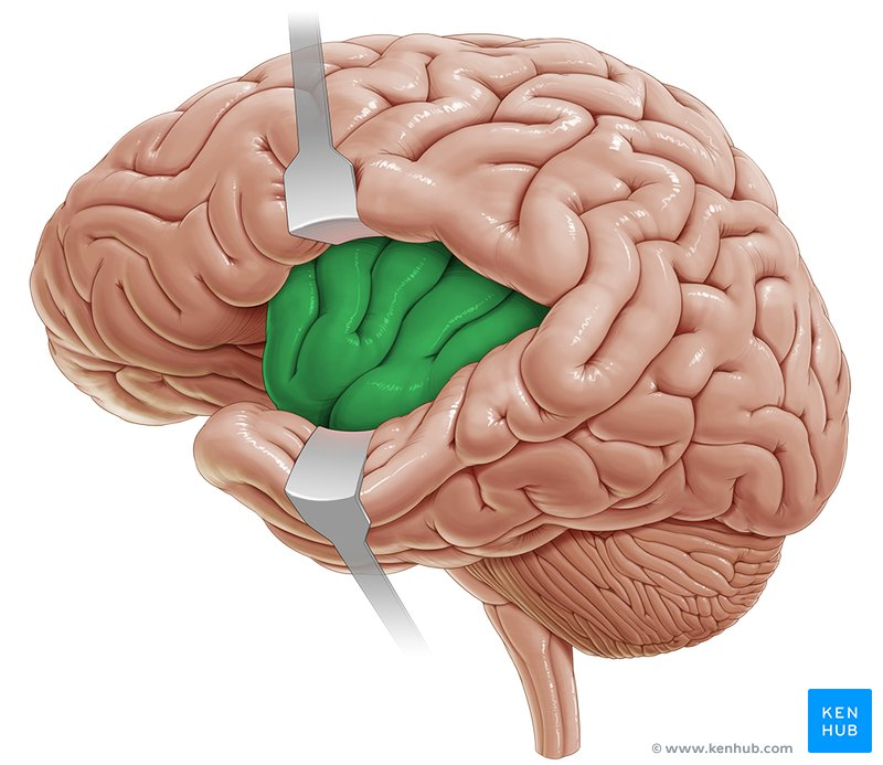
Insula
a specialized brain region crucial for self-awareness, emotional experience, and interoception. Located deep in the lateral fissure.
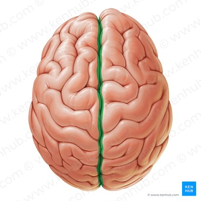
Longitudinal fissure
the deep, prominent groove that runs along the midline of the brain, dividing the cerebrum into the left and right cerebral hemispheres
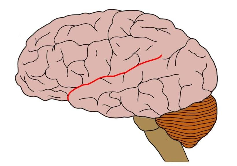
Lateral sulcus
Fissure that is located on the side and separates the temporal lobe from the parietal lobes. Also called the lateral fissure or Sylvian fissure.
Diencephalon
Consists of the thalamus and hypothalamus. Lies between the cerebrum and the brain stem. Contains the subthalamus and epithalamus.
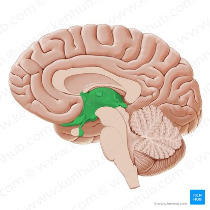
Thalamus
The largest part of the Diencephalon. It consists of two lateral portions connected by a stalk called the interthalamic adhesion, which is sometimes referred to as the intermediate mass. Carries all sensory information to the cerebral cortex, with the exception of the sense of smell, which is carried directly to the frontal lobe of the cerebral cortex by the olfactory nerves.
Sometimes referred to as a relay station for sensory information. Also intimately involved in emotions due to its connections to the limbic system.
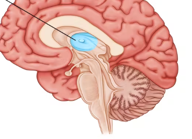
Hypothalamus
Lies inferior and anterior to the thalamus. Contains mamillary bodies on its anterior surface. The mamillary bodies process info associated with smell and emotions. Also regulates body temperature, thirst, hunger, and sexual drive, and is involved in processing emotions, mood, and sleep along with the reticular activating system.
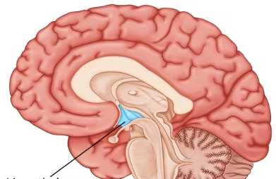
Pituitary Gland
a pea-sized endocrine organ located at the base of the brain. Known as the "master gland," it regulates vital bodily functions—including growth, metabolism, and stress responses—by producing essential hormones and directing other glands in the body.
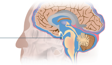
Pineal Gland
A small, pinecone-shaped gland located between the cerebral hemispheres. It attaches to the posterior portion of the thalamus, and secretes melatonin.
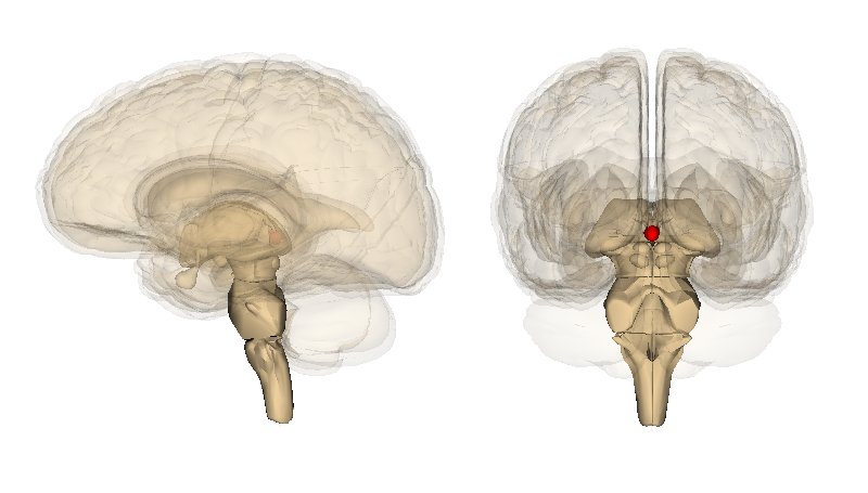
Corpus callosum
A white matter “bridge” that connects the two hemispheres of the brain
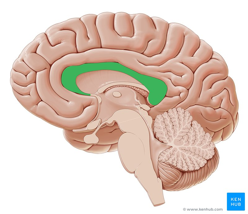
Brain Stem
3 part structure that lies between the cerebral cortex and the spinal cord. It consists of the midbrain, pons, and medulla oblongata. It connects the cerebrum to the spinal cord.
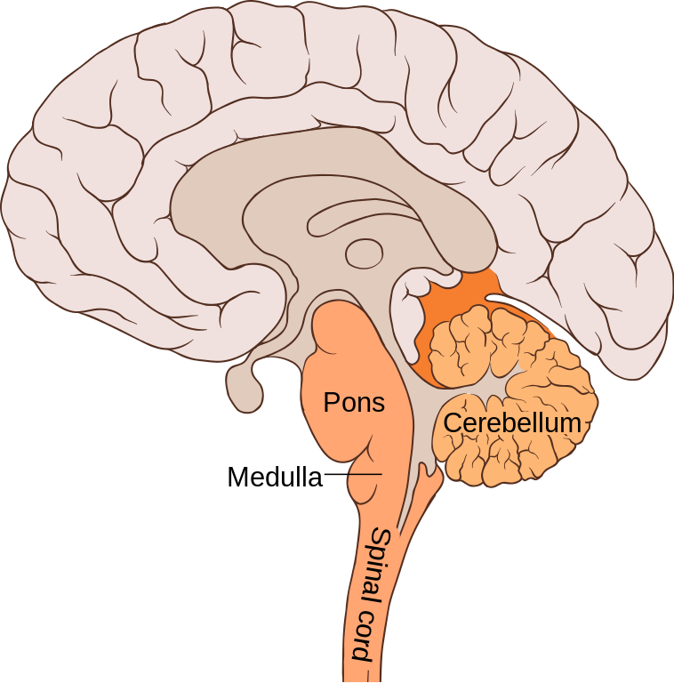
Midbrain
The most superior portion of the brainstem. Contains a roof (tectum) that contains four bumps (nuclei) called the corpora quadrigemina. The two superior nuclei are the superior colliculi, and same for the inferior colliculi.
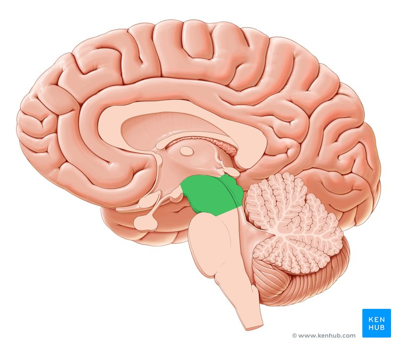
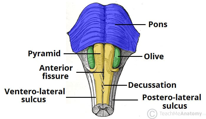
Pons
The middle section of the brainstem. Similar to the medulla oblongata, it contains spinal cord tracts as well as nuclei, that help control respiration and sleep.
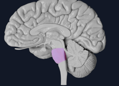
Medulla oblongata
The most inferior portion of the brainstem. Contains a number of centers for controlling heart rate, respiration, swallowing, vomiting, and blood vessel diameter. The centers consist of nuclei; clusters of neuron cell bodies. The spinal tracts also continue through the medulla, connecting the spinal cord with the brain.
Cerebrum
The largest portion of the nervous system. Consists of two hemispheres (right and left) connected by a white matter bridge called the corpus callosum. On the surface are folds called gyri and grooves called sulci. Deep grooves are known as fissures. Each hemisphere is divided into lobes. The lobes are the frontal, parietal, temporal, and occipital.
Lateral Ventricle
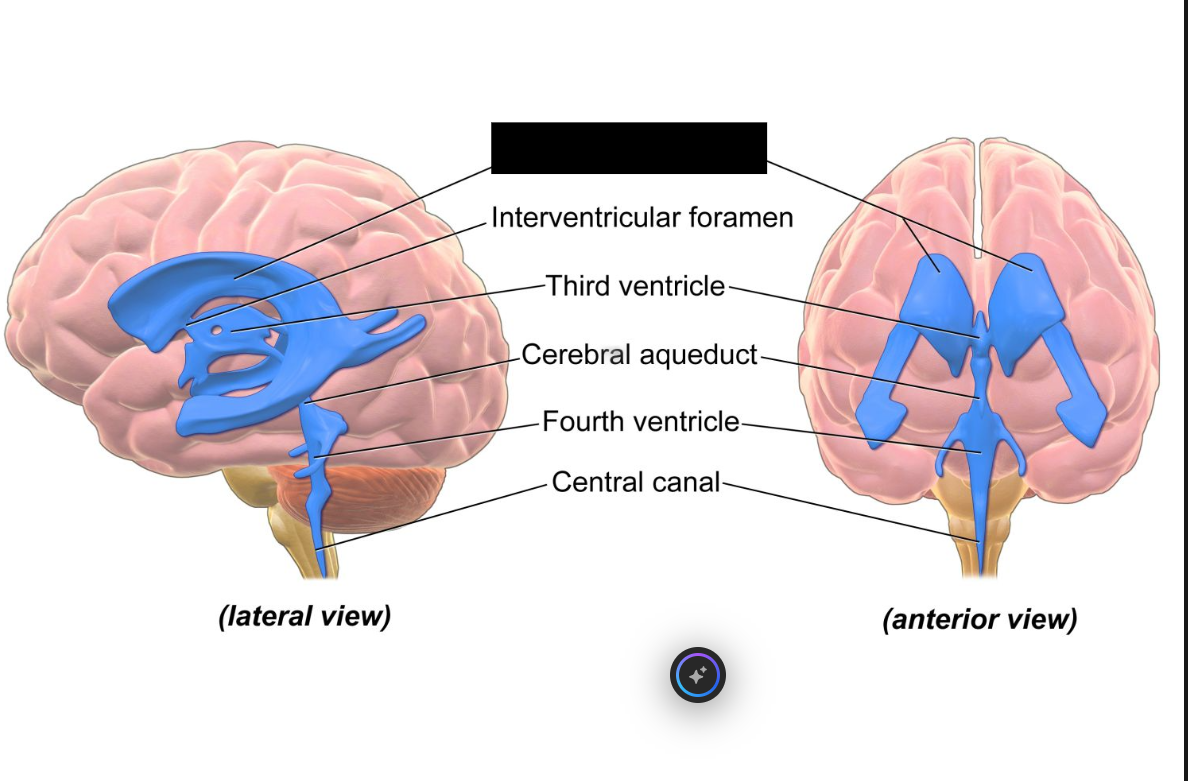
two C-shaped, fluid-filled cavities located within the cerebral hemispheres of the brain. As the largest components of the brain's ventricular system, they house the choroid plexus which produces the majority of the body's cerebrospinal fluid (CSF)
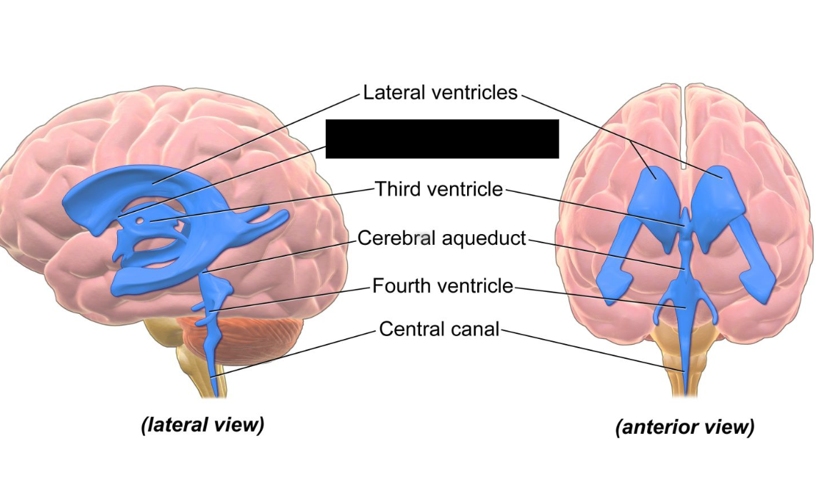
3rd Ventricle
a narrow, fluid-filled midline cavity deep within the brain. It is a vital part of the ventricular system, producing and circulating cerebrospinal fluid (CSF) to protect and cushion the brain.
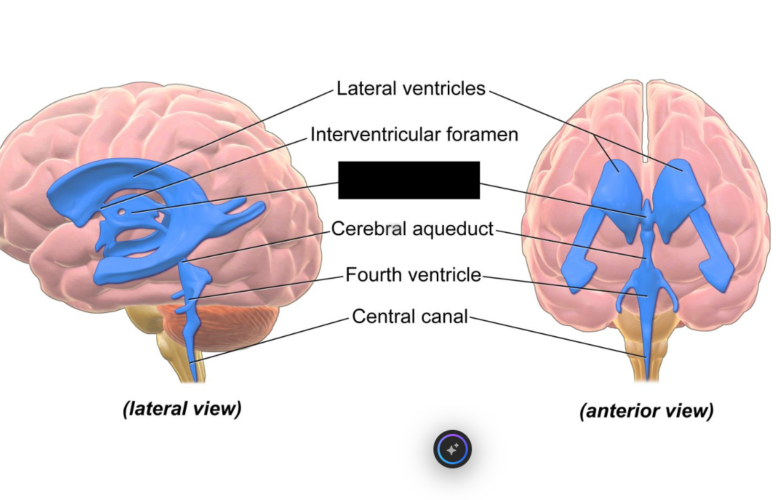
4th ventricle
a diamond-shaped cavity in the brainstem. It is a crucial part of the ventricular system that stores and circulates cerebrospinal fluid (CSF). It delivers CSF to the brain's surface and the spinal cord, and protects the brain from injury by acting as a cushion
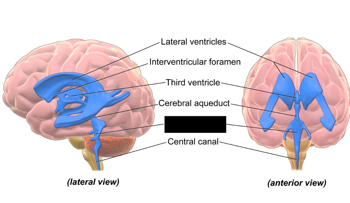
Cerebral Aqueduct
connection of the ventricular system between the third and fourth ventricles located in the midbrain
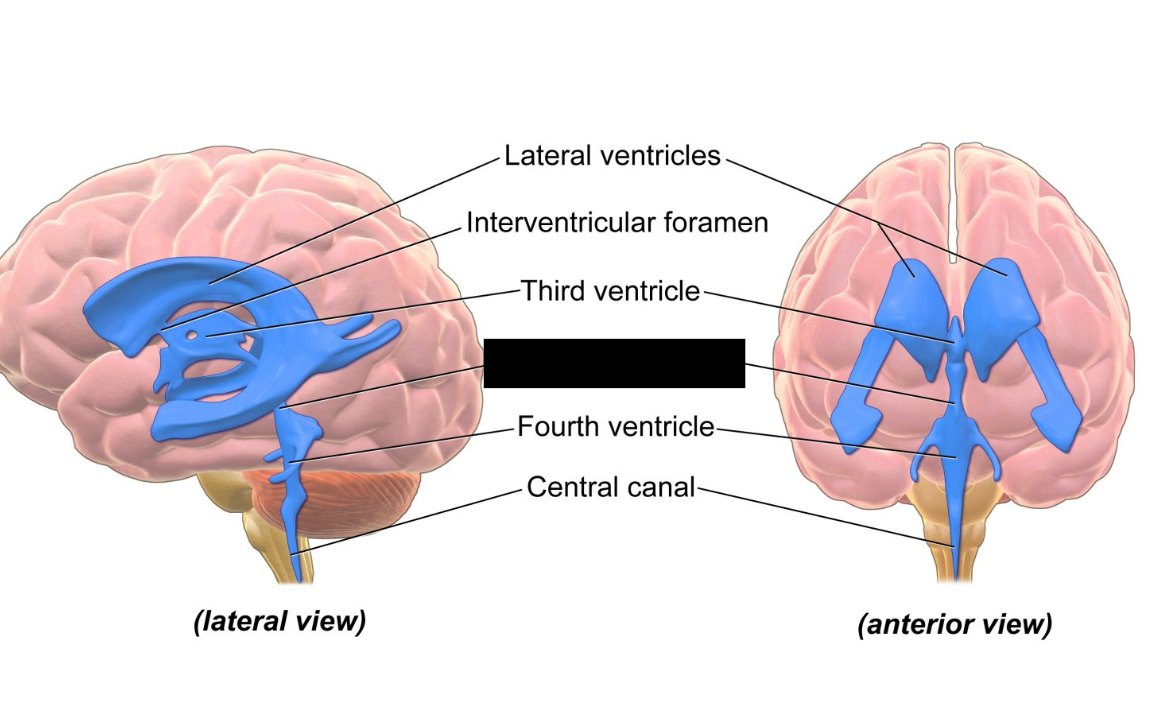
Interventricular Foramen
paired, slit-like channels in the brain that connect the two lateral ventricles to the third ventricle. They are a critical component of the brain's ventricular system, allowing cerebrospinal fluid (CSF) to flow and circulate.
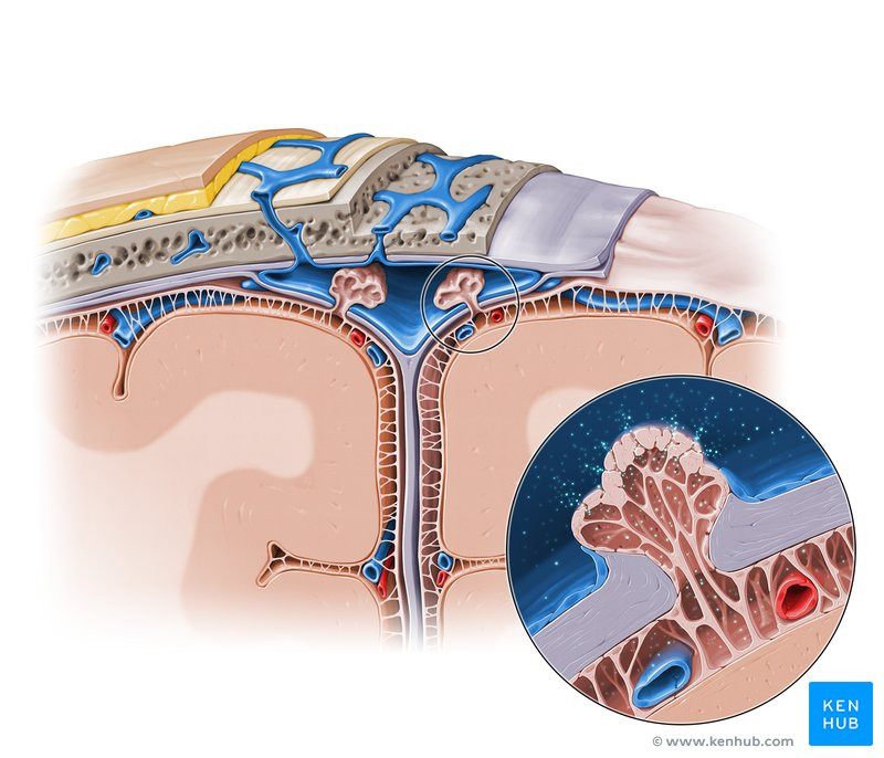
Arachnoid Granulations
small, cerebrospinal fluid (CSF)-filled outpouchings of the arachnoid membrane that project into the dural venous sinuses of the brain. Their primary function is to mediate the drainage of CSF from the subarachnoid space back into the bloodstream.
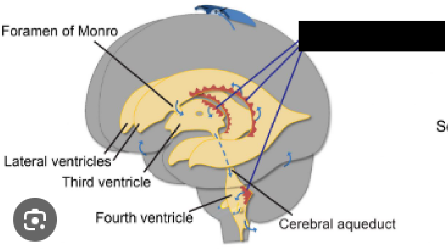
Choroid Plexus
a network of specialized, vascularized cells located within the brain's ventricles (lateral, third, and fourth) that is primarily responsible for producing cerebrospinal fluid (CSF). It functions as a crucial blood-CSF barrier and regulates brain homeostasis, protecting the brain and spinal cord by delivering nutrients, removing waste, and providing structural buoyancy.
Found within all 4 brain ventricles.
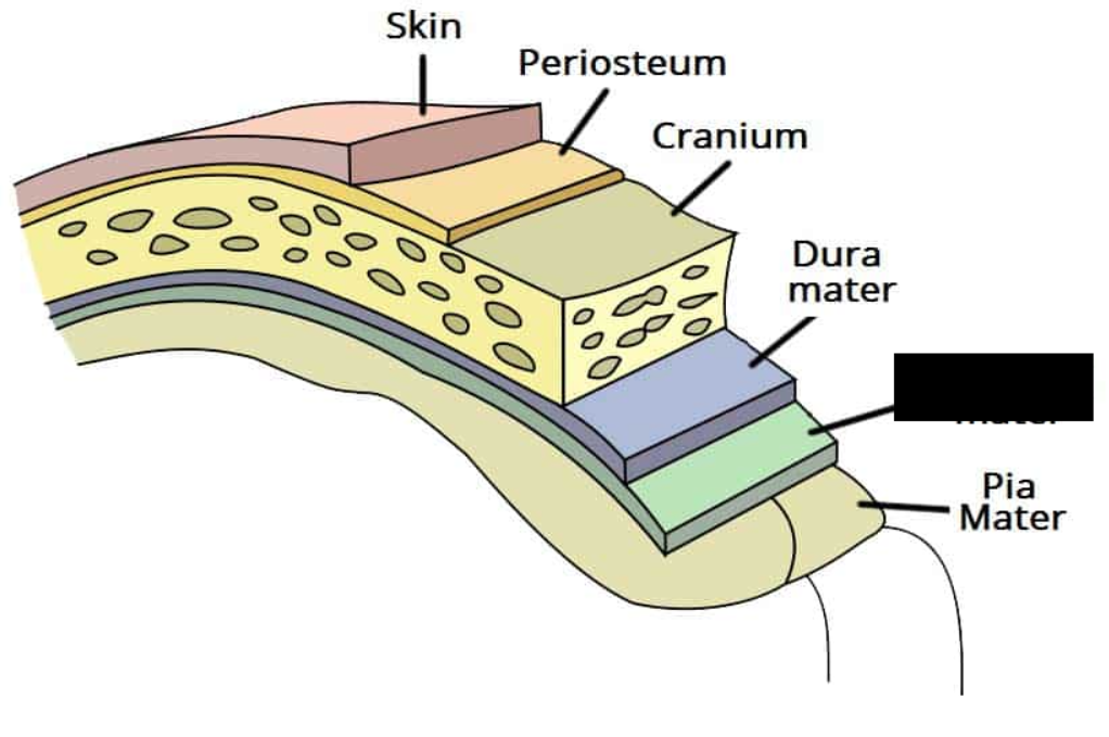
Arachnoid Mater
The middle layer of the meninges is known as the _________ _____, which is a thin layer consisting of simple squamous epithelum. It adheres to the inner portion of the dura mater.
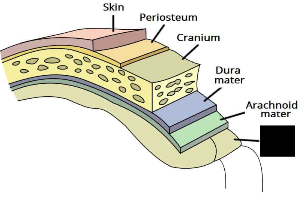
Pia mater
The innermost membrane of the meninges, and is closely attached to the spinal cord as a thin membrane. It continues inferiorly to produce the filum terminale. It also extends laterally to the dura mater at points along the spine to produce the dentate ligaments, which work to anchor the cord in place.
Dura mater
the most superficial layer of the meninges. Forms a sac known as the thecal sac that encases the spinal cord
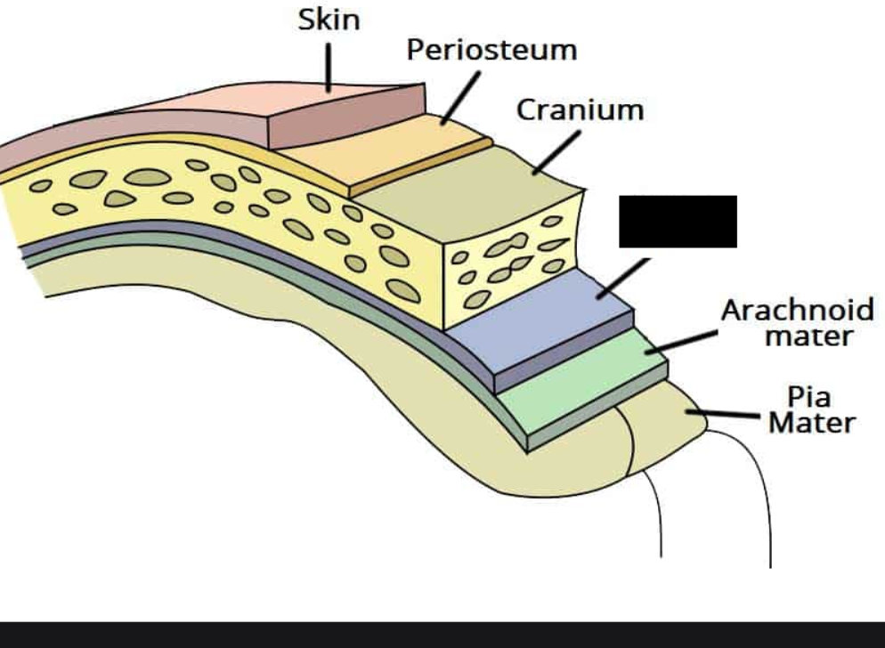
Subarachnoid space
The space between the arachnoid and pia mater, filled with cerebrospinal fluid.
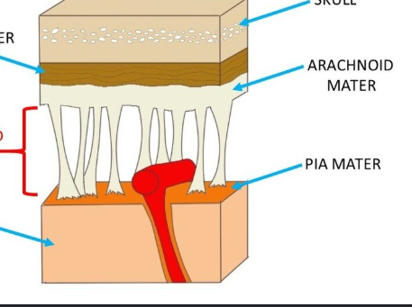
Meninges
The _______ are three protective membranes that envelop and support the brain and spinal cord.
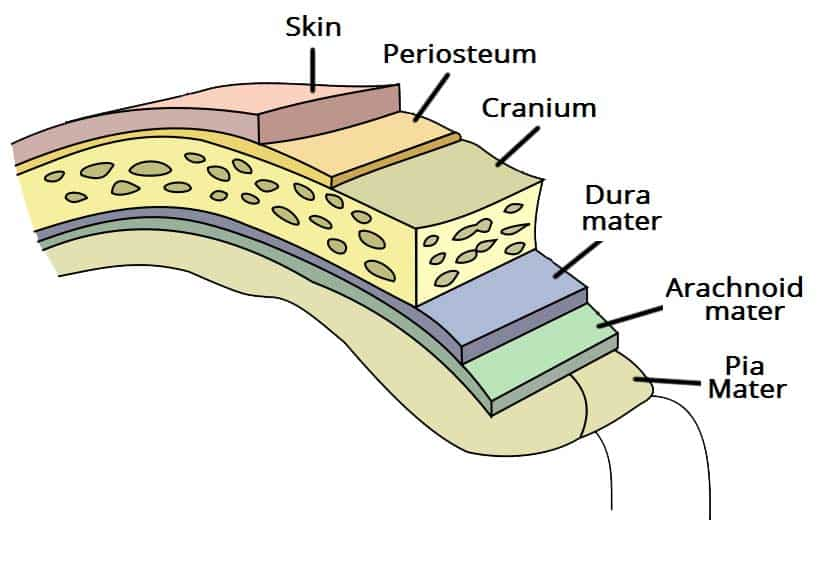
Conus medullaris
the tapered, cone-shaped lower end of the spinal cord. It typically ends near the first or second lumbar vertebrae (L1-L2) in adults. Below this point, the spinal cord transitions into the cauda equina, a bundle of nerve roots that controls pelvic organs and the lower limbs
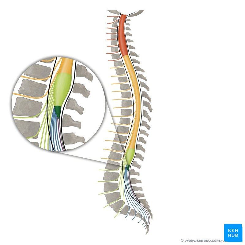
Filum Terminale
a ~20 cm long, delicate connective tissue strand extending from the conus medullaris (end of the spinal cord) to the coccyx, acting as a stabilizer that anchors the spinal cord. Produced by the Pia mater.
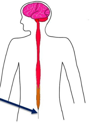
Cauda Equina
a bundle of nerve roots located at the base of the spinal cord (in the lower back) that controls sensation and movement in the legs, as well as bladder and bowel functions. Its name derives from Latin, translating to "horse's tail," which perfectly describes its visual appearance.
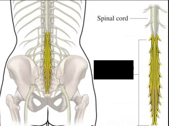
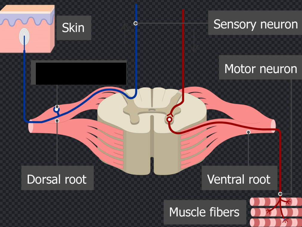
Dorsal root
The nerve root exiting the back of the spinal cord.
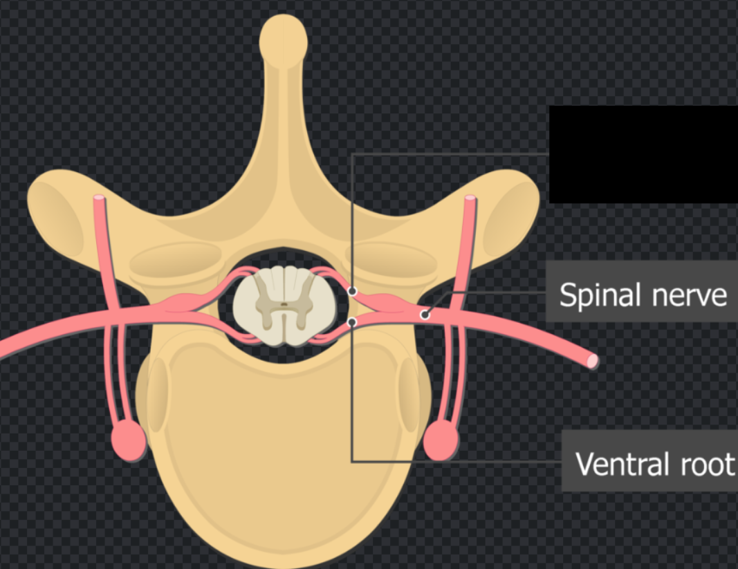
Ventral root
The nerve root exiting the front of the spinal cord.
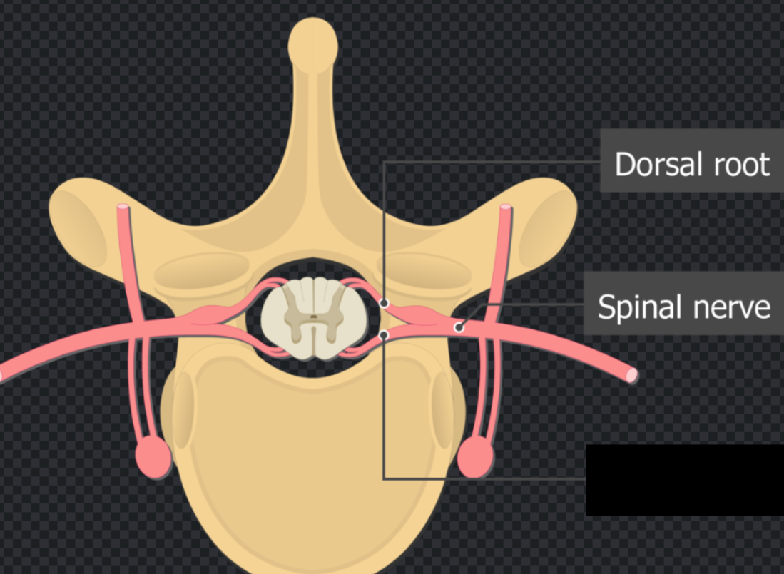
Dorsal Root Ganglion
a cluster of sensory nerve cell bodies located along the spine. Acting as a critical relay station, it transmits sensory information—such as touch, temperature, and pain—from the body's peripheral to the central nervous system.
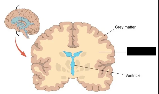
Gray Matter
a major component of the central nervous system, consisting mainly of neuronal cell bodies, dendrites, and unmyelinated axons. It is primarily responsible for processing information, controlling movement, and managing cognitive functions like memory, emotion, and decision-making
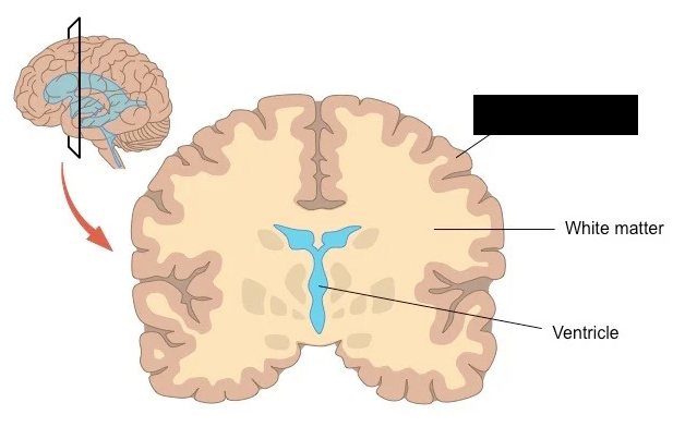
White Matter
a primary tissue of the central nervous system composed of myelinated nerve fibers (axons) which makes up it’s color. It makes up about half the human brain and acts as the communication network, relaying electrical signals between different regions of the brain and spinal cord
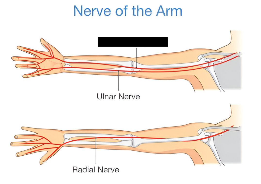
Median Nerve
a major peripheral nerve originating from the brachial plexus. It runs down the arm, forearm, and through the carpal tunnel in the wrist. It is responsible for the motor control of most forearm flexors and thumb movements, as well as sensory innervation to the palm, thumb, index, middle, and half of the ring finger.
Sciatic Nerve
the longest and largest nerve in the human body. It originates in the lower back, runs through the hips and buttocks, and branches down the back of each leg to the feet and toes.
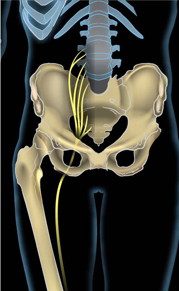
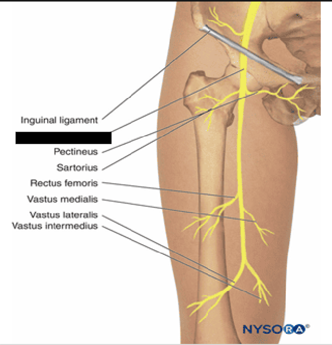
Femoral Nerve
the largest nerve in the lumbar plexus, originating in the lower back (L2–L4) and running down into the leg. It is a mixed nerve, meaning it controls both motor functions (muscle movement) and sensory functions (feeling)
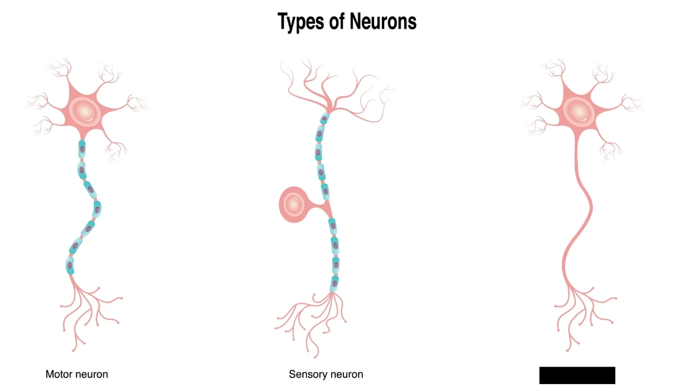
Sensory Neurons
Neurons that transmit sensory information from the body (skin, eyes, ears, organs) toward the central nervous system (CNS). Also called Afferent Neurons.
Motor Neurons
Neurons that carry signals away from the CNS to command muscles, organs, and glands to react. Also called Efferent Neurons.
Interneurons
Neurons that connect sensory and motor neurons exclusively within the CNS, playing a vital role in thinking, learning, and reflexes.
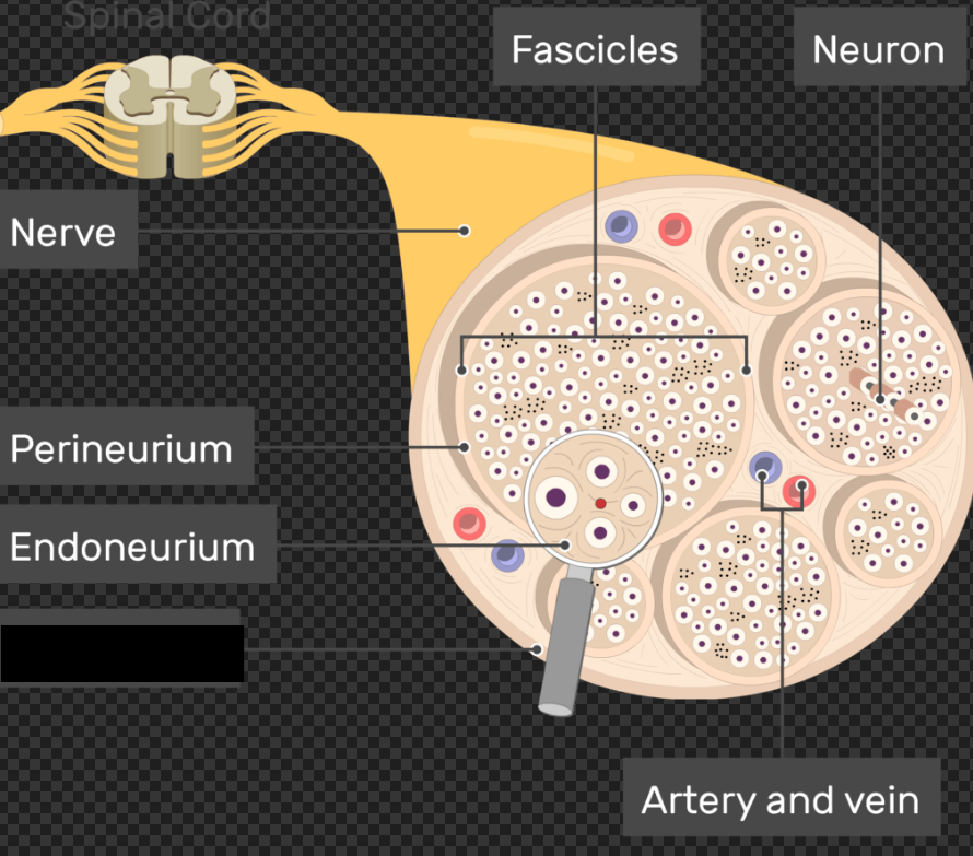
Perineurium
a protective, specialized connective tissue sheath that encloses bundles of nerve fibers (called nerve fascicles) in the peripheral nervous system. It acts as a selective diffusion barrier and provides crucial mechanical strength and structural integrity to the nerve.
Middle layer bundling groups of axons into fascicles.
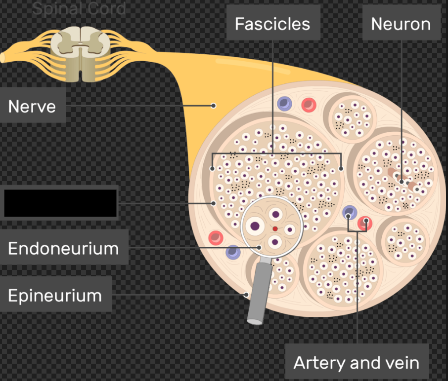
Endoneurium
the innermost, delicate layer of connective tissue that surrounds individual nerve fibers (axons) and their myelin sheaths in the peripheral nervous system
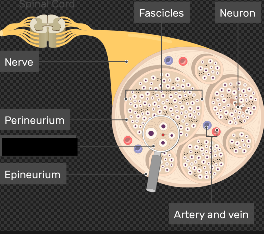
Epineurium
the outermost layer of dense connective tissue surrounding a peripheral nerve. It encapsulates entire nerve trunks, bundles smaller groups of nerve fibers (fascicles) together, and provides vital physical cushioning and structural protection against stretching or compression
Brain Spinal Cord
The main structures of the CNS are the _____ and the ______ ____.
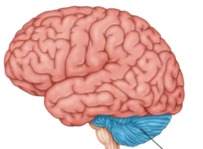
Cerebellum
Processes information for balance, coordination, fine motor movements, and joint position. Located in the back of and below the cerebrum, and connected to the brainstem via three cerebellar peduncles (superior, middle, and inferior).
Sympathetic Nervous System
Often referred to as the fight or flight system, located in the thoracic and lumbar spines, and sends fibers to the viscera. It primarily utilizes two major hormones—epinephrine and norepinephrine—which are produced in the adrenal medulla and circulated through the bloodstream to rapidly prepare the body for quick physical action and survival. Also produces Acetylcholine, which carries signals between nerve cells and is critical for processes like voluntary muscle control, memory formation, attention, and arousal.
norepinephrine
a neurotransmitter and stress hormone that increases alertness, focus, blood pressure, and heart rate as part of the body's "fight-or-flight" response
epinephrine
Hormone that initiates the body's "fight-or-flight" response, released by the CNS. Also called Adrenaline
Acetylcholine
The neurotransmitter that promotes skeletal muscle contraction. Also plays a role in the parasympathetic nervous system and is responsible fore effects of the parasympathetic nervous system on the body.
Parasympathetic Nervous System
Begins in the cervical and lower lumbar spines, and sends fibers to the same viscera as the sympathetic. Division of the autonomic nervous system responsible for "rest-and-digest" activities, acting as the body's calming system.
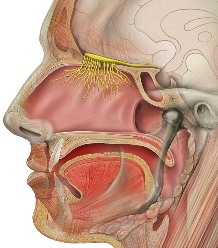
Olfactory Cranial Nerve
The first cranial nerve. A sensory nerve that carries info for the sense of smell. The only nerve that originates in the frontal lobe of the brain; its fibers pass through the cribriform plate of the ethmoid bone to reach the upper nasal passages.
Optic Cranial Nerve
The second cranial nerve. Sensory nerves that carry information relating to vision from the retina of the eyes, and pass through the optic canals of the sphenoid bone. This nerve is checked by testing a patient with an eye chart.
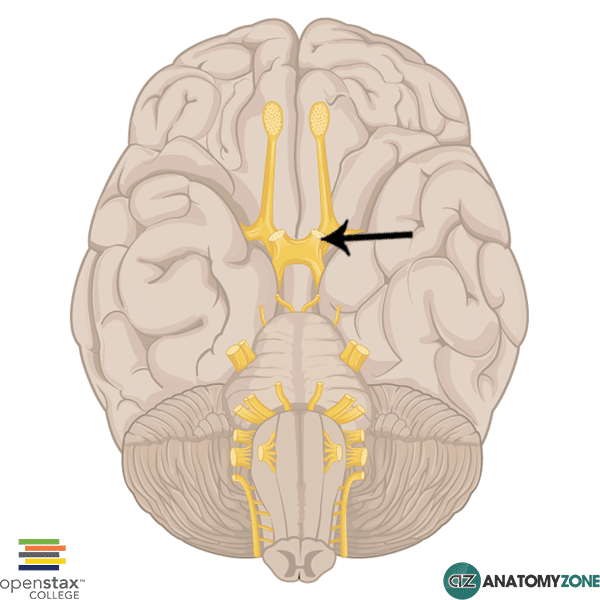
Occulomotor Cranial Nerve
The third cranial nerve. Motor nerves that innervate some of the muscles of the eye, including the superior, inferior, and medial rectus, as well as the inferior oblique. They also innervate the levator palpebrae superioris muscles that move the eyelids. This nerve is checked by testing the patients horizontal eye movement.
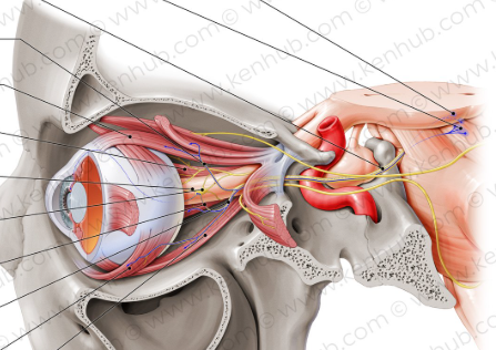
Trochlear Cranial Nerve
The fourth cranial nerve. Motor nerves that innervate the superior oblique muscles of the eyes. Responsible for downward vertical and inward rotation of the eye. This nerve is checked by asking a patient to follow an object with their eyes.
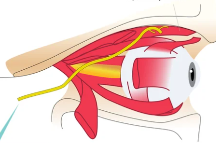
Trigeminal Cranial Nerve
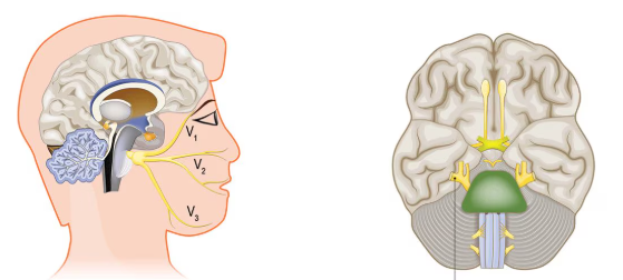
The fifth cranial nerve. Mixed nerves carrying both sensory and motor info. They originate in the pons.
The superior ophthalmic branch carries sensory information from the upper portion of the face (above the eyelids).
The middle maxillary branch carries sensory information from the middle portion of the face (below the lower eyelid to the upper lip).
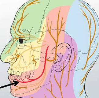
The lower mandibular branch carries sensory information from the mandible. The mandibular branch also carries motor information to the muscles of mastication, including the masseter and temporalis.
Abducens Cranial Nerve
The sixth cranial nerve. Motor nerves carrying information to the lateral rectus muscles of the eyes
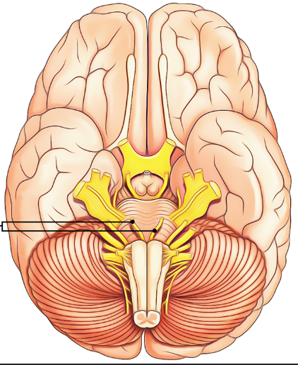
Facial Cranial Nerve
The seventh cranial nerve. Mixed nerves that carry motor information to the muscles of the face and are responsible for producing facial expressions. The information consists of taste from the anterior 2/3rds of the tongue, along with proprioception of the facial muscles and deep pressure in the face. The motor function is tested by asking the patient to make multiple facial expressions, while the sensory function is tested by asking the patient to identify some tastes.
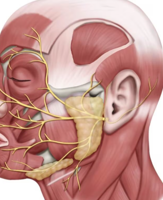
Vestibulochochlear Cranial Nerve
The eighth cranial nerve. Sensory nerves that carry information regarding hearing, balance, and equilibrium from the inner ear. The nerves form 2 branches. A vestibular branch innervates the vestibule and semicircular canals of the ear, and carries information related to balance and equilibrium. A cochlear branch carries hearing information from the cochlea of the inner ear. Tested with hearing and balance tests.
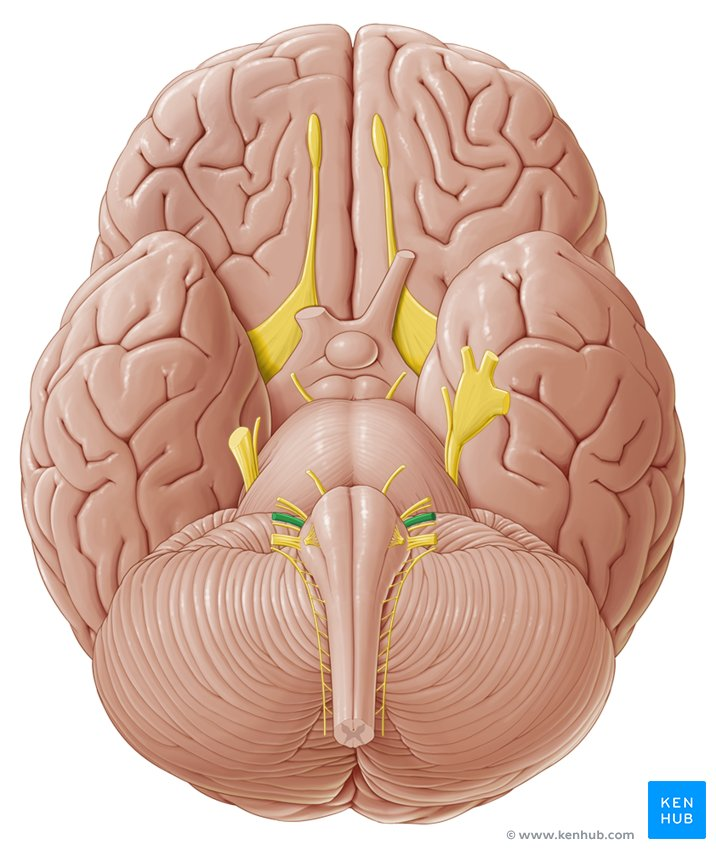
Glossopharyngeal Cranial Nerve
The ninth cranial nerve. Mixed nerves that carry sensory information regarding taste from the posterior 1/3rd of the tongue, as well as motor information to the muscles in the pharynx for swallowing. Tested by observing the patient’s soft palate, along with Cranial Nerve X.
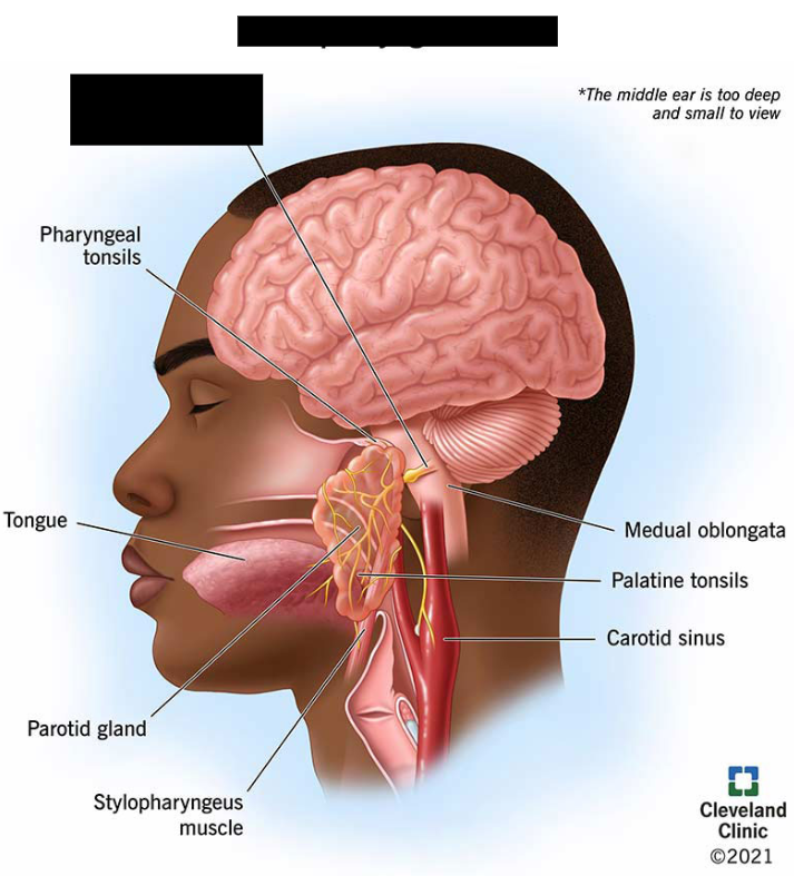
Vagus Cranial Nerve
The tenth cranial nerve. Mixed nerves that carry sensory information from the viscera of the esophagus, respiratory tract, and abdomen. They carry motor information to the heart, stomach, intestines, and gallbladder. They also carry information for the coordination of swallowing. Integral to the autonomic nervous system. Originates from inside the skull (brainstem). Tested by observing the patient’s soft palate, along with Cranial Nerve IX.
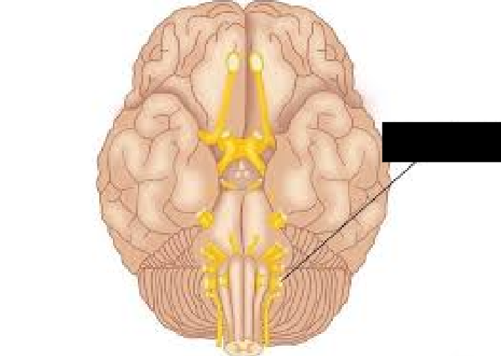
Spinal Accessory Cranial Nerve
The eleventh cranial nerve. Motor nerves that carry information to the muscles of the neck and upper back, including the sternocleidomastoid and trapezius. Tested by asking the patient to shrug their shoulders and turn their head.
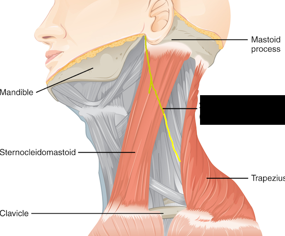
Hypoglossal Cranial Nerve
The twelfth cranial nerve. Motor nerves that primarily carry motor information to the muscles that move the tongue. Checking these nerves requires sticking one’s tongue out. Deviation of the tongue from one side to the other indicates a problem with the nerve.
Cervical Plexus
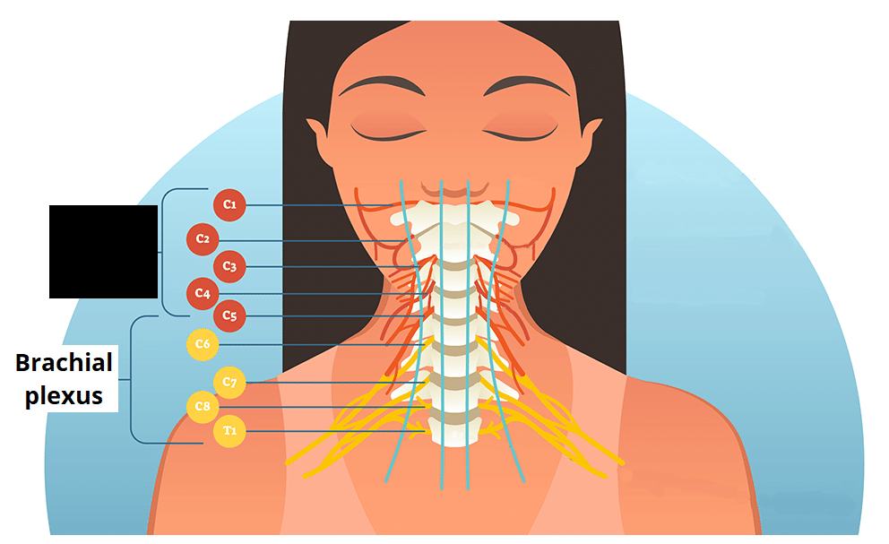
Nerve plexus located in the posterior triangle of the neck, deep within the prevertebral layer of fascia (C1-C4). It serves the head, neck, and shoulders, and provides the phrenic nerve for the diaphragm.
Brachial Plexus
C5-T1. Consists of the ventral rami from spinal nerves C5-T1. The rami form three trunks, and the trunks become six divisions, which again join to form three cords.
Five branches emerge from the three cords, which constitute the major nerves of the upper extremity. These include the axillary, radial, musculocutaneous, ulnar, and median nerves.
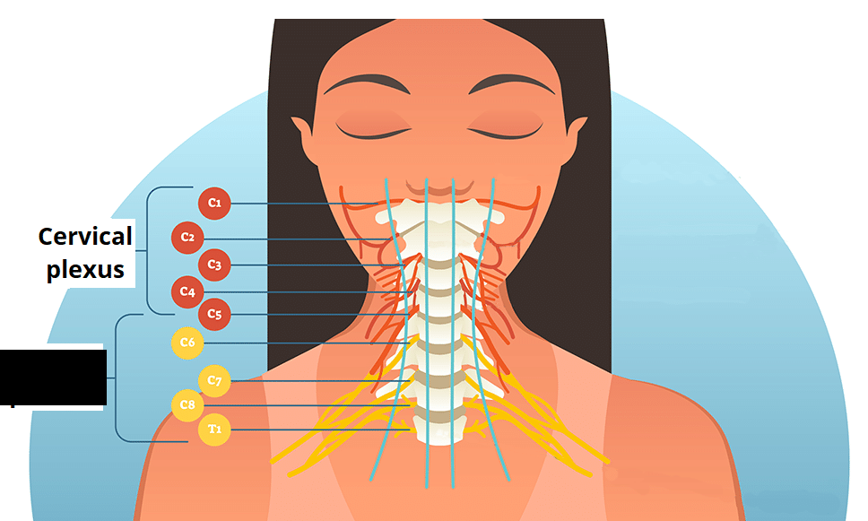
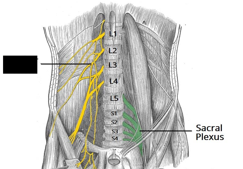
Lumbar Plexus
Consists of the ventral rami from spinal nerves L1-L4. It provides motor and sensory innervation to the lower abdomen, pelvis, and most of the front of the legs
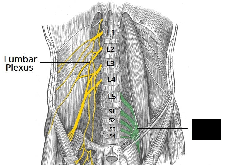
Sacral Plexus
Consists of the ventral rami from spinal nerves L4-S4. It provides motor and sensory function to the pelvis, perineum, buttocks, thighs, and feet.
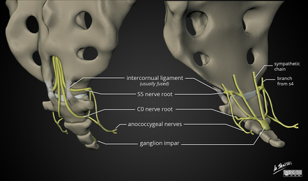
coccygeal plexus
a small network of nerves located on the pelvic floor. It supplies sensory and motor innervation to the tailbone, surrounding muscles, and nearby skin. Formed by the anterior (ventral) rami of the 4th and 5th sacral nerves (S4,S5 ) and the coccygeal nerve.
-60 -70
The resting membrane potential of a neuron is the electrical voltage difference across its cell membrane when it is not actively sending signals, typically ranging between ___ and ___ Mv.
6 4 2
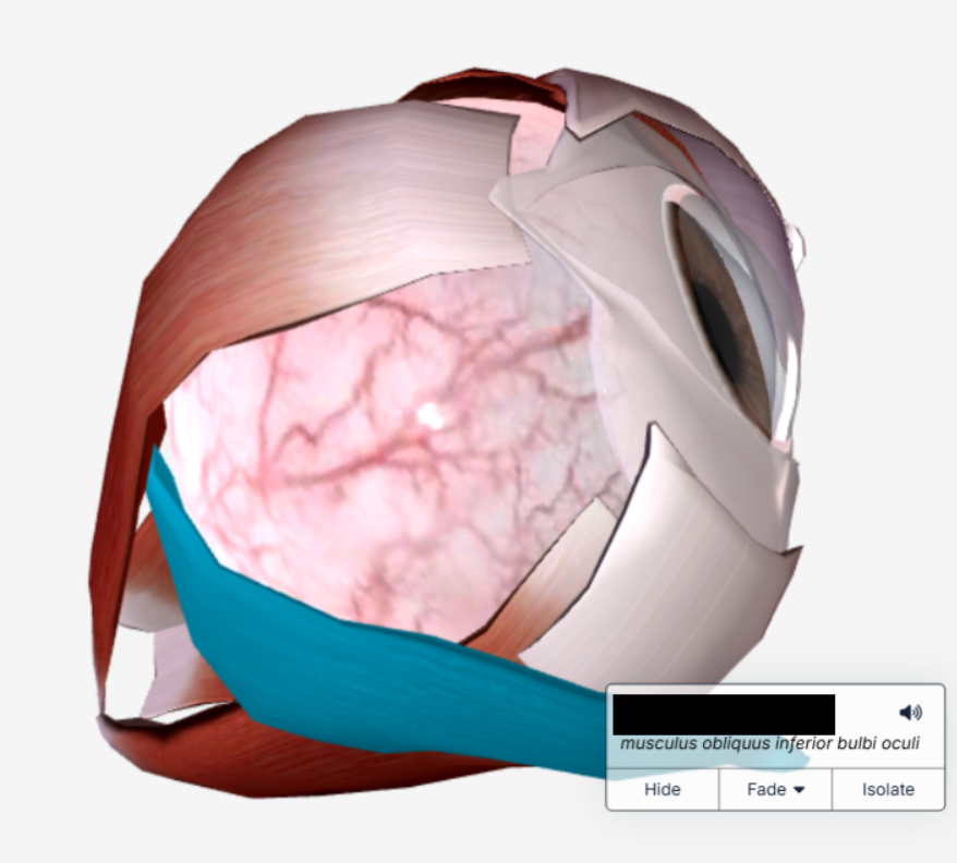
There are ___ extrinsic eye muscles that move the eyeball. There are ___ rectus muscles (superior, inferior, medial, lateral) and ___ obliques (the superior and inferior oblique)
Inferior Oblique
The _____ _____ extends from the medial wall of the orbit of the eye to its attachment on the inferolateral aspect of the eyeball.
Outer fibrous tunic
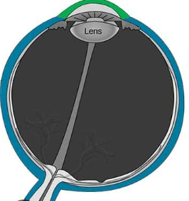
The eye layer/tunic that consists of the cornea and sclera. The sclera (white portion) consists of dense connective tissue containing blood vessels and nerves. The cornea is the transparent portion of the anterior aspect of the eye.
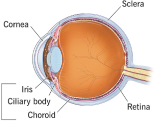
Middle vascular tunic
The eye layer/tunic that consists of the choroid coat, ciliary body, and iris. Also called the uvea because of its similarity to a peeled grape.
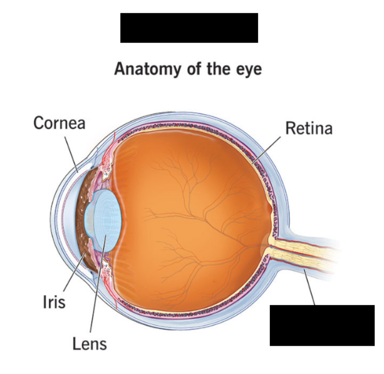
Inner tunic
The eye layer/tunic that consists of the retina and a portion of the optic nerve
optic disc ora serrata
The retina attaches to the eye at only two points: the _____ ____ and the ___ _______.
Optic Nerve
Lies medial to the fovea centralis. Nerve fibers from the eye converge and exit the eye at the optic disc.
Pupillary constrictor
The ________ __________muscle narrows the pupil to decrease the amount of light entering the eye. This muscle is innervated by the parasympathetic nervous system.
Superior Oblique
The _____ _____ attaches to the medial surface of orbit. Its tendon passes through a trochlea.
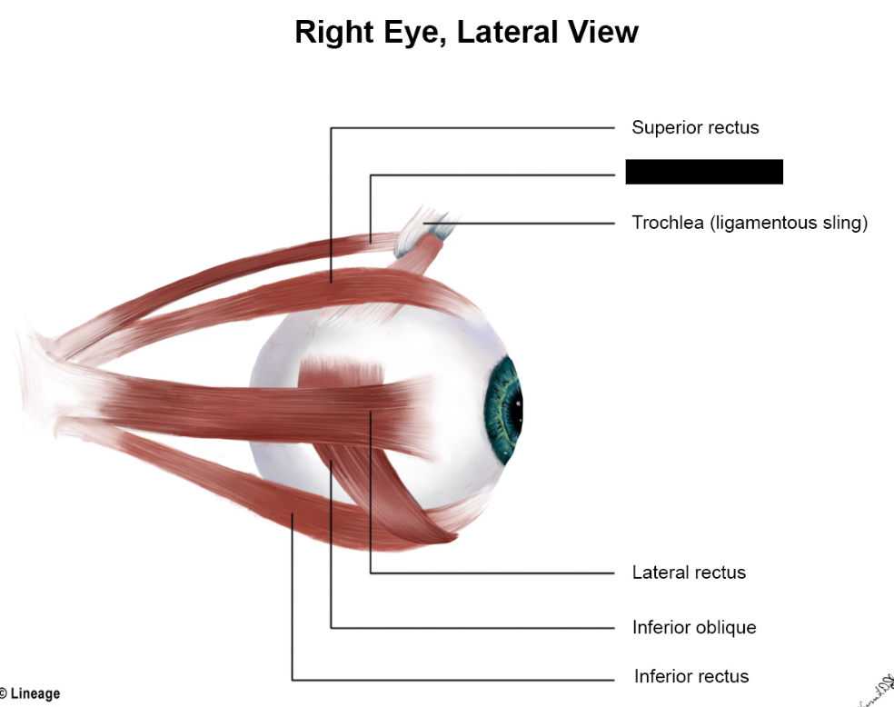
oculomotor
What cranial nerve innervates the superior rectus, inferior rectus, medial rectus, and inferior oblique?
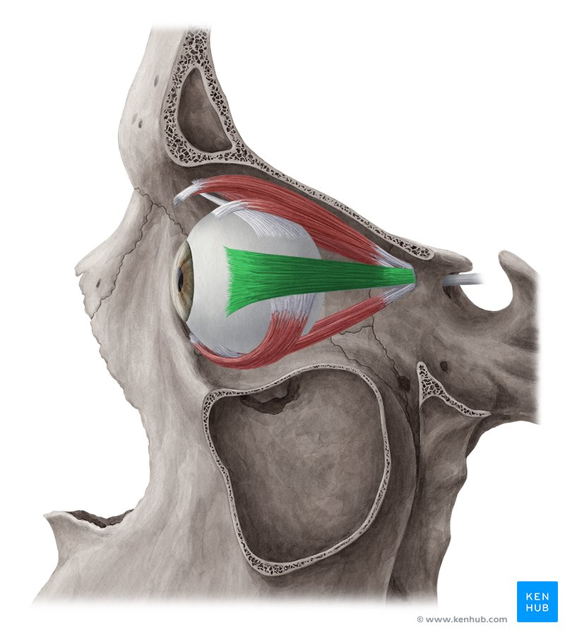
abducens
What cranial nerve innervates the superior oblique?
trochlear
What cranial nerve innervates the lateral rectus?
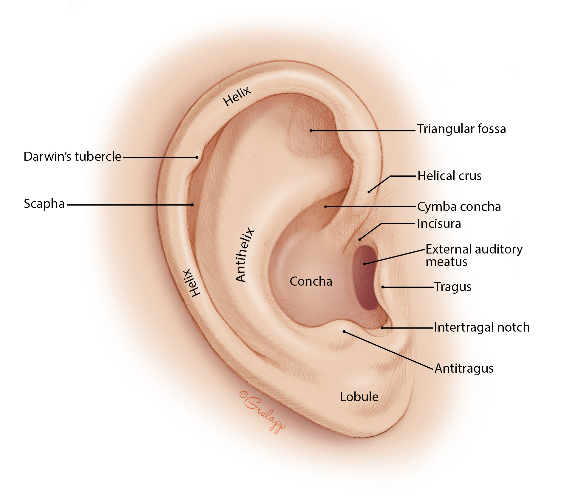
External ear
The section of the ear that consists of the auricle (pinna) and the external auditory meatus (canal).
The auricle is the outer portion of the ear, consisting of elastic cartilage covered by skin. Its oval rim is called the helix, and the earlobe is also known as the lobule.
The external auditory meatus is a canal that extends from the outside to the tympanic membrane. It is lined with skin containing sebaceous glands, hair, and ceruminous glands that secrete a waxy substance called cerumen (earwax). Cerumen traps foreign particles and helps to protect the canal.
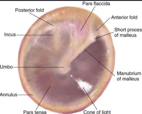
Tympanic membrane
the boundary between the external and middle ear. It consists of a thin layer of connective tissue. It has a layer of skin on its external surface and a mucous membrane on its internal surface. It is slightly cone-shaped with the apex pointing toward the middle ear. Also reffered to as the eardrum.
Middle ear
Resides in a hollow chamber called the tympanic cavity. The cavity is lined with a mucous membrane. The cavity contains a canal called the Eustachian tube (pharyngotympanic tube) that connects with the nasopharynx. The tube is normally closed but opens with chewing, yawning, or swallowing. The tube opens briefly to equalize pressure between the tympanic cavity and the outside. Changes in pressure can disrupt the vibrations of the tympanic membrane and produce muffled sounds.
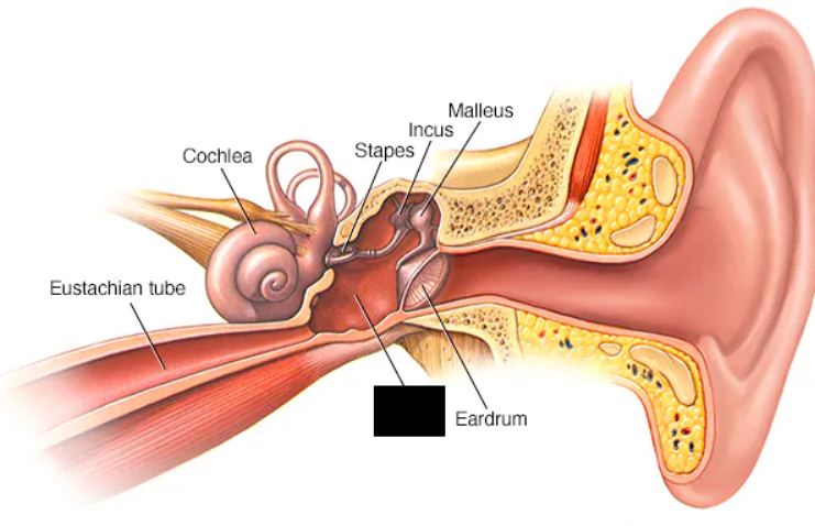
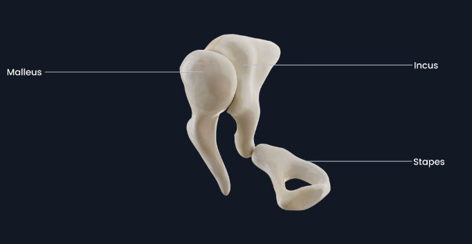
Auditory ossicles
Three small bones that transmit vibrations from the tympanic membrane to the oval window of the inner ear. These are the malleus, incus, and stapes (hammer, anvil, and stirrup).
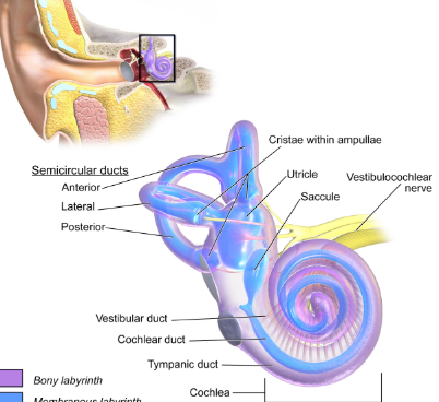
Inner ear
resides within a cavity inside the temporal bone. It consists of the cochlea, vestibule, and semicircular canals. Sometimes referred to as the bony labyrinth. On the inside esides a membranous labyrinth that contains fluid.
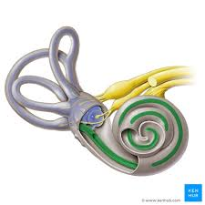
Cochlear Duct
The innerost chamber of the cochlea, and contains the corti organ (spiral organ) which senses hearing.
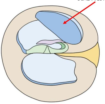
Scala Vestibule
Chamber of the cochlea that is superior to the cochlear duct.
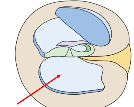
Scala Tympani
Chamber of the cochlea that is inferior to the cochlear duct.
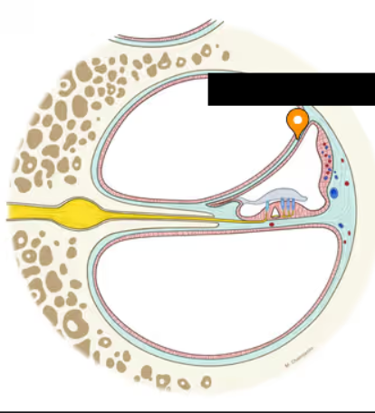
Vestibular Membrane
A thin, fluid-secreting membrane that produces a fluid called endolymph. Located on the superior aspect of the cochlear duct.
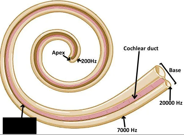
Basilar Membrane
Membrane located on the inferior aspect of the cochlear duct, important in producing hearing.
Mechanoreceptors
Receptors that sense mechanical deformation of tissue
Sensory receptors
______ _________ collect information in various forms from the environment, and convert them to electrochemical impulses (action potentials) for processing by the CNS.
Chemoreceptors
Receptors that sense changes in chemical concentration
Pain Receptors
Also called nociceptors; receptors that sense tissue damage
Thermoreceptors
Receptors that sense changes in temperature.