Morph Corr Nuc Med 2 (ALL EXAM ANSWERS FOR FINAL)
 Call Kai
 Learn
Learn Practice Test
 Spaced Repetition
 Match
Match Flashcards
 Knowt Play
1/190

There's no tags or description
Looks like no tags are added yet.
Name | Mastery | Learn | Test | Matching | Spaced | Call with Kai |
|---|
No analytics yet
Send a link to your students to track their progress
191 Terms
Both non-blood brain barrier (non-BBB) crossing and blood brain barrier (BBB) crossing radiopharmaceuticals can be used for diagnosing brain death. Which class of tracers are considered to be the most sensitive and accurate for brain death?
neither, both are equally as good
the BBB crossing
the non-BBB crossing
neither, amyloid labeling agents should be used
the BBB crossing
What are the main differences between HMPAO and ECD?
ECD clears the blood stream quicker and can be imaged sooner after injection
HMPAO is a more stable tracer and has a longer shelp life after reconstituting
ECD shows both white and grey matter where as HMPAO mainly shows Grey matter
HMPAO is considered to be better in diagnosing changes in metabolic activity
ECD clears the blood stream quicker and can be imaged sooner after injection
When performing a brain death study using a BBB crossing radiopharmaceutical, the main sign of brain death is:
absence of uptake in the brain
visualized superior sagital sinus without seeing the brain stem
increase in the activity flowing into the circle of Willis from the internal carotid arteries
a large area of uptake where the BBB has been disrupted
Absence of uptake in the brain
The purpose of placing an elastic band (tourniquet) around a patient's head just above the orbits during a radionuclide angiogram (primarily for brain death evaluation) is:
To decrease the activity from the orbits
To increase intracranial pressure
To immobilize the patient
To decrease blood supply to small scalp vessels
To decrease blood supply to small scalp vessels
The venous phase of a cerebral blood flow study is signaled by:
Visualization of the jugular veins
Appearance of the radiopharmaceutical in the superior sagittal sinus
Disappearance of radiopharmaceutical from the carotid arteries
Appearance of radiopharmaceutical in the middle cerebral arteries
Appearance of the radiopharmaceutical in the superior sagittal sinus
The localization of 99mTc HMPAO is related to:
Cerebral blood flow
A breakdown of the blood-brain barrier
Glucose metabolism
Distribution of neuroreceptors
Cerebral blood flow
Which of the following is true regarding injection of the radiopharmaceuticals for SPECT brain imaging?
Family members should be allowed to talk to the patient during injection to keep them calm.
The injection should be made immediately after venous puncture to avoid clot formation.
Lights should be dimmed (or dark) during injection.
The patient should be encouraged to read or watch TV during injection to take his or her mind off any pain.
Lights should be dimmed (or dark) during injection.
Advantages of using ECD over HMPAO for SPECT imaging of the brain include:
More rapid clearance from the bloodstream
Better target to background ratio
All of these are correct
Can be injected up to 6 h after preparation
All of these are correct
Ictal SPECT and PET studies require an injection of radiopharmaceutical:
During a seizure
Immediately after a seizure
All of these answers are correct
Between seizures
During a seizure
When using F-18 FDG to evaluate a patient with epilepsy, which of the following statements are true:
If injected when the seizure starts, the epileptic foci will be cold
If injected immediately after the seizure finishes, the epileptic foci will be hot
If injected when the seizure starts, the epileptic foci will be hot
if injected immediately before, during, or after the seizure stops, the entire brain will have increased uptake
If injected when the seizure starts, the epileptic foci will be hot
In a patient with a parkinsonian syndrome, the DaTscan does not deposit in the caudate nucleus and the putamen as it should. It is said that the images show a period sign instead of a comma sign.
True
False
True
To prepare a patient for DaTscan study, which of the following is correct?
NPO overnight or at least 4 hours
Thyroid blockage by SSKI prior to the study
Injection must occur in a dimly lit or dark room
No Caffeine
Thyroid blockage by SSKI prior to the study
For DaTscan study, which of the following is correct?
It detects beta-amyloid deposition.
It detects brain infarction.
It detects brain neurofibrillary tangles.
It detects brain presynaptic dopamine transporter.
It detects brain presynaptic dopamine transporter.
To perform a DaTscan® study, which of the following is correct?
Imaging can be obtained 3-6 h after tracer injection.
A cereberal blood flow image should be obtained at the time of injection.
The camera shouldn't be brought too close to the patients head to avoid discomfort
Patients must remain quiet (no talking) and avoid moving to avoid uptake in the motor cortex
Imaging can be obtained 3-6 h after tracer injection.
The primary purpose of I-123 Ioflupane brain imaging is
find epileptic foci
diagnose Alzheimer's disease
to differentate parkinsonian disease from essential tremors
detect primary brain malignancy
to differentate parkinsonian disease from essential tremors
For Amyvid PET study, which of the following is correct?
none of these answers are correct
Arms should be above shoulders during imaging.
Blood sugar <200 mg/dl.
NPO for 4 h before the study is needed.
Imaging can be started as soon as 30 to 50 min after tracer injection.
Imaging can be started as soon as 30 to 50 min after tracer injection.
When using F-18 Amyloid imaging agents, which of the following is correct?
They detect brain Lewy bodies
They detect brain beta-amyloid deposition
They detect brain neurofibrillary tangles
They detect brain atrophy
They detect brain ischemia
They detect brain beta-amyloid deposition
Amyloid PET imaging is most helpful in patients with which of the following conditions:
Clinically diagnosed Parkinson's disease, to confirm with imaging findings
Clinically diagnosed Alzheimer's disease, to confirm with imaging findings
Suspected Alzheimer's disease but without definite diagnosis
Suspected Parkinson's disease but without definite diagnosis
Suspected Alzheimer's disease but without definite diagnosis
Amyloid PET is indicated in patients with the following conditions except:
Clinically progressive dementia with an early onset (65 years or less in age)
Persistent or progressive MCI with uncertain clinical diagnosis
Diagnosed Alzheimer’s disease, to determine the severity of dementia
Clinically suspected possible Alzheimer’s disease but with atypical clinical course
Diagnosed Alzheimer's disease, to determine the severity of dementia
Until recently, the biggest challenge with doing beta amyloid imaging for Alzheimer's disease was:
The exams were not positive until there was significant cognitive impairment
The sensitivity for diagnosing the disease is very low
There was little to no payment available because of a lack of treatment for the disease
Other tests, like MRI and cognitive exams were more accurate in diagnosing the disease
There was little to no payment available because of a lack of treatment for the disease
Which of the following statements is true when performing a Cerebral Spinal Fluid (CSF) scan
Immediately after the injection, the patient should sit up to allow for normal spinal fluid flow
The pharmaceutical of choice is In-111 Oxime
Since you are using In-111 you will have best results if you acquire the image using a PET scanner
The injection should be performed intrathecally by a physician between L3-L4
The injection should be performed intrathecally by a physician between L3-L4
Which of the following statements is incorrect (false)when performing a Cerebral Spinal Fluid (CSF) scan
At 6 hours after the injection you should see a Vikings Helmet sign
On the immediate post injection image you should see the isotope beginning to spread out in the spinal column
Imaging should occur at 2 hours, 6 hours, 24 hours, and optionally 48 hours after the injection
At 24 hours after the injection you should see a tulip sign
At 24 hours after the injection you should see a tulip sign
In patients who have suffered head trauma, they may be evaluated for a CSF leak. Although imaging may demonstrate this, pledgets (cotton fabric balls) are the preferred method. How are they used?
After patients are injected for a CSF study, they are taped to the injection site to see if any tracer is leaking out
After patients are injected for a CSF study, they are inserted in the patient's nasal cavity and ears to see if they become radioactive
After patients are injected for a CSF study, the pledgets are coated with the patients blood to see if there is radioactivity in their bloodstream
After patients are injected for a CSF study, the pledgets are placed in the patients mouth to see if their oral cavity becomes radioactive
After patients are injected for a CSF study, they are inserted in the patient's nasal cavity and ears to see if they become radioactive
What is the purpose of a CSF shunt study?
To make sure that the shunt is removing the excess CSF from the patients brain
To quantify the amount of CSF that is being produced by the brain
To make sure that the CSF is not being shunted to the wrong area of the body
To confirm the correct placement of the shunt in the brain's ventricles while surgery is being performed
To make sure that the shunt is removing the excess CSF from the patients brain
Why is 111In DTPA preferred over 99mTc DTPA for cisternography in adults?
Because it has a superior biodistribution
Because it has a longer half-life allowing delayed imaging
Because it has lower-energy photons
Because it has two photopeaks
Because it has a longer half-life allowing delayed imaging
The injection for a CSF shunt patency study is:
Intrathecal
Intravenous
Into the shunt reservoir or tubing
Subcutaneous
Into the shunt reservoir or tubing
START OF TEST TWO (GI)
50 Questions (Spring 2026)
The patient preparation for a gastric emptying study is __________.
that the patient must void before imaging
NPO for 4 - 8 hours
the patient should have a barium enema within the 24 hours before this exam
no patient preparation is required
NPO for 4 - 8 hours
In performing a gastric-emptying study, imaging should begin ___________.
immediately after meal consumption, and continue for at least 2 to 4 hours
immediately after meal consumption, and continue for 1 hours
15 minutes after meal consumption and continue for 1 hours
1 hour after meal consumption then once an hour for 6 hours
immediately after meal consumption, and continue for at least 2 to 4 hours
When performing a gastric emptying study, what is the purpose of giving Reglan (Metoclopramide)?
this stimulates the production of bile to increase the rate of digestion
this increases the amount of acid production to prevent an obstruction from forming out of the food
this increases the rate of gastric emptying in attempt to determine if the the gastroparesis is treatable
this drug is used for gall bladder imaging not gastric emptying
This increases the rate of gastric emptying in attempt to determine if the the gastroparesis is treatable
When administering Reglan (Metoclopramide) what special precautions should be taken?
there are no precaustions and this drug has no known side effects
do not quickly bolus inject the pharmaceutical because it can lead to a rapid drop in blood pressure
have a antidote pharmaceutical available in case the drug causes respiratory arrest
do not administer this drug until after the patient has received an injection of CCK (Kinevac)
Do not quickly bolus inject the pharmaceutical because it can lead to a rapid drop in blood pressure
When performing a gastric emptying study with a solid meal, what is considered to be the minimum normal range?
30% of the meal should have left the stomach by 1 hours after eating the meal
95% of the meal should have left the stomach by 2 hours after eating the meal
50% of the meal should have left the stomach by 3 hours after eating the meal
60% of the meal should have left the stomach by 2 hours after eating the meal
60% of the meal should have left the stomach by 2 hours after eating the meal
Which of the following is the reason it is important to have a standardized meal size and composition in a gastric-emptying scan?
It can affect the radiation dose to the patient
It can cause gastric tissue necrosis
It can affect the ability to have gastric emptying
It can affect the rate of gastric emptying
It can affect the rate of gastric emptying
A gastric-emptying study uses which of the following radiopharmaceuticals?
Tc-99m sulfur colloid
Tc-99m Macroaggregated Albumin
Tc-99m medronate
Tc-99m disida
Tc-99m sulfur colloid
When performing a gastric empty scan with a dual head camera, the most accurate measurement of emptying can be calculated by:
average the counts in the anterior and posterior views
divide the counts measured in the anterior views by the counts measured in the posterior views
just image from the anterior view since the posterior view is to far away from the stomach
performing a calculation of the geometric mean of the anterior and posterio views
performing a calculation of the geometric mean of the anterior and posterio views
A technologist decided to use a single-head camera in the anterior position for a normal solid-phase gastric emptying study. For the first 10 minutes, the counts will __________.
remain unchanged
decrease linearly
increase
decline exponentially
increase
Eggs are preferred over semi-solid meals like oatmeal because ___________.
it is more cost-effective and less likely to spoil
the exam is more reproducible and it has a higher binding percentage and is less likely to disintegrate in gastric fluid
None of the above
it has a higher fat content and results in a stronger stomache contraction
The exam is more reproducible and it has a higher binding percentage and is less likely to disintegrate in gastric fluid
The advantage of performing gastric emptying with a solid instead of a liquid is __________.
solids are emptied faster
the labeling procedure is easier (faster)
the patient does not need to be fasting
imaging with solids is more sensitive and accurate for abnormalities
Imaging with solids is more sensitive and accurate for abnormalities
Images acquired during a gastric-emptying study are primarily used for __________.
determination of an effective gastric plasma flow
determining when the tracer first exits the stomach
quantifying the rate at which the tracer is leaving the stomach
detecting the presence of Heliobacter-Pylori infection
quantifying the rate at which the tracer is leaving the stomach
A normal esophageal transit study result is __________.
more than 90% has traversed the esophagus by the end of 15 seconds
small localized hot spots throughout the esophagus
no activity reaching the esophagus
a clearance half-time of 4-6 minutes
more than 90% has traversed the esophagus by the end of 15 seconds
Which imaging study is acquired while an abdominal binder (resembling a large blood pressure cuff) is inflated to increasing pressures?
Gastrointestinal bleeding
Esophageal reflux study
Esophageal transit study
Gastric emptying study
Esophageal reflux study
In infants, 24-hour images are sometimes performed over what area to demonstrate gastroesophageal reflux?
Lung Fields
Upper small intestine
Spleen
Lower esophagus
Lung Fields
What would be considered a normal result for an esophageal reflux study in an adult?
no activity in the stomach
significant activity in the esophagus only after 50 mm of pressure
no activity in the esophagus
significant activity in the esophagus within 5 minutes of administration
no activity in the esophagus
In assessing gastric reflux of a communicative non-pediatric patient, ROIs should be placed over the __________.
esophagus and stomach
esophagus, small bowel, stomach, and liver/gallbladder
esophagus, small bowel, and stomach
esophagus and lungs
esophagus and stomach
What procedure should you follow in performing the infant equivalent of an adult esophageal reflux study?
image the lungs
reduce the pressure on the abdomen by about one-half
inject reduced activity according to the size of the child
image sooner than in adults
image the lungs
Patient preparation for a Meckel's study on an infant includes ___________.
written informed consent from the patient receiving the exam must be obtained
perform a barium study in conjunction with this study
CCK administration 15 minutes before starting the exam
NPO for 6 - 12 hours and consider administering ranatidine or glucagon
NPO for 6 - 12 hours and consider administering ranatidine or glucagon
What patient population is most commonly associated with Meckel's diverticulum scanning?
it is commonly used in all age populations
young adults
children younger than 2 years old
the elderly
children younger than 2 years old
A positive Meckel's diverticulum study demonstrates __________.
diffuse activity by 20 minutes after the injection in the bowel that fades with time
focal area of increased activity in the upper left quadrant that moves with time
unusualy increased activity in the stomach
focal area of increased activity in the right lower quadrant or mid-abdomen
Focal area of increased activity in the right lower quadrant or mid-abdomen
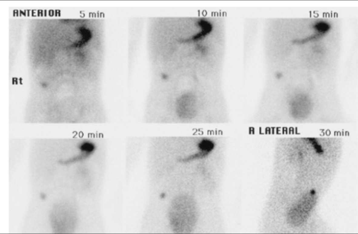
After acquiring the images below on a patient suspected of having a Meckel's diverticulum, the technologist should ___________.
image again in 1 - 2 hours
administer a different radiopharmaceutical and reimage
submit the images for reporting, the exam is complete
check the radiochemical purity
submit the images for reporting, the exam is complete
Localization of Meckel's diverticulum can be accomplished with which of the following radiopharmaceuticals?
Tc-99m sulfur colloid
Tc-99m mebrofenin
Tc-99m pertechnetate
Tc-99m MAA
Tc-99m pertechnetate
When performing Meckel's diverticulum imaging, which of the following would be visualized in a positive study?
Malignant colon tissue
Ectopic stomach mucosa
Hepatic uptake
Ectopic endometrial tissue
Ectopic stomach mucosa
One disadvantage of using sulfur colloid for assessing a GI bleed is __________.
the persistent high body background from circulating Tc S.C. in the blood stream
its lack of sensitivity
it takes a relatively long time to perform the exam
it is only good if the patient is actively bleeding
It is only good if the patient is actively bleeding
To perform a sulfur colloid GI bleed study, __________.
Tc99m Sulfur Colloid can not be used to perform GI Bleed studies
perform a flow study of the abdome
ensure the entire liver and spleen are visualized by placing them in the center of the field of view
do a static image every 10 minutes for 90 - 120 minutes
perform a flow study of the abdome
Which of the following is the initial positioning for visualizing gastrointestinal bleeding with Tc99m labeled red blood cells?
Lower border of liver and spleen at top of field of view and pubic symhasis at the bottom of the field of view
Symphysis pubis (lower pelvis) in middle of field of view
Lower esophagus at top of field of view
Xiphoid (center of the chest) in middle of field of view
Lower border of liver and spleen at top of field of view and pubic symhasis at the bottom of the field of view
When labeling RBCs with Tc-99m, the stannous ions (tin form a cold PYP vial) __________.
is not required to label RBCs and is primarily used for bone imaging.
stabilize the RBCs so that they live longer
allows the Tc-99m to enter the RBCs by oxidizing it
lower the oxidation state of the Tc-99m that enters the cells trapping it inside of the cells
lower the oxidation state of the Tc-99m that enters the cells trapping it inside of the cells
A nuclear medicine GI bleed study is used to __________.
detect the presence of ulcers
quantitate the amount of blood from the gastrointestinal tract
determine if the bowel is obstructed
localize the bleeding site
localize the bleeding site
The advantage of a nuclear medicine GI bleed study over other modalities is it __________.
is able to locate bleeds precisely
it usually takes very little time
is very sensitive for low-volume bleeds
rarely produces false-positive or false-negative results.
is very sensitive for low-volume bleeds
During a GI bleed study, lateral or oblique views are sometimes acquired to:
distinguish between a bleed and a hemangioma
lateral and oblique views are never necessary for this study
quantitate the bleed
more accurately localize the bleed
more accurately localize the bleed
After imaging a GI bleed study with Tc99m labeled RBCs for 60 - 90 minutes, no focus of activity has appeared in the abdomen. The technologist should __________.
administer cimetidene
perform a SPECT study
administer 10 mg IV reglan to increase gastric motility
discontinue the study or obtain delayed images of intervals from 2-6 hours and/or 18-24 hours
discontinue the study or obtain delayed images of intervals from 2-6 hours and/or 18-24 hours
Which radiopharmaceutical is commonly used to image cavernous hemangioma?
Tc-99m sulfur colloid
Tc-99m RBCs
Tc-99m albumin colloid
Tc-99m pertechnetate
Tc-99m RBCs
A Tc99m labeled RBC liver scan demonstrates normal blood flow image and a focally increased blood pool on the delayed images. The most likely explanation for these findings is a(n):
cavernous hemangioma
abscess
focal nodular hyperplasia (FNH)
cavernous hemangioma
What is the preferred radiopharmacutical used when imaging spleen tissue?
In111 White Blood Cells
Tc99m Sulfur Colloid
Tc99m Heat treated Red Blood Cells
Tc99m Labeled Red Blood Cells
Tc99m Heat treated Red Blood Cells
What is the normal biodistribution of sulfur colloid?
85% liver, 10% spleen, 5% bone marrow
30% liver, 60% spleen, 5% bone marrow, 5% lung
60% liver, 30% spleen, 5% bone marrow, 5% lung
70% spleen, 20% Liver, 10% bone marrow
85% liver, 10% spleen, 5% bone marrow
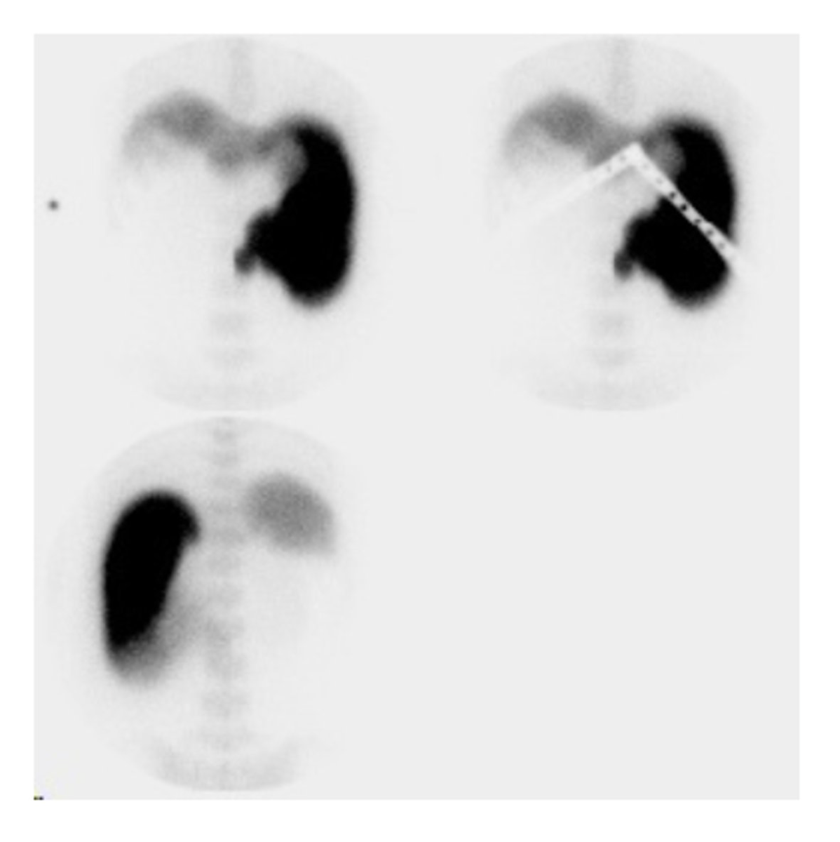
What best describes the liver/spleen images below?
Top 2 images are from the anterior and the bottom image is from the posterior
This study shows colloidal shift with decreased liver function
The patient has abnormal stomach uptake
This is a normal appearing study
This study shows an infarcted spleen
This study shows colloidal shift with decreased liver function
What appearance is most common with the presence of metastasis on a liver/spleen sulfur colloid scan?
multiple hot spots
increased flow to the entire lobe containing the affected area
hepatomegaly
multiple cold spots
multiple cold spots
You have a patient whom you have just performed a liver spleen scan on but no spleen was visualized even though their spleen is visible on the CT scan. When you perform a spleen scan, again no spleen is visualized. What is this most likely cause of this anomaly?
the patient has a spleen but uptake of the pharmaceutical is being blocked by the contrast used for the CT scan
the patient does not have a spleen and the CT scan was read in error
the patient has a spleen but it is not functioning
the patient most likely has a Meckels diverticulitis
the patient has a spleen but it is not functioning
In SPECT liver imaging with Tc-99m sulfur colloid, how long after the injection should liver imaging typically begin?
30 minutes - 1 hour
3 - 4 hours
10 to 20 minutes
1 - 2 hours
10 to 20 minutes
What is the major advantage of performing a SPECT liver/spleen study?
decrease in imaging time
improved sensitivity
decrease the dose of radiopharmaceutical
improved spatial resolution
improved sensitivity
Typical appearance(s) of hepatitis on a liver/spleen sulfur colloid scan include(s) ___________.
hepatomegaly and a homogeneous increased of activity in the liver, and increased visualization of the spleen
hepatomegaly, increased activity in the spleen, and decreased activity in the liver
homogeneous increased activity in the liver with normal uptake in the spleen
hepatomegaly and a decrease in activity in both the liver and the spleen
hepatomegaly, increased activity in the spleen, and decreased activity in the liver
Which of the following should NOT be performed immediately before liver/spleen imaging with Tc-99m sulfur colloid?
Radiographic upper GI series using barium
CT of the abdomen using iodinated contrast
Gallbladder examination with ultrasound
Thyroid uptake with I-123 sodium iodide 24 hours earlier
Radiographic upper GI series using barium
A liver spleen image using Tc-S.C. indicative of a liver abscess will demonstrate which of the following characteristics?
single focal hot spot
multiple blotchy cold spots (Swiss cheese) with significant colloidal shift
single (possible multiple) focal cold spots usually without colloidal shift
multiple blotchy hot spots
single (possible multiple) focal cold spots usually without colloidal shift
Sulfur colloid uptake in the lungs usually means ___________.
there is increased lung function
a lung tumor is present
the patient has severe liver disease or the particles injected are too large
the spleen is missing or has been removed
the patient has severe liver disease or the particles injected are too large
Finding >20% of sulfur colloid activity administered, depositing in the bone marrow may indicate which of the following?
Acute avascular necrosis
Particles are too small or the patient has severe liver disease
Invasive metastatic disease in the bone marrow
Anemia
Particles are too small or the patient has severe liver disease
LeVeen shunts are placed to relieve _________.
hepatomegaly
bronchitis
reflux
ascites
ascites
The patency of a LeVeen shunt may be demonstrated with which of the following radiopharmaceuticals?
Tc-99m pertechnetate or Tc-99m labeled red blood cells
Tc-99m MAA or Tc-99m sulfur colloid
Tc-99m HDP or Tc-99m MDP
Tc-99m mebrofenin or Tc-99m disofenin
Tc-99m MAA or Tc-99m sulfur colloid
When imaging to determine LeVeen shunt patency, the camera initially is positioned anteriorly over the abdomen, and delayed images can be acquired __________.
over the gastrointestinal tract
over the brain
over the bladder
over the lung or liver fields (depending on the radiopharmaceutical chosen)
Over the lung or liver fields (depending on the radiopharmaceutical chosen)
You have a patient that has just been diagnosed with multiple small masses in their abdomen on a CT scan. They are 5 years post motor vehicle accident (MVA) and surgical removal of their spleen and you really suspect that this is remnant spleen tissue. How would you best determine (least invasive, simple, cheap, accurate) if the masses are remnant spleen tissue or some form of malignancy.
In other words choose the best option even though multiple options are somewhat correct:
Do a spleen scan with denatured red blood cells
Do a liver spleen scan with sulfur colloid
Do a whole body bone scan looking for metastatic disease
Do a biopsy of multiple nodules
Do a spleen scan with denatured red blood cells
START EXAM THREE (CARDIAC)
CARDIAC TEST SPRING 2026 42 questions
Myocardial perfusion imaging is primarily performed to:
determine ejection fraction
record the rate of delivery of a bolus injection
detect the location and extent of ischemia
evaluate wall motion of the ventricles
Detect the location and extent of ischemia
When performing a SPECT myocardial perfusion exam, the best imaging acquisition would be:
A 360 degree acquisition around the patient
A 180 degree acquisition around the patient starting at the anterior view and ending on the posterior view while moving around the patients left side
A 180 degree acquisition around the patient starting at the anterior view and ending on the posterior view while moving around the patients right side
A 180 degree acquisition around the patient starting at the right anterior oblique view and ending on the left posterior oblique view while moving around the patients left side
A 180 degree acquisition around the patient starting at the right anterior oblique view and ending on the left posterior oblique view while moving around the patients left side
Calculate the target heart rate when performing a Bruce protocol stress test on a 53-year-old male.
115 BPM
125 BPM
136 BPM
142 BPM
142 BPM
For myocardial SPECT imaging, the technologist will typically position the patient:
supine with the left arm above the head
on their right side with the left arm above the head
on their left side with the right arm above the head
prone with the right arm above the head
Supine with the left arm above the head
In the 1 day protocol for myocardial perfusion imaging performed with a 99mTc-labeled tracer, the larger amount of activity is administered __________.
for the rest study
for the study performed second
for the study performed first
for the stress study
For the study performed second
An exercise/treadmill cardiac stress test requires the placement of how many electrodes on the patient's body?
6
10
12
15
10
The inferior wall of the heart is predominantly perfused by which artery?
Left Main Artery
Left Anterior Descending
Left Circumflex
Right Coronary Artery
Right Coronary Artery
When performing a myocardial perfusion exercise stress test, ideally the radiopharmaceutical should be injected at:
the beginning of stress
85% of anticipated maximum stress and the exercise continued for 1 to 2 minutes
peak stress and exercise terminated immediately
once peak stress has been achieved and then waiting 2-3 minutes after the exercise treadmill has been stopped
85% of anticipated maximum stress and the exercise continued for 1 to 2 minutes
Stress-rest myocardial imaging performed with 99mTc-sestamibi or 99mTc-tetrofosmin requires two administrations of the radio-pharmaceutical, a low dose then a higher dose, because the tracer __________.
does not redistribute once it has been taken up by the myocardium
rapidly washes out of the myocardium after administration
has too short a half-life to permit delayed imaging
has a fast biological half-life
Does not redistribute once it has been taken up by the myocardium
When dual-radionuclide myocardial perfusion imaging is performed, which of the following tracers are used for stress and which for rest?
Stress - Tc-99m sestamibi; Rest - Tc-99m Teboroxime
Stress - Tl-201 Thallous chloride; Rest - Tc-99m sestamibi
Stress - Tc-99m sestamibi; Rest - Tl-201 Thallous chloride
Stress - Tc-99m sestamibi; Rest - labeled red blood cells
Stress - Tc-99m sestamibi; Rest - Tl-201 Thallous chloride
Treatment of the adverse effects induced by dipyridamole (persantine) involves the administration of __________.
acetazolamide
adenosine
aminophylline
Esmolol
Aminophylline
Which of the following drugs will reverse the effects of adenosine (adenocard)?
Aminophylline
Triptophyline
Furosemide
Metoclopramide hydrochloride
Breviblock
Aminophylline
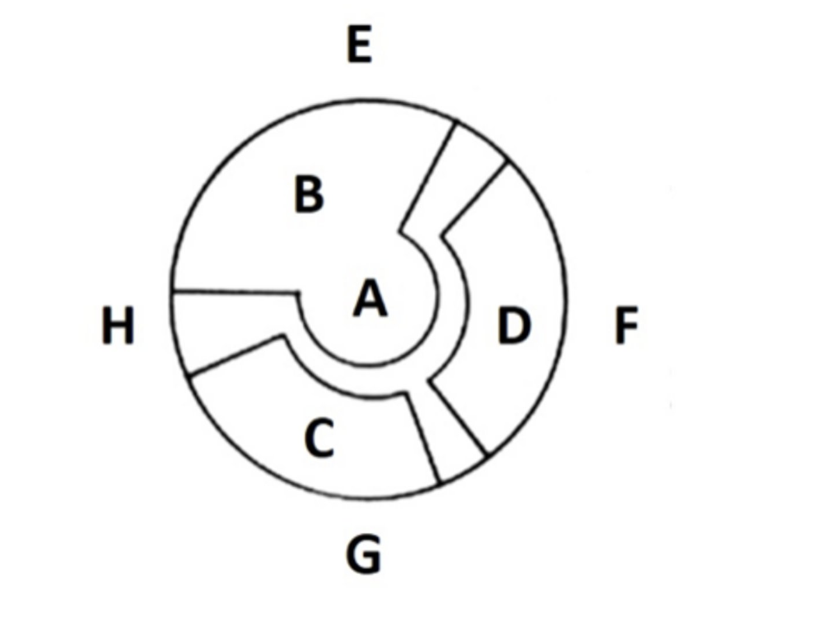
Which coronary artery primarily supplies region "D" ?
a. Left Anterior Descending (LAD) artery
b. Right Coronary Artery (RCA)
c. Left Circuflex (LCx) artery
d. Obtuse Marginal (OM) artery
Left Circuflex (LCx) artery
A patient with a history of bronchospastic disease (asthma) and wheezing arrives for a pharmacological myocardial stress test. The technologist will most likely perform the study using:
exercise stress
dipyridamole
adenosine
dobutamine
dobutamine
The advantage Regadenosine has over Adenosine and Dipyridamole is:
a. It has no side effects
b. It increases the amount of vasodilation in comparison to the other agents
c. It has a decreased risk of respiratory attacks because it only targets A2a receptors
d. It is not effected by methylxanthines
It has a decreased risk of respiratory attacks because it only targets A2a receptors
After stress myocardial imaging with 201Thallous chloride, a second dose of 1-3 mCi of 201Thallous chloride may be administered after the stress images and before the rest myocardial images to __________.
improve patient throughput
demonstrate reversible ischemia more readily or detect stunned/hybernating myocardial tissue
demonstrate infarct size more precisely
minimize visualization of attenuation artifacts
demonstrate reversible ischemia more readily or detect stunned/hybernating myocardial tissue
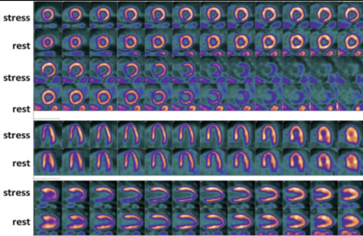
The image below best demonstrates
a. A normal myocardial perfusion study
b. a narrowed left anterior descending artery
c. a narrowed right coronary artery
d. a 100% occluded / blocked left anterior descending artery
a narrowed right coronary artery
Which of the following must be discontinued for at least 24-36 hours before the administration of dipyridamole?
Iodine containing foods
Insulin
Caffeine
Blood Pressure Meds (ACE Inhibitors)
Caffeine
To help clear the liver and bowel when performing the imaging of the stress portion of a myocardial perfusion study with 99mTc-sestamibi, a technologist should give the patient which of the following before imaging?
Fatty meal
Dobutamine
Aminophylline
CCK
Fatty meal
Each of the following is a (relative or absolute) contraindication for adenosine stress testing EXCEPT:
a. recent myocardial infarction (< 4 days)
b. hypertension ( > 160mm systolic)
c. hypotension (< 100mm systolic)
d. allergy to adenosine or aminophylline
e. severe COPD and asthma
Hypertension ( > 160mm systolic)
What is the reason for performing a 4 hour delayed redistribution thallium image
Must wait for the heart rate to return to normal
Because ischemic tissue redistribute faster than normal tissue
4 hours must be waited to allow pulmonary uptake to deminish
Because the tracer does not remain fixed in the myocardium, by 4 hours most of the redistribution has taken place showing viable tissue.
Because the tracer does not remain fixed in the myocardium, by 4 hours most of the redistribution has taken place showing viable tissue.
A bull's-eye display of myocardial perfusion data is constructed by stacking then flattening which reconstructed/reoriented slices?
none, this comes from the 2d anterior planar view
vertical long-axis slices
horizontal long-axis slices
short-axis slices
short-axis slices