Ch. 9 - Muscles
1/24
Name | Mastery | Learn | Test | Matching | Spaced | Call with Kai |
|---|
No analytics yet
Send a link to your students to track their progress
25 Terms
3 Types of muscle tissue (where, functions, striations, voluntary)
Skeletal muscle- striated, voluntary (somatic), on bones of skeleton
Cardiac muscle - striated, involuntary (autonomic), in heart
Smooth muscle - non-striated, involuntary, walls of internal organs
Tissues found in muscle?
Muscle tissue, connective tissue
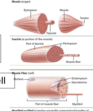
Epimysium, perimysium, endomysium (where, made of?)
Epimysium - out-most wrapping of muscle, CT, continuous with tendon
Perimysium - wraps each fascicle
Endomysium - between/wraps each muscle fiber (cell)
Tendons vs. aponeuroses?
Aponeurosis is a sheet like structure, and covers a broader area
Gross anatomy of skeletal muscle (whole muscle - cell)
Whole muscle - a lot of muscle cells, CT wrapping, blood vessels, nerve fibers (organ)
Fascicle - bundles of muscle fibers, has its own CT sheath
Muscle Fiber - muscle cell, elongated multi-nucleate cell, striated
Structural/functional characteristics of muscle fibers? How are structure specialized for function?
Sarcolemma - plasma membrane
Sarcoplasm - cytoplasm
Myofibrils - 80% of volume, rod-like, sarcomeres for contraction (for contraction and recoil)
Sarcoplasmic reticulum - Smooth ER that stores and releases calcium (relaxed vs. contracting)
T tubules - conduct nerve impulses to all myofibrils (“rapid telegraph system”)
Structure of sacromere? What causes striations? What happens during contraction? Does sarcomere or myofilaments shorten?
Thin filaments - actin (blue), binding sites
Thick filaments - myosin (red), bind to actin and needs energy
Elastic filaments - titin (springs), connects myosin to z-disc
Tropomysoin - string (orange)
Troponin - clamp (yellow)
Striations are caused due to the sarcomere and where the actin and myosin make it thick
The sarcomere shortens, but the myofilaments just overlap
Why doesn’t smooth muscle have striations? Does it have actin and myosin?
It does not have the special arrangement of myosin and actin that the other types of muscle have. It still has both myosin and actin.
Describe the structure of myosin (thick) and actin (thin) - how does their structure relate to their function in sarcomere?
Myosin has heads and tails, heads have ATP and actin binding sites, as they need energy
Actin has troponin and tropomyosin, and myosin binding sites, for contraction block and to connect to myosin
Fascicle
Each bundle in muscle
Cross-bridge
Myosin binding sites
What is resting membrane potential? How does resting membrane potential change during depolarization?
Resting membrane potential is the potential inside the cell versus outside (-70 mV)… it becomes positive during depolarization because Ca+ flood in
Describe how a neural stimulation leads to a muscle contraction.
The definition and role of the neuromuscular junction
The sequence of events at the neuromuscular junction
How a muscle cell action potential is generated
How the electrical signal of a muscle cell action potential is coupled to an increase in the Ca2+ concentration of the sarcoplasm
The structure and function of T tubules and the sarcoplasmic reticulum
How the signal for muscle contraction is shut off
The NMJ is the somatic neuron axon terminal, the synaptic cleft, and the sarcolemma.
Electrical signals arrive at the axon terminal, the Ca+ channels open and Ca+ floord into the terminal, Ca+ enters and signals Ach in vesicles to be released (exocytosis), Ach diffuses across cleft and binds to Ach receptors of sarcolemma, ion channels open and depolarise the membrane potential
T tubules let the signal head towards the SR, so it can release Ca+
Shut off by: Ca+ getting pumped back into SR, troponin lock turns back on, neuron stops firing, and acetylcholinesterase breakes down remained acetylcholine
Describe how troponin, tropomyosin and calcium are used to regulate muscle contractions.
Calcium when released attaches to troponin, which causes the lock to turn off, and the tropomyosin no longer covers the myosin-binding sites of actin
Describe the myofilament cross-bridge cycle, including:
What is the source of chemical energy used to “spring load” the myosin heads?
How are myosin heads released from actin
What is and what causes rigor mortis?
Energy is used to spring load the myosin head. Myosin heads are released from actin using ATP. Rigor mortis happens because there is no ATP, so the myosin heads stay attached since ATP is required to release them.
What is a muscle motor unit? What is the difference between small, medium, and large?
One motor neuron and all the muscle fibers it supplies - small control precise movements (and are attached to less) while large control powerful movements
Describe sequence of events that occur during a muscle twitch.
Latent: no movement, signal speads and calcium is released
Contraction: tension rises/peaks and regulatory proteins are unlocked, sarcomere shortens
Relaxation: tension decreases, calcium is brought back to SR and myosin detaches from actin
Describe what happens when muscles are subjected into multiple stimulations, at low and high frequences:
wave/temporal summation
tetanus (fused and un-fused)
why does the force (tension) of muscle contraction increase with an increase in stimulus frequency?
Wave/temporal: stimulus is applied before muscle fully relaxes, “ride on the wave”
Tetanus (unfused): multiple stimuli with only partial relaxation in between (sustained but wavering)
Tetanus (fused): multiple stimui with no relaxation in between (steady!)
It increases because as more units are stimulated, and repeatedly, the tension will increase
Describe how the force (tension) of muscle contraction is regulated by the recruitment of motor units.
As more units are stimulated/recruited, the tension increases
Which type of motor unit are recruited first?
Small
What is the difference between an isotonic and isometric contraction?
Isotonic involves the muscle shortening to move the load, while isometric does not shorten since tension isn’t enough to move the load (filaments don’t slide)
Describe the three mechanisms a muscle can use to generate ATP, including:
how fast is each
how long can each be useful
advantages/disadvantages
what fuels are used for each
Direct phosphorylation (ATP/creatine phosphate)
stored, ~10 seconds, small duration (weight-lifting, sprinting)
Anaerobic pathway
fast, ~2 minutes, uses glycogen and no oxygen for 2 ATP, short duration (tennis, soccer, short swim)
Aerobic pathway
slow, 1 glucose = 32 ATP, prolonged duration exercises (jogging, marathon), requires oxygen and glucose (could also use fatty/amino acids)
Compare/contrast the three different types of muscle fibers. Include:
speed of contraction
primary pathway for ATP synthesis
myoglobin content (high/low)
color
mitochondria
blood capillaries
what kind of activity each type of muscle fiber is best used for
Slow Oxidative
Slow, aerobic, high, dark red, many, many, endurance and posture
Fast Oxidative
Fast, aerobic (and some anaerobic), high, red, many, many, sprinting/walking
Fast Glycolytic
Fast, anaerobic, low, white, few, few, short term/intense movements
Compare and contrast muscle adaptations in response to aerobic (endurance) and resistance exercise.
Aerobic (endurance)
more capillary density (delivery of oxygen), mitochondria (ATP), and myoglobin (SO fibers, and FOG can change into SO)
Resistance exercise
muscle hypertropy/bigger muscles (more actin/myosin), store more glycogen ((fg fibers)
What is muscle growth due to?
muscle fibers getting bigger in size, not more of them