Physiology Ch.4 Membrane Transport
1/44
There's no tags or description
Looks like no tags are added yet.
Name | Mastery | Learn | Test | Matching | Spaced | Call with Kai |
|---|
No analytics yet
Send a link to your students to track their progress
45 Terms
Body fluid compartments
Subdivided into intracellular and extracellular fluid
- Separated by cell membrane
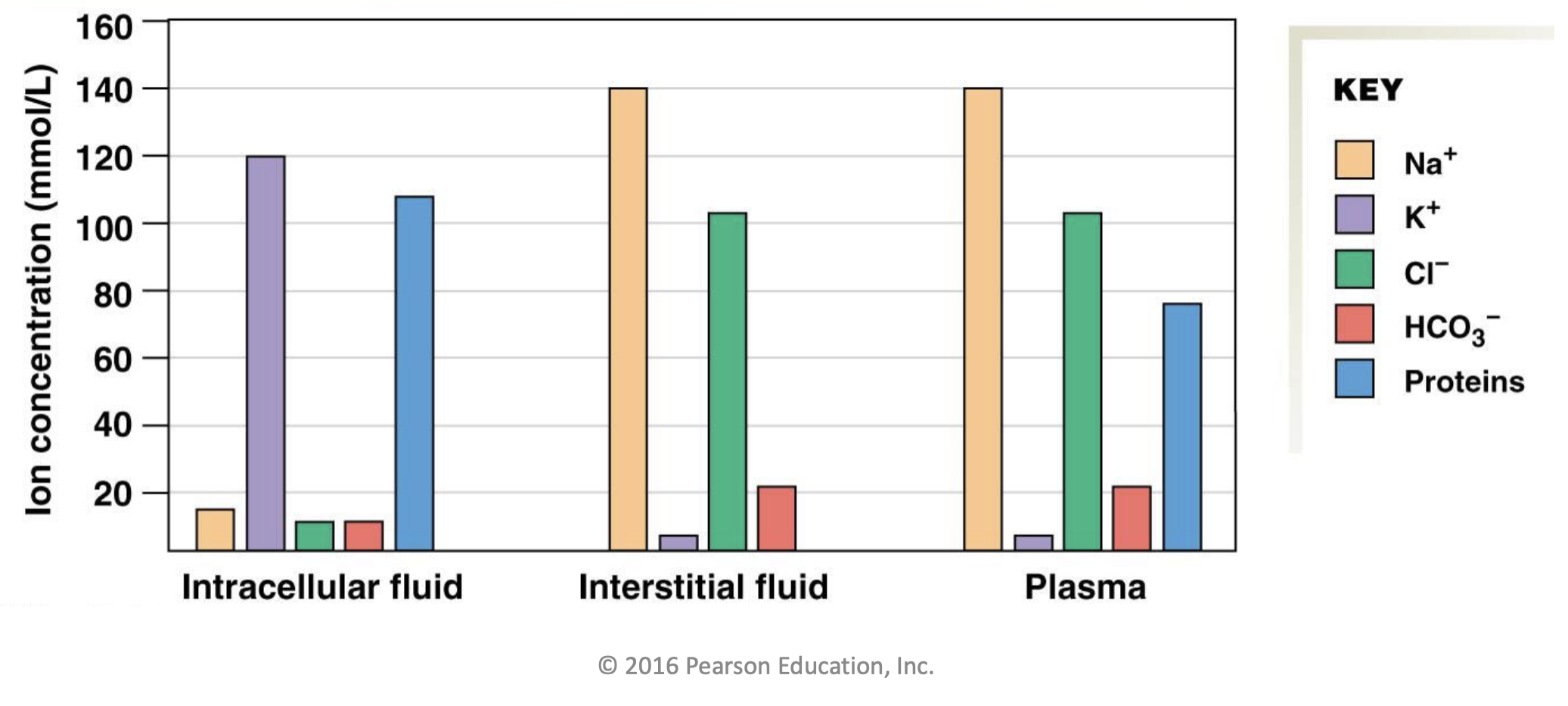
Contains Na+, K+, Cl-, HCO3-, Proteins
Intracellular fluid
Fluid inside of the cells
Majority subdivision of body fluid
- 66% of the body’s fluid
Very low in Na+ and high in K+
- Caused by sodium (Na+) potassium (K+) pump
Very low in Cl2+
- Caused by calcium (Cl2+) pump
Very low in HCO3-
High in proteins
- Caused by protein synthesis in the cells

Extracellular fluid
Fluid on the outsides of the cells, between cells
33% of the body’s fluid
Subdivided into interstitial fluid and plasma
- Separated by leaky exchange epithelial of capillary wall
Regulated by body to being the internal environment back to normal

Interstitial fluid
The majority subdivision of extracellular fluid
- 93% of extracellular volume
Very high in Na+
Very low in K+
High in Cl-
Very low in HCO3-
No protein

Plasm
The minority subdivision of extracellular fluid
- 7% of volume
Very high in Na+ and very low in K+
- Caused by permeability of capillary tissue
High in Cl-
- Caused by permeability of capillary tissue
Very low in HCO3-
Moderate in protein
- Caused by ingestion and digestion of protein
Passive transport
Moves solutes between body fluids without using ATP
- No need for ATP because movement is down the concentration gradient, the way particles want to move naturally
Subdivided into simple and facilitated diffusion Simple
Active transport
Moves solutes between body fluids with the use of ATP
- ATP needed because movement is up the concentration gradient, against the way particles want to move naturally
Subdivided into primary and secondary active transport
Simple diffusion
Type of passive transport
Plasma membrane does not hinder movement of solute in any way
- Solute passes through as if the plasma membrane does not exist
Used for movement of O2, CO2, fatty acids, steroid hormones, thyroid hormones, fat-soluble vitamins, ethanol
Facilitated diffusion
Type of passive transport that uses proteins to help diffusion
Solutes must move with the help of a channel or carrier / transport protein
Rate of diffusion affected by
- Number of channels or transport protein
- Open or closes channels
- Set transport rate of channels
Net diffusion
The way that most of a substance is diffusion
- Diffusion is always done in both directions but one is normally greater than the other
Permeable membrane
A membrane that allows the passage of other substances
Driving force
Power solvents to move across a plasma membrane
Subdivided into chemical, electrical, and electrochemical forces
Chemical driving force
Caused by solvents’ concentration gradient
Affects all solvents
Electrical driving force
Caused by the separation of charges across the plasma membrane
Only affects charged solvents
Electrochemical driving force
Caused by both the concentration gradient and electrical difference in the plasm membrane
Only affects ions
The ion moves with the net effect of the two kinds of forces
- Sometimes they are in opposite direction, in that case the stronger one wins
Membrane potential
Separation of electrical charges across plasma membrane
Results in a negative 70mV charge on the inside of the cell
Necessary for the electrical force
Rate of diffusion
How many particles are diffusing across a membrane in a set time
Mean diffusion time
Average amount of time it takes for a solute to diffuse
When there is less distance to travel this process is done faster
Affecting factors
- Mass of the molecule: Smaller moves faster
- Surface area: Larger surface are moves faster
- Medium of diffusion: Faster in gas than liquid
- Concentration difference: Higher difference causes a higher driving force and faster diffusion
- Permeability: Higher means faster diffusion
- Thickness: Thiner means faster
- Temperature: Hotter means faster
Ligand-gated ion channels
An ion channel that is opened or closed based on a ligand binding to it
- Binding changes that shape of the protein, therefore it’s function
Voltage-gated ion channels
An ion channel that is opened or closed based on an electrical charge
- Charge change changes shape of the protein, therefore it’s function
Mechanically-gated ion channels
An ion channel that is opened or closed by a physical stimuli
- Physical stimuli changes the shape of the partition, therefore the function
Aquaporins
Chanels that move water past the cell membrane
Abundant in nearly all cells
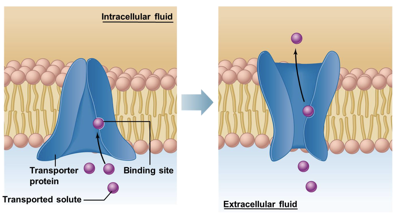
Facilitated diffusion via transport protein
Used for large and polar molecules
Solute binds to transport protein, causing change in protein
Change in protein causes the protein to flip and expose the bound solute to the opposite side of the cell membrane
Factors effecting rate
- Transport rate of protein
- Number of carrier proteins
- Magnitude of concentration gradients
GLUT4
Transport protein that comes from the inside of the cell and connects to the cell membrane
Allows for the ingestion of glucose from the extracellular fluid
Stimulated by insulin
Clinical application: Diabetes does not allow for these proteins to attach, glucose never gets into the cells
Primary Active Transport
Active transport that directly uses ATP
- Uses an enzyme that catalyzes an ATP molecule through hydrolysis
Secondary Active Transport
Uses an electrochemical gradient that drives the transport of a solvent
Sodium (Na+) Potassium (K+) Pump
Located in almost every cell in the body
Uses active transport to move Na and K against their concentration gradient
- 3 Na moved out of the cell, intracellular to extracellular
- 2 K moved into the cell, extracellular to intracellular fluid
Process
- Step 1: 3 Na and 1 ATP bind to intracellular side of pump
- Step 2: Protein changes shape and flips intracellular side to face the extracellular fluid
- Step 3: 2 K bind to the originally intracellular side of the pump
- Step 4: Protein changes shape and flips back to original configuration
- Step 5: K is released into inside of the cell
Ca2+ pump
Located in the plasma membrane of cells in the endoplasmic reticulum
Removed Ca2+ from a cytoplasm and dumps it into the extra cellular fluid or endoplasmic reticulum
Used in muscle cells for fast communication
Secondary Active Transport
A substance moving down its concentration gradient is coupled with a substance moving up it’s concentration gradient
- Substance moving from high to low concentration gradient powers the movement of the substance from low concentration to high concentration
Subdivided into cotransport and anti port
Cotransport
Type of secondary active transport
Both substances are moving from the same side of the plasma membrane to the other side of the plasma membrane

Antiport
Type of secondary active transport
The two substances involved in the transport start on opposite ends of the membrane and move in opposite direction

Osmosis
Diffusion that involves water
- Often facilitated but sometimes simple
Osmolarity
The total amount of solute dissolved in a solution
When osmolarity raises water concentration falls
- Water concentration falling triggers water to diffuse to the area from a high concentration water area
Normal osmolarity 300mOsm
Osmotic Pressure
Indirect measurement of a solution’s total solute concentration
Total osmotic pressure increases then osmotic pressure increases
Tonicity
The effect the osmolarity of a solution has on cells that are submerged in it
Isotonic
Related to tonicity
When the non penetrating solutes on the intracellular and extracellular sides are the same

Hypotonic
Related to tonicity
When the nonpenetrating solutes on the intracellular fluid is lower than the extracellular
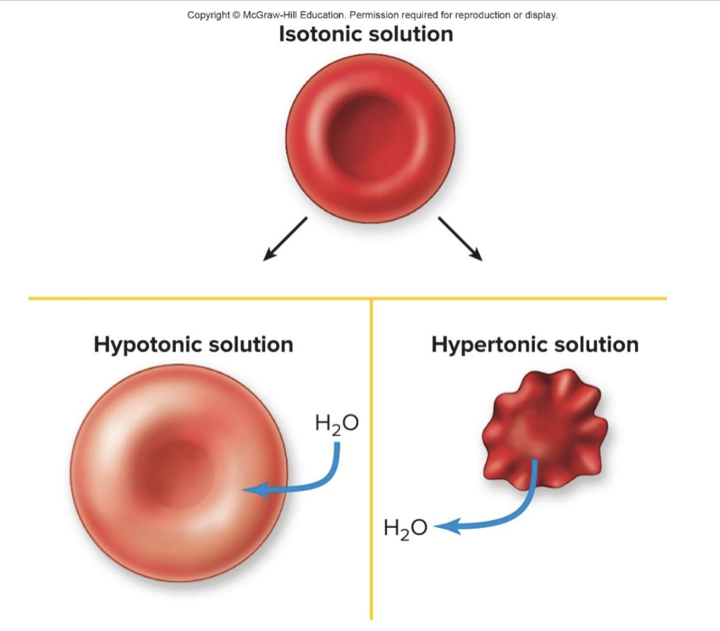
Hypertonic
More solute in the solution, compared to another solution
In respect to red blood cells a hypertonic solution caused blood to be pulled out of the cell

Endocytosis
Region of the plasma membrane folds into the cell
Small pockets are formed that then pinch into the cells
An intracellular membrane bound vesicle is born
Exocytosis
Region of the plasma membrane pushes out into the extracellular fluid
Small pockets are formed that then pinch into extracellular fluid
An extracellular membrane is born
Pinocytosis
Type of endocytosis
Non-specific
ECF + dissolved solutes (water, ions, nutrients, small molecules)
Phagocytosis
Type of endocytosis
Ingests bacteria or large particles
Fuse with lysosomes in cytoplasm
Receptor-mediated endocytosis
Type of endocytosis
Allows for cells to take specific molecules in
- Certain molecules bind to plasma membrane receptors, concentrated area becomes an endocytosis
Paracellular transport
Diffusion between epithelial cells
Transcellular transport
Substances move into epithelial cells at the apical or basal surface then exits on the opposite surface
- Moves through epithelial cell