Pathology (copy)
1/99
Earn XP
Description and Tags
Name | Mastery | Learn | Test | Matching | Spaced | Call with Kai |
|---|
No analytics yet
Send a link to your students to track their progress
100 Terms
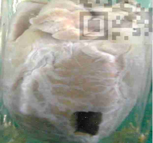
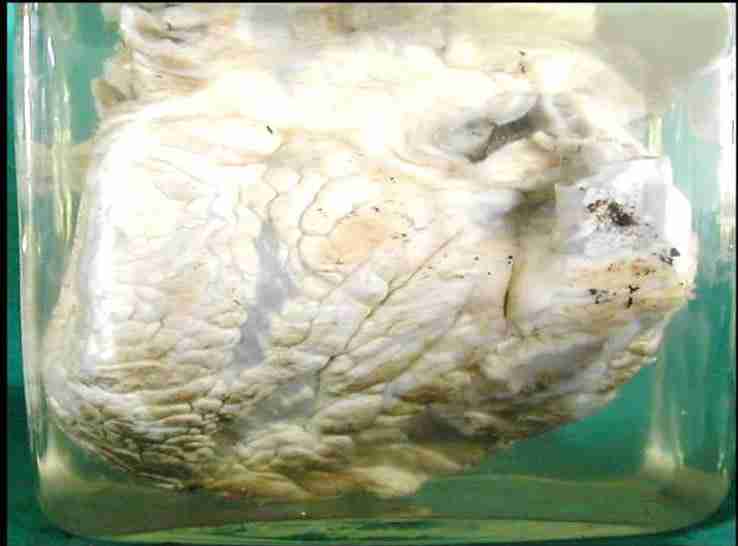
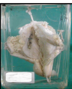
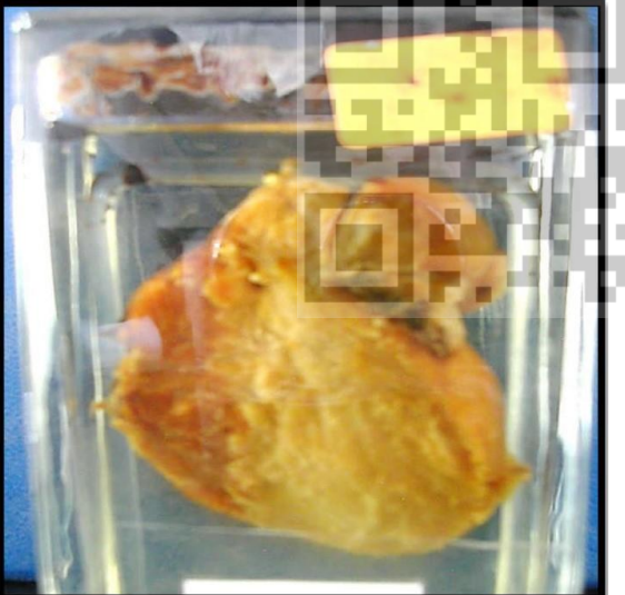
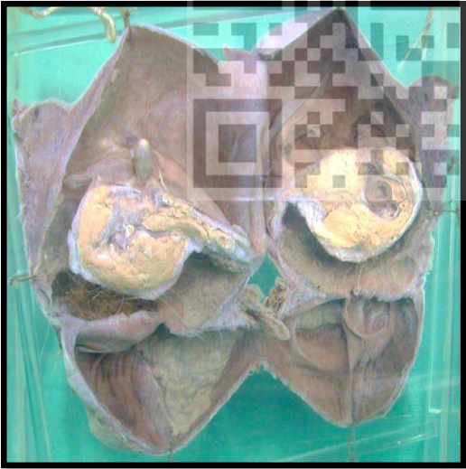
Brown atrophy of heart
Tissse adaptation
A heart of an adult showing:
• Diminished size of the heart in relation to the aorta and pulmonary vessels.
• Tortuous coronary arteries.
• Brown colour of the thinned myocardium seen through an opened window in the cardiac walls
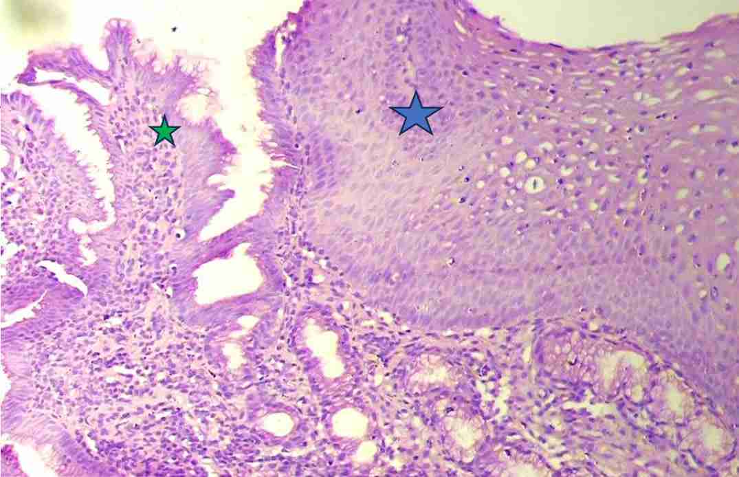
Glandular metaplesia
Tisse adaptation
Blue St sq of esophagus
Green columnar with goblet of intestinal mucosa
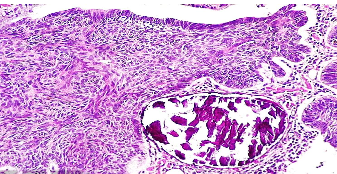
Dystrophic calcification in a tumor:
Pathological process: Cell injury and accumulations in a benign tumor.
-Basophilic deposits of calcification in a tumor
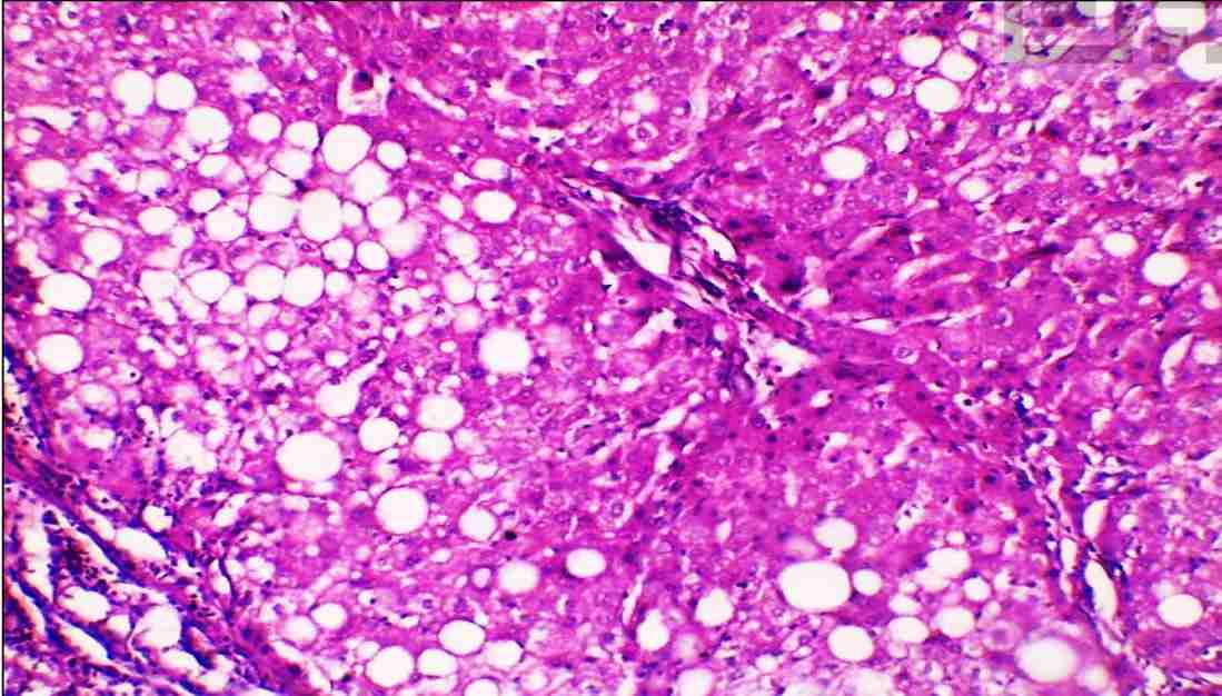
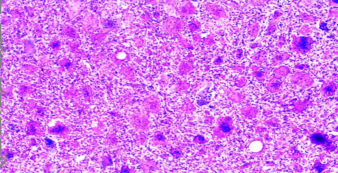
liver steatosis (Fatty liver):
Pathological process: Cell injury and accumulation
Fatty infiltration of the heart
* Pathological process: Cell injury and accumulations
A thick yellowish fatty material is deposited under the visceral layer of the pericardium.:
Fatty change of uterine leiomyoma
Pathological process: Cell injury and accumulations
*A jar contains one half of enlarged uterus cut longitudinally
• Large interstitial leiomyoma showing: -Bundles of smooth muscle fibers separated by bands of whitish fibrous tissue arranged in concentric manner taking whorlly appearance
. -Yellowish areas in the tumor tissue denoting fatty leiomyoma. change of uterine
• Uterine cavity: Distorted compressed.
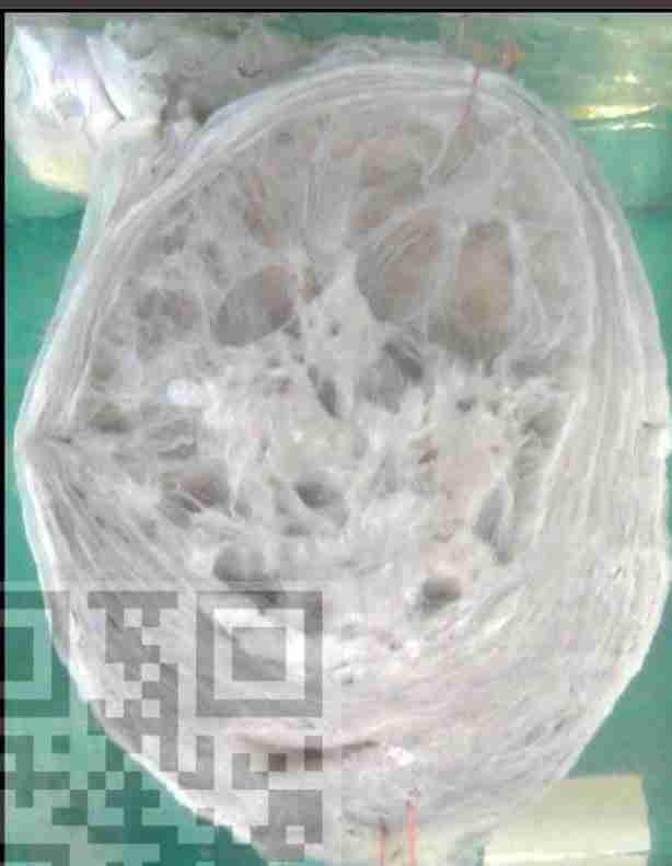
Myxomatous degeneration of uterine leiomyoma: * Pathological process: Cell injury and accumulations
Large interstitial leiomyoma showing:
-Bundles of smooth muscle fibers separated by bands of whitish fibrous tissue arranged in concentric manner taking whorlly appearance.
- Myxomatous degeneration replacing most of the uterine leiomyoma formed of: Multiple large cystic spaces; variable in size and shape containing jelly-like semi translucent mucoid material.
• Uterine cavity: Distorted compressed.
Myxoid degeneration in a tumor
* Pathological process: Cell injury and accumulations
Areas of bluish myxoid material separating tumor tissue.
Hyaline degeneration in a tumor:
Pathological process: Cell injury and accumulations
-Extracellular deposition of homogeneous pink hyaline material in a tumor.
Russel bodies in rhinoscleroma:
Pathological process: Cell injury and accumulations
-Type of intracellular hyaline degeneration.
Intracellular deposition of homogeneous pink hyaline material within plasma cells in a case of rhinoscleroma.
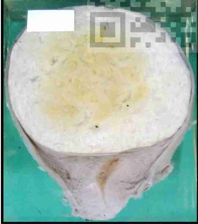
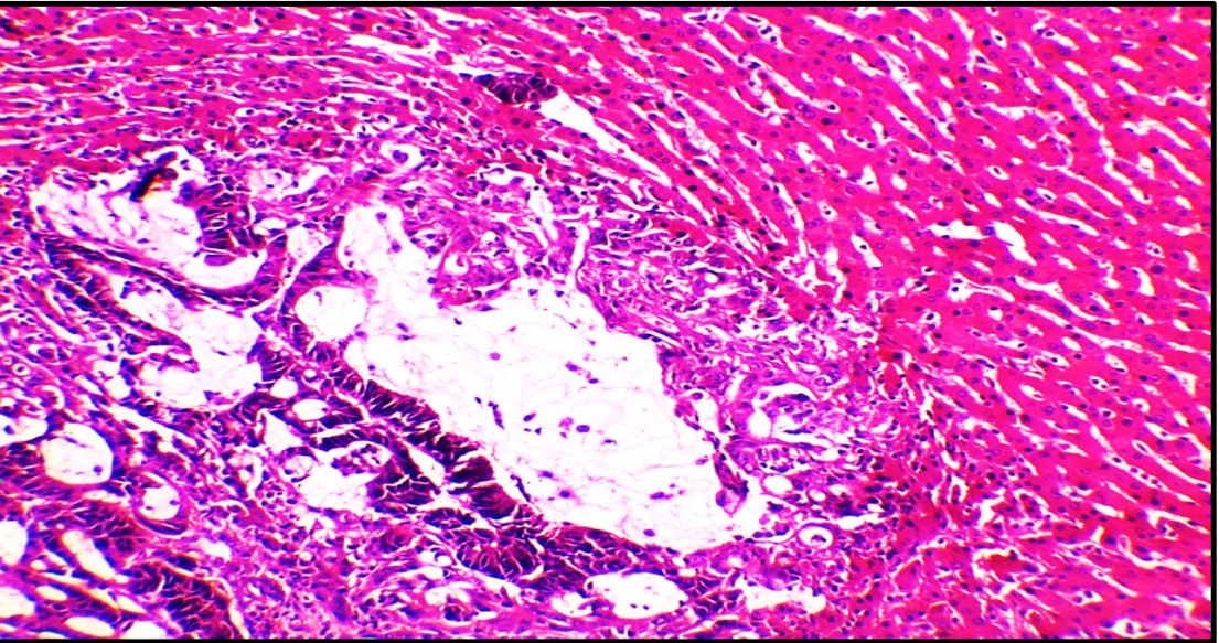
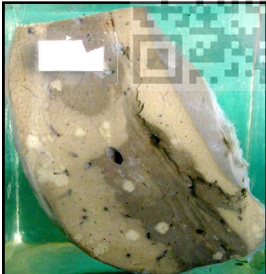
Amyloid degeneration of the liver
Pathological process: Cell injury and accumulations
A jar contains a slice of liver tissue
• The cut surface is waxy with light brown areas (amyloid) alternating with yellowish areas (fatty changes) giving the mosaic appearance.
• The borders are sharp.
• Outer surface is smooth.
• The capsule is tense and stretched.
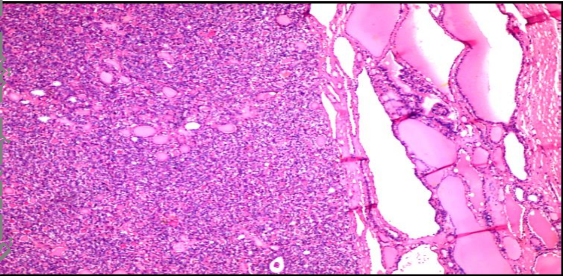
: Diffuse amyloid degeneration of the spleen:
* Pathological process: Cell injury and accumulations
Section in splenic tissue showing:
• Diffuse deposition of homogenous pink amyloid substance in basement membrane of sinusoids of red pulp & central arterioles of white pulp.
• Atrophy of white pulp (lymphoid follicles)
• Pattern is preserved.
Diffuse amyloid degeneration of the spleen: ( lymph node
* Pathological process: Cell injury and accumulations Section in splenic tissue showing:
• Diffuse deposition of homogenous pink amyloid substance in basement membrane of sinusoids of red pulp & central arterioles of white pulp.
• Atrophy of white pulp (lymphoid follicles)
• Pattern is preserved.
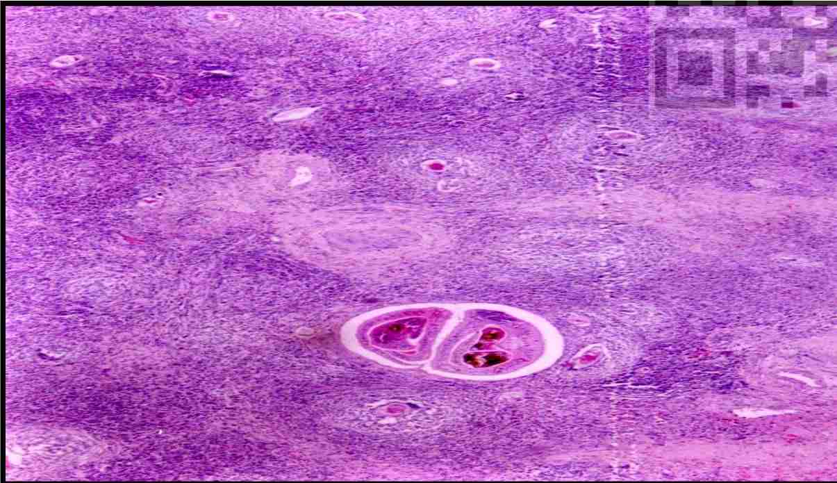
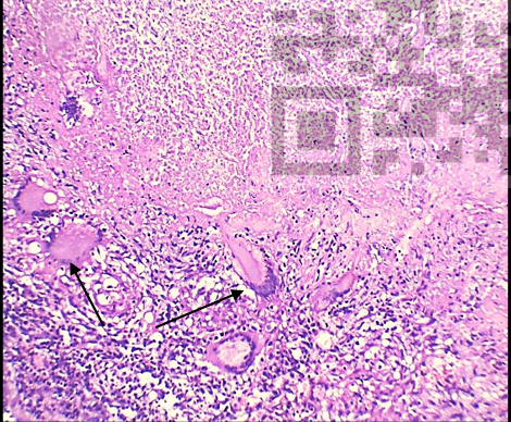
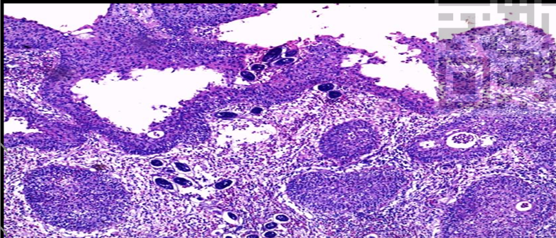
Bilharzial pigment in colonic bilharzial polyp:
* Pathological process: Pigment deposition in a granulomatous reaction. Section in colonic polyp: -
Bilharzial worms are seen inside the blood vessels showing brownish bilharzial pigments. - - Bilharzial pigments are haematin pigment formed by bilharzial worms due to ingestion and digestion of RBCs from portal blood.
Multiple bilharzial ova are seen, surrounded by granulomatous reaction (bilharzioma) formed of macrophages, plasma cells, eosinophils, lymphocytes, and giant cells.
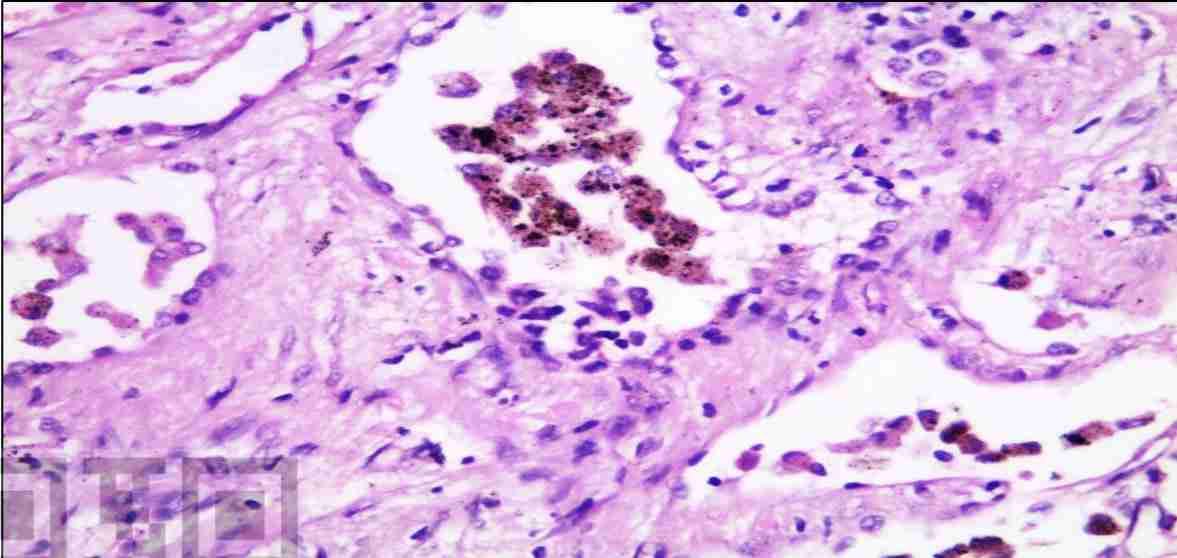
Melanin pigment in melanoma:
* Pathological process: Pigmentation in malignant tumor
. -Intracellular and extracellular deposition of brownish melanin pigment in a case of melanoma.
Note: rounded to polyhedral malignant melanocytes showing malignant criteria.
Hemosiderin pigment in chronic venous congestion of the lung: -
Heart failure cells: They are haemosiderin-laden macrophages found in the alveolar spaces in a case of chronic venous congestion of the lung.
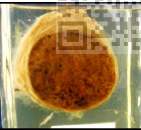
Thyroid adenoma with dystrophic calcification: * Pathological process: Cell injury and accumulations in a benign tumor.
Solitary (single) thyroid nodule cut into two halves
The nodule is:
Well encapsulated by fibrous tissue capsule • Brownish in color
he cut surface shows chalky white area of dystrophic calcification
Reactive follicular hyperplasia
*Pathologic process: Tissue adaptation • Hyperplastic lymphoid follicles: -
Variable in size - Germinal center lightly stained than the mantle.
• Preserved nodal architecture.
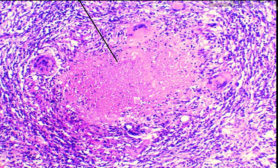
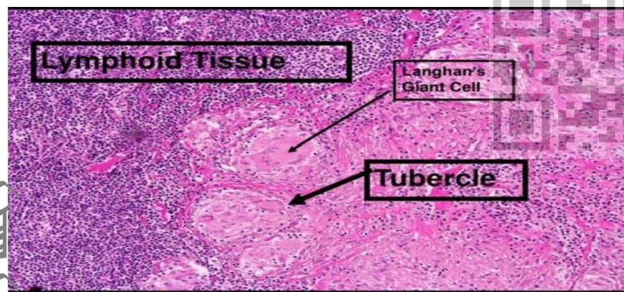
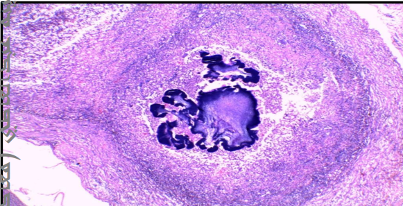
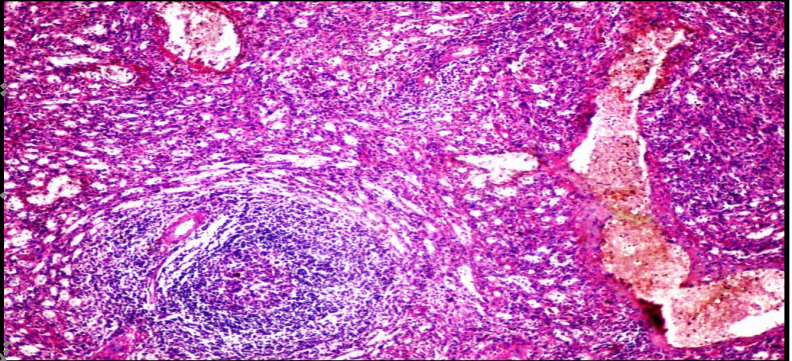
Tuberculous granuloma with central caseation.
*Pathologic process: Cell death in a granuloma
Granulomatous reaction consisting of epithelioid cells, Langhans giant cells with peripheral rim of lymphocytes and fibroblasts
. - The Centre of the granuloma showed caseating material (caseous necrosis) appears as structureless homogenous pale eosinophilic material.
Coagulative necrosis:
Pathologic process: Cell death -
It is a type of cell death caused by ischemia or infarction.
- The outline of tissues is preserved with loss of cellular details (no nuclei).
- Only ghost of cells could be identified.
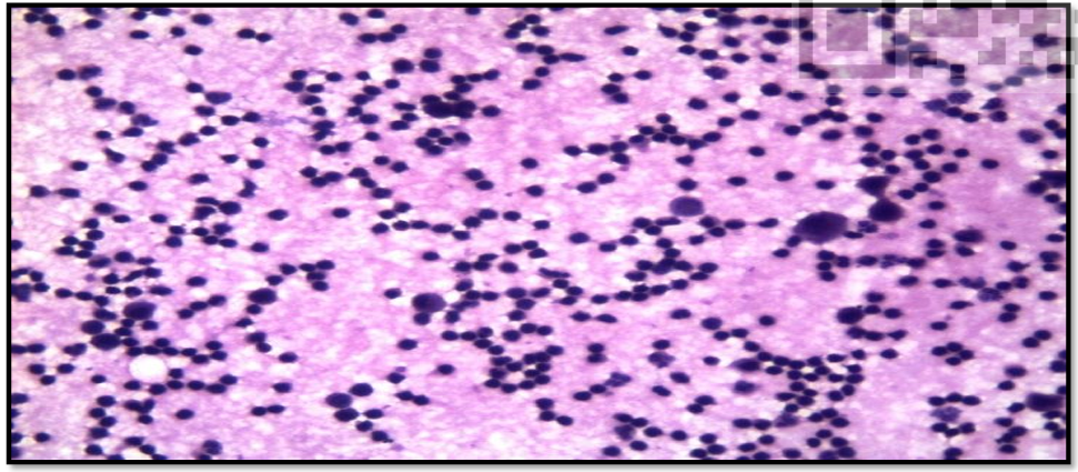
Lymphocytic effusion
Cytological smears from pleural effusion showing collection of lymphocytes. Lymphocytes: -
Dark stained nucleus with thin rim of minimal cytoplasm. - Seen in chronic inflammation
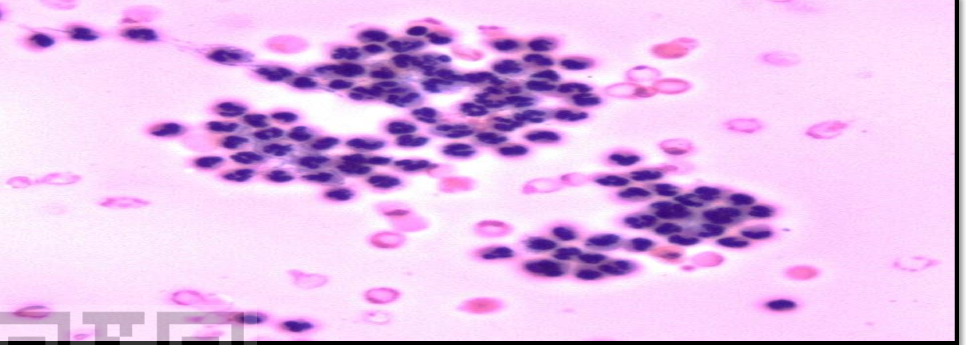
Pyogenic effusion
Cytological smears from pleural effusion showing collection of neutrophils. Neutrophils: -
- Characteristic cells of acute inflammations. They have multilobed nucleus with neutrophilic cytoplasm.
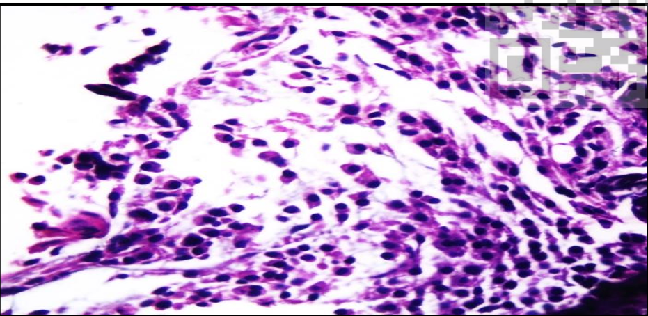
Plasma cells
• Inflammatory cells with abundant cytoplasm and eccentric nucleus.
• The are the most characteristic cells in chronic inflammation.
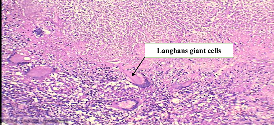
Langhans giant cells:
Langhans giant cells are large cells with eosinophilic cytoplasm and multiple nuclei forming a circle or an arc of a circle at the periphery of the cytoplasm. They are seen in granuloma of Tuberculosis.
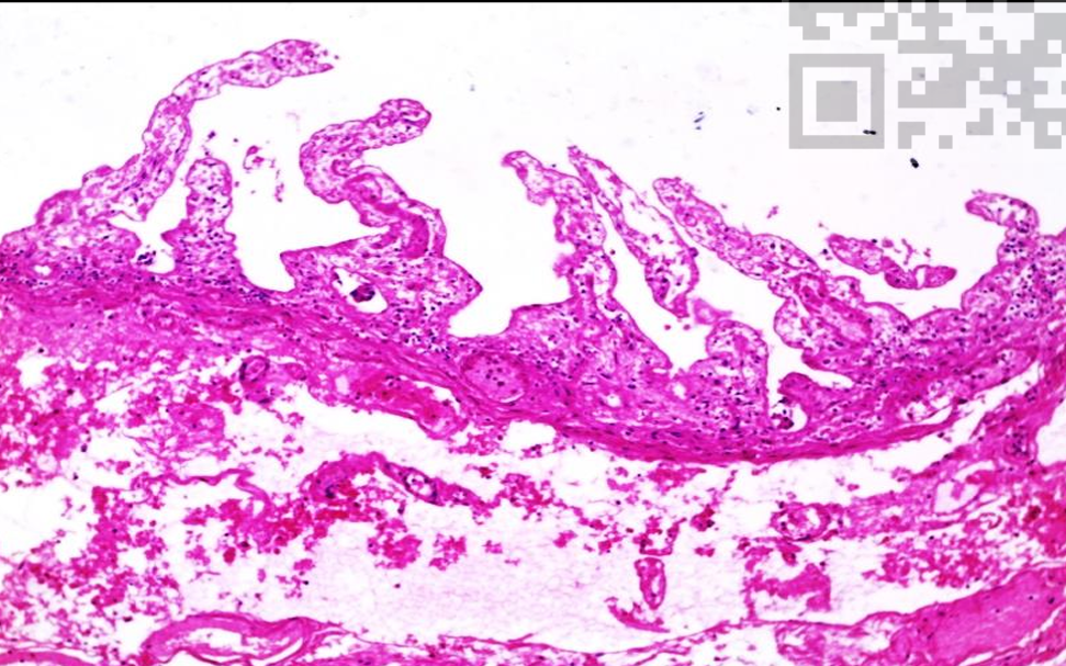
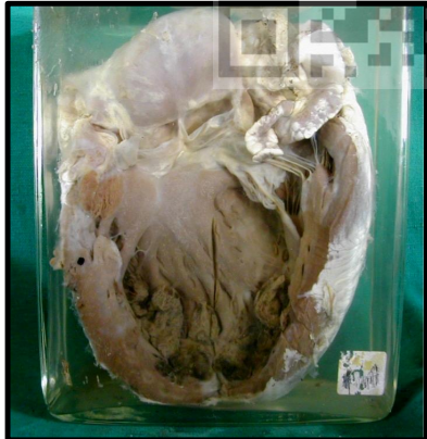
Serofibrinous pericarditis
Pathologic process: Non-suppurative inflammation
Heart of child evidenced by small size of the heart in relation to the small size of the great vessels
• The visceral pericardium is covered by fluffy fibrinous exudate giving the bread and butter appearance.
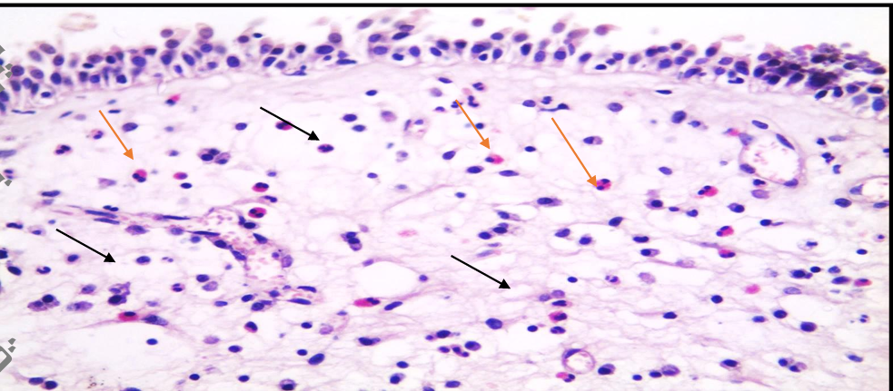
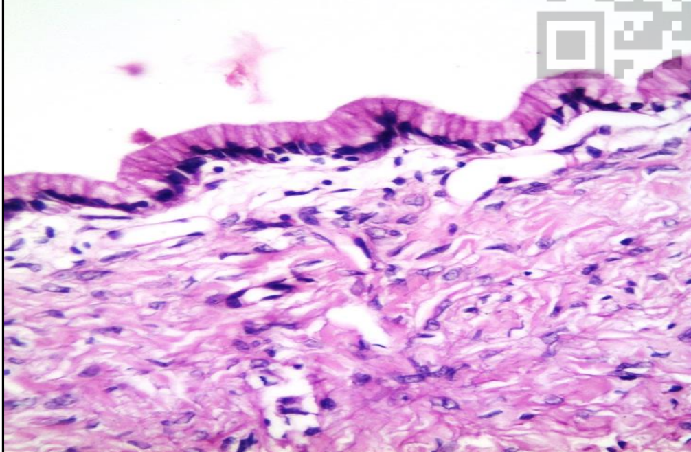
Allergic rhinitis '
Pathologic process: Non-suppurative inflammation Section in nasal mucosa showing
• Oedematous stroma covered by respiratory epithelium. (Tissue Oedema means abnormal accumulation of fluid within the interstitial tissue (Black arrows)
• Subepithelial infiltration by mixed inflammatory cells in the form of lymphocytes, plasma cells and many eosinophils.
• Eosinophils have bi-lobed nucleus and eosinophilic granular cytoplasm. - They are seen in allergic reactions and parasitic infections (Orange arrows)
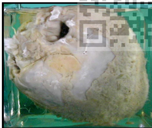
Acute suppurative appendicitis
Pathologic process: acute inflammation Vermiform appendix with constriction in the middle:
• Proximal half shows glistering serosa.
• Distal half shows: - Swelling - Opaque serosa. suppurative Yellow areas due to suppuration.
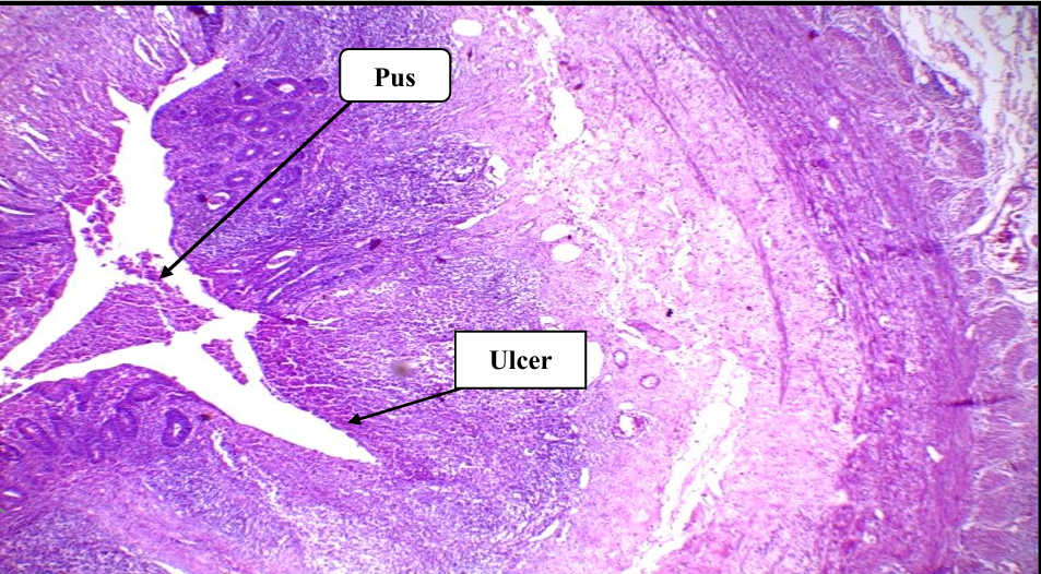
Acute suppurative appendicitis
Pathologic process: acute suppurative inflammation.
Section in vermiform appendix showing:
• Lumen: filled with pus formed of necrotic debris, neutrophils and pus cells.
• Transmural inflammation: Infiltration of all the layers (mucosa, submucosa, muscle, and serosa) by acute inflammatory cells mainly neutrophils along with lymphocytes and macrophages.
• The mucosa shows ulceration which defined as discontinuity of surface epithelium
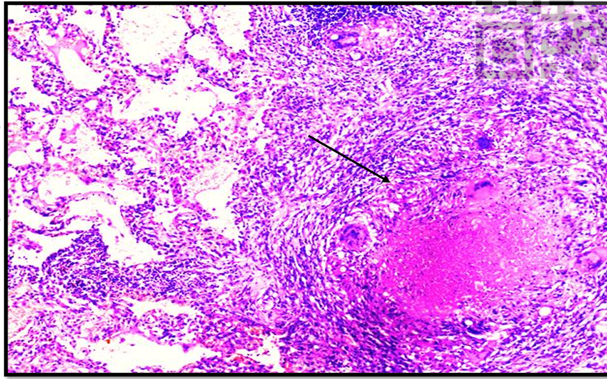
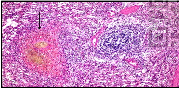
Caseating pulmonary tuberculosis:
Pathological process: Granuloma (Chronic inflammation).
Section in the lung showing:
• The lung tissue is infiltrated by multiple tubercles (arrow) with central caseation. • The tubercle consists of epithelioid cells, Langhans giant cells and lymphocytes.
- Epithelioid cells are histicoytes that resemble epithelial cells (hence named epithelioid cells). They have vacuolated cytoplasm (lipid content) with ill defined cell borders and oval vesicular nuclei.
- Langhans giant cells are large cells with multiple nuclei forming a circle or an arc of a circle at the periphery of the cytoplasm.
- A peripheral rim of lymphocytes and fibroblasts is also seen.
• Caseation appears structureless pale homogenous eosinophilic material.
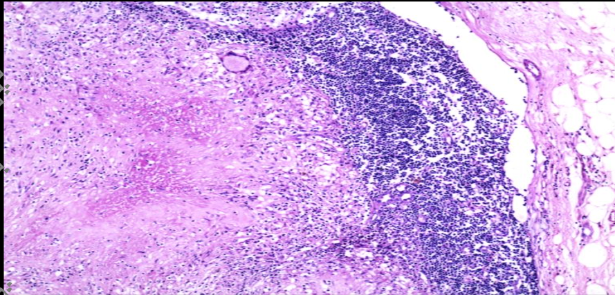
Caseating tuberculous lymphadenitis Pathological process: Granuloma (Chronic inflammation).
Section in lymph node showing:
• The lymph node is infiltrated by multiple tubercles with central caseation
. • The tubercle consists epithelioid cells, langhans giant cells and lymphocytes. - Epithelioid cells are histicoytes that resemble epithelial cells (hence named epithelioid cells). They have vacuolated cytoplasm (lipid content) with ill- defined cell borders and oval vesicular nuclei. - Langhans giant cells are large cells with multiple nuclei forming a circle or an arc of a circle at the periphery of the cytoplasm. - A peripheral rim of lymphocytes and fibroblasts. • The caseation appears structureless pale homogenous eosinophilic material.
Caseating tuberculous lymphadenitis Pathological process: Granuloma (Chronic inflammation).
Section in lymph node showing:
• The lymph node is infiltrated by multiple tubercles with central caseation
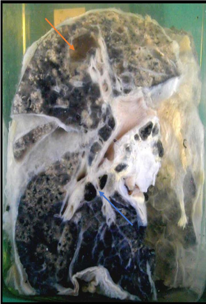
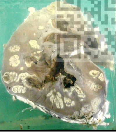
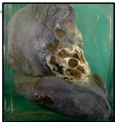
Chronic fibrocaseous pulmonary tuberculosis with cavitation and secondary miliary spread.
Pathological process: Granuloma (Chronic inflammation)
A jar containing lung tissue showing: • Tuberculous cavity (red arrow), which is: - Solitary - Apical - Irregular yellowish wall (caseation) surrounded by greyish white fibrosis.
• The rest of lung tissue shows caseous foci: - Minute pale yellow. - Homogenously distributed over the cut surface. - Some of them show secondary cavitation.
• The hilar lymph nodes (blue arrow) are minimally enlarged and show black colour (anthracosis).
LANGHAN
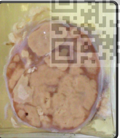
Chronic Tuberculosis of the kidney (surgical type): ‘
Pathological process: Granuloma (Chronic inflammation).
A jar contains half of destroyed kidney.
• The cut section shows multiple, pale-yellow caseating foci at the cortex and the cortico medullary junction, reaching the calyces leading to their destruction.
• Some of these caseous areas coalesce together. • The caseous material is shed leaving multiple cavities.

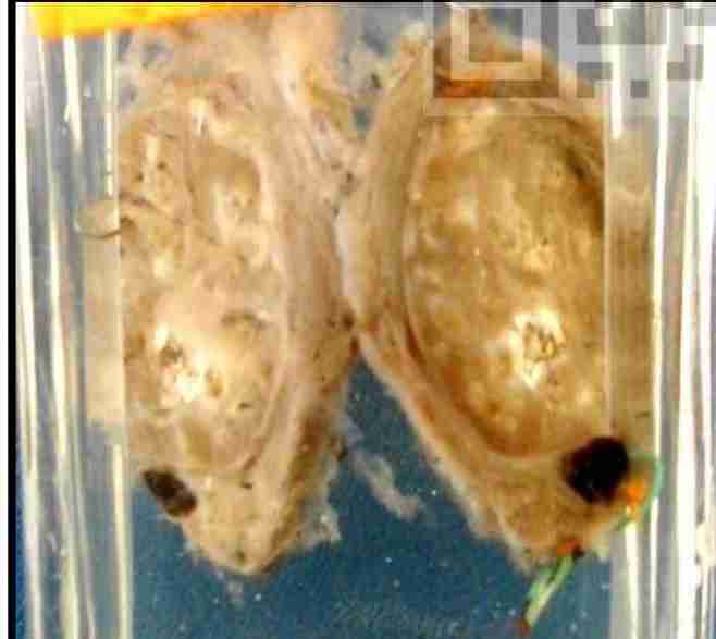
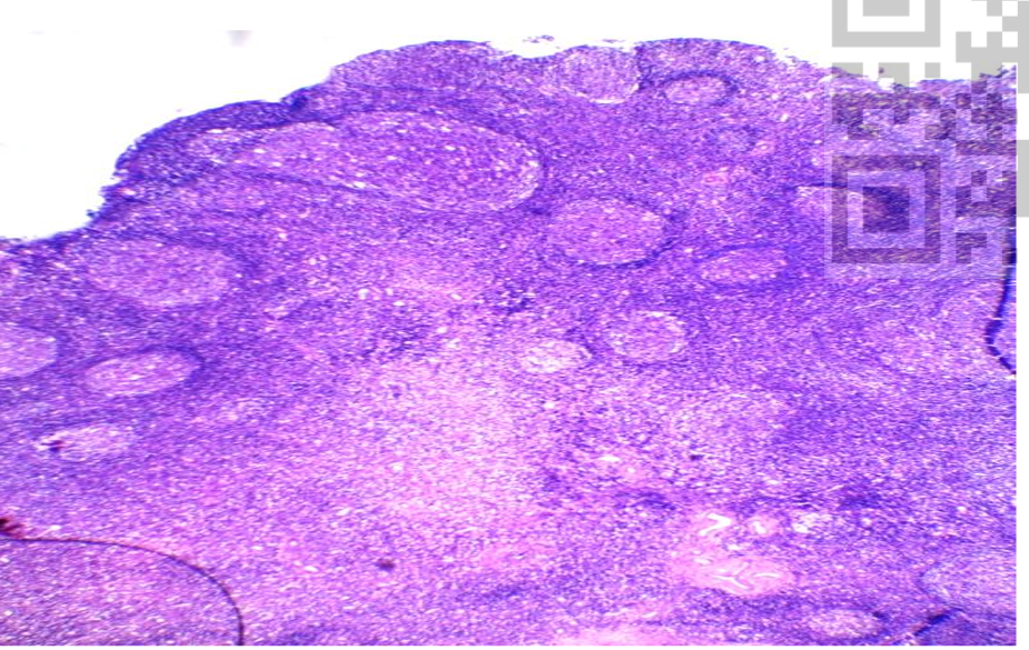
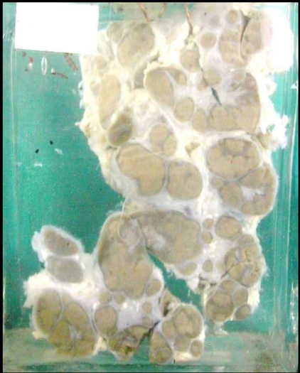
Miliary tuberculosis of the spleen
*Patholog'ical process: Granuloma (Chronic inflammation).
A jar contains two halves of a spleen showing: -
The cut surface is studded with small yellowish foci of caseation, 0.1-0.3 cm in size, homogenously distributed all-over the surface, - - Some nodules are coalesced together giving larger nodule.
Miliary TB results from blood spread of tuberculosis.
Caseating tuberculous lymphadenitis * Pathological process: Granuloma (Chronic inflammation).
A jar containing enlarged lymph nodes cut into two halves showing:
-Multiple yellowish caseating foci replacing a major part of nodal tissue.
- Some of these foci coalesced together forming geographic areas of caseation.
Tabes mesenterica:
Pathological process: Granuloma (Chronic inflammation).
A jar contains a group of enlarged mesenteric lymph nodes showing:
- Multiple yellowish caseating foci replacing a major part of the nodal substance.
- Some of these nodes are fused together "matted" forming a single mass.
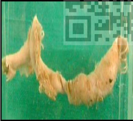
Bilharzial polypi of the colon with bilharzial pericolic mass.
Pathological process: Granuloma (chronic inflammation).
A jar contains a part of the colon opened longitudinally.
• Multiple bilharzial polypi projecting in the lumen (black arrow) which are:
- Variable in size and shape.
- Some are sessile and others are pedunculated.
• The muscle layer is continuous all over the length of the colonic segment, with no interruption indicating that the lesion is benign (blue arrow).
• The pericolic tissue shows large greyish white mass (bilharzial granulation tissue) (red arrow).
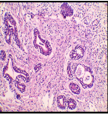
Bilharzial polyp in colon
Pathological process: Granuloma (chronic inflammation).
Section in bilharzial colonic polyp showing:
- Central fibrovascular core of connective tissue formed from the submucosa showing pink degenerated or blue calcified ova.(Black arrows) -
The ova are surrounded by bilharzioma (bilharzial granuloma) formed of macrophages, plasma cells, eosinophils, lymphocytes, and giant cells (Blue arrows).
- Bilharzial worms may be seen inside blood vessels (yellow arrows).
- The covering mucosa shows hyperplastic glands (increase in number) that are regular and uniform.

Bilharzial polyp in colon
Pathological process: Granuloma (chronic inflammation).
Section in bilharzial colonic polyp showing: - Central fibrovascular core of connective tissue formed from the submucosa showing pink degenerated or blue calcified ova.(Black arrows) - The ova are surrounded by bilharzioma (bilharzial granuloma) formed of macrophages, plasma cells, eosinophils, lymphocytes, and giant cells (Blue arrows).
- Bilharzial worms may be seen inside blood vessels (yellow arrows).
- The covering mucosa shows hyperplastic glands (increase in number) that are regular and uniform.

Bilharzial polyp in colon
Pathological process: Granuloma (chronic inflammation).
Section in bilharzial colonic polyp showing: -
Central fibrovascular core of connective tissue formed from the submucosa showing pink degenerated or blue calcified ova.(Black arrows) - The ova are surrounded by bilharzioma (bilharzial granuloma) formed of macrophages, plasma cells, eosinophils, lymphocytes, and giant cells (Blue arrows).
- Bilharzial worms may be seen inside blood vessels (yellow arrows).
- The covering mucosa shows hyperplastic glands (increase in number) that are regular and uniform.
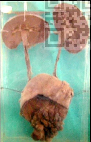
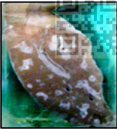
Bilharzial polypi of the urinary bladder with bilateral mild hvdronephrosis and hydroureter:
Pathological process: Granuloma (chronic inflammation).
A jar contains the urinary system of a child evidenced by the small size.
• The trigone shows multiple bilharzial polypi that show the following:
- Variable in size and shape.
- Some are sessile and others are pedunculated. - Obstructing vesico-ureteric orifices.
• The ureters are slightly dilated with mild hydroureter.
• The kidneys show mild dilatation in the pelvicalyceal system (hydronephrosis).
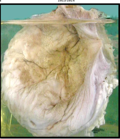
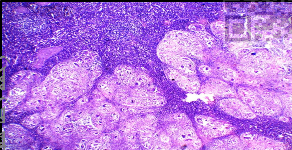
Bilharziasis of the urinary bladder (Sandv patches):
Pathological process: Granuloma (chronic inflammation).
A jar contains a urinary bladder, which is opened to show the mucosa
. • The mucosa showed dirty greyish yellow granular areas (called sandy patches) due to the presence of calcified ova under the atrophic mucosa.
The wall of the bladder is markedly thickened, with small narrow lumen (both are due to fibrosis).
Bilharziasis in urinary bladder Pathological process:
Granuloma (chronic inflammation). Section in the urinary bladder showing:
▪ The urinary bladder is lined with hyperplastic transitional epithelium.
- This hyperplastic epithelium dips down in the submucosa forming Brunn's nests.
- Some of these nests show cystic degeneration giving rise to cystitis cystica.
- Others undergo glandular metaplasia, named cystitis glandularis
. ▪ The submucosa is infiltrated by large number of fresh and calcified bilharzia ova.
▪ Viable ova are surrounded by granulomatous reaction consisting mainly of histiocytes, eosinophils, plasma cells, lymphocytes and giant cells.
cystitis cystica and cystitis glandularis in urinary bladder bilharziasis
Pathological process: Granuloma (chronic inflammation).
Section in the urinary bladder showing: ▪ Nests of transitional epithelium in the submucosa. (Brunn's nests) ▪ Some of these nests show cystic degeneration giving rise to cystitis cystica. ▪ Other nests show glandular metaplasia
Coarse periportal bilharzial fibrosis of the liver: Pathological process: Granuloma (chronic inflammation).
A jar contains a part of the liver showing:
• The liver is shrunken.
• Thickened capsule.
• Irregular surface
. • The cut surface shows the following:
➢ It is pale brown due to anaemia, brownish bilharzial pigment and fibrosis.
➢ The portal tracts are thickened and surrounded by greyish white fibrous tissue (periportal fibrosis) giving pipe stem appearance.
Sarcoidosis of spleen:
Pathologic Process: Granuloma (Chronic inflammation)
Section in spleen showing:
Multiple non-caseating granulomas replacing splenic tissue, composed of: ⚫ Epithelioid cells, langhans giant cells. ⚫ Asteroid Bodies: giant cells with stellate inclusions in their cytoplasm. ⚫ Schaumann Bodies: Calcified laminated concretions.

Asteroid Bodies:
Giant cells with stellate inclusions in their cytoplasm in a case of Sarcoidosis.
Rhinoscleroma:
Pathologic Process: Granuloma (Chronic inflammation)
Section from nasal tissue showing:
⚫ Granuloma composed of: -Extensive infiltration by Mikulicz cells: it is a large macrophage with foamy clear cytoplasm and central nucleus (diagnostic cells)
-Lymphocytes and plasma cells.
-Russel Bodies: plasma cells with hyaline degeneration
⚫ The overlying mucous membrane usually shows squamous metaplasia
Mikulicz cells in Rhinoscleroma:
⚫ They are large macrophages with central nucleus and foamy clear cytoplasm containing the organism.
⚫ Diagnostic cells of rhinoscleroma
Actinomycosis:
Pathologic Process: Granuloma (Chronic inflammation)
Chronic suppurative granuloma composed of 3 zones:
⚫ Central suppuration made of bacterial colony surrounded by heavy infiltrate of neutrophils and pus cells.
⚫ Middle zone of granulation tissue and inflammatory cells (macrophages, plasma cells and lymphocytes)
⚫Peripheral zone of extensive fibrosis
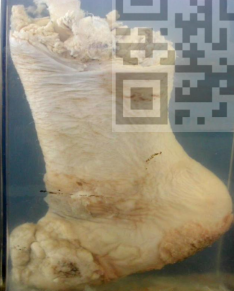
Leprosy
Pathologic Process: Granuloma (Chronic inflammation) Jar containing Foot and part of leg shows:
⚫ Dropping of all toes.
⚫ Thin atrophic skin.
⚫ A wide area of hypopigmentation.
Foreign Body Granuloma in a toe
Pathologic Process: Granuloma (Chronic inflammation)
⚫ A whitish firm rounded mass in the middle of the toe.
⚫ A foreign body (a piece of wood) is seen at the site of such mass.
Myocardial infarction with mural thrombosis:
Pathologic Process: Hemodynamic disorder (infarction and thrombosis)
Left side of the heart showing:
⚫ Area of infarction at the left ventricle:
- Pale healed infarction.
- Thinning of the ventricular wall at the apex.
⚫ Large brownish thrombi adherent to the endocardium at the area of infarction (mural thrombi).
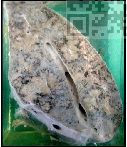
Pulmonary Embolism:
Pathologic Process: Hemodynamic disorder (Embolism)
⚫ Pulmonary arterial branches are obstructed by large brown embolus (detached thrombus) ⚫ Rest of lung is healthy (due to sudden death of respiratory failure)

Tumor emboli (Vascular invasion)
• A blood vessel lined by endothelial cells showing a group of malignant epithelial cells attached to the endothelium denoting tumor emboli.
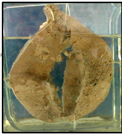
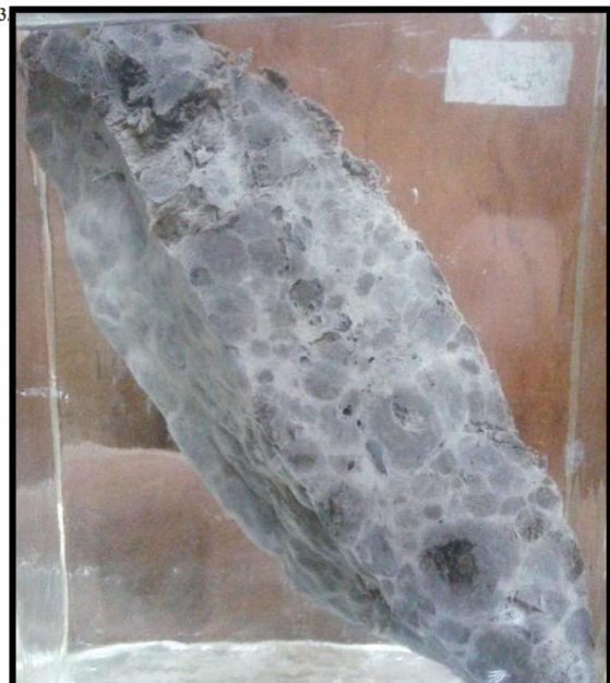
Splenic Infarction:
Pathologic Process: Hemodynamic disorder (infarction).
Cut surface shows 2 areas of infarction in spleen having the following characters:
⚫ Whitish in color (Pale infarction).
⚫ Triangular in shape; with its apex towards the hilum and its base towards the free border (due to fan distribution of blood vessels).
Chronic venous congestion (CVC) of the liver “Nut meg liver”
Pathologic Process: Hemodynamic disorder (venous congestion).
⚫ Enlarged liver
⚫ Tense capsule.
⚫ Dilated hepatic veins.
⚫ Mottled cut surface with yellow and brown colors due to alteration of blood and fat.
Congested Vessels:
Dilated vessels filled with blood
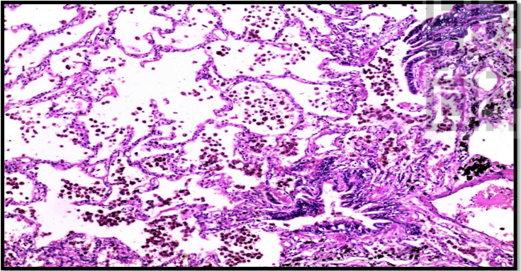
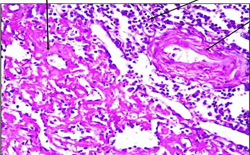
Chronic Venous Congestion of the lung (CVC lung):
Pathologic process: Hemodynamic disorder (venous congestion)
⚫ The alveolar spaces contain:
- Intact and hemolysed RBCs
- Heart failure cells (hemosiderin laden macrophages)
⚫ Alveolar walls are thickened.
⚫ Alveolar capillaries are dilated and congested.
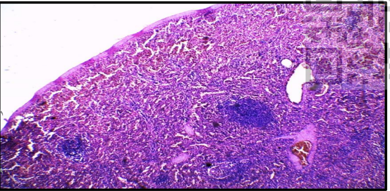
Congestive Splenomegaly:
Pathologic process: Hemodynamic disorder (venous congestion)
Section in spleen showing:
⚫ Red pulp shows:
✓ Marked dilatation of sinusoids filled with RBCs with accumulation of hemosiderin containing macrophages.
✓ Gandy-Gamna bodies “siderofibrotic nodules”: Areas of fibrosis surrounding hemosiderin deposition.
⚫ white pulp shows: Inconspicuous atrophic lymphoid folliccles.
preserved pattern of spleen
Gamna Gandy Body:
⚫ It is a siderofibrotic nodule formed of fibrosis surrounding hemosiderin deposition.
⚫ It is seen in congested spleen
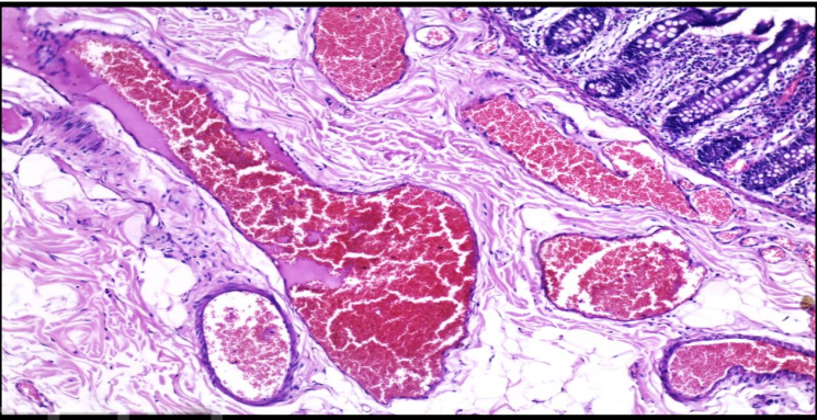
Interstitial hemorrhage:
Collections of blood within the interstitial tissue, that do not conform to the shape of tubules or capillary networks
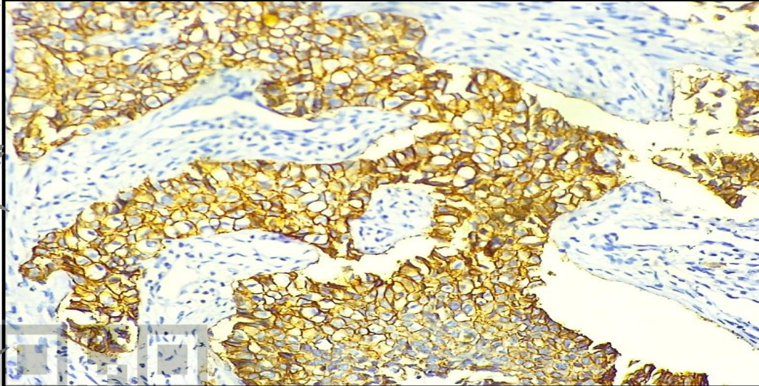
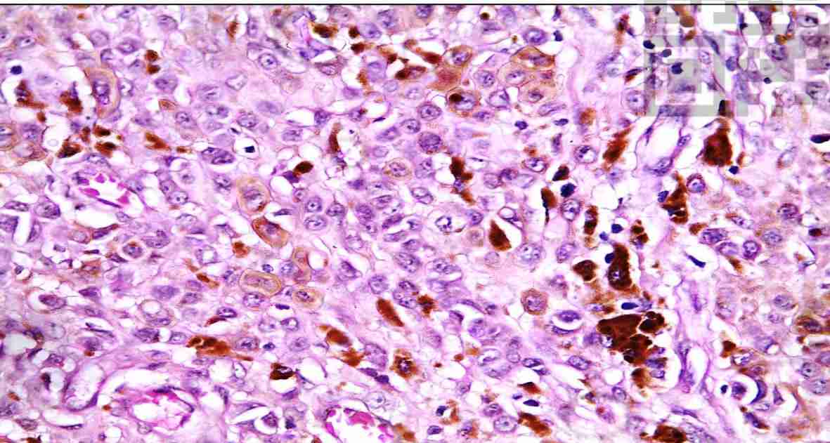
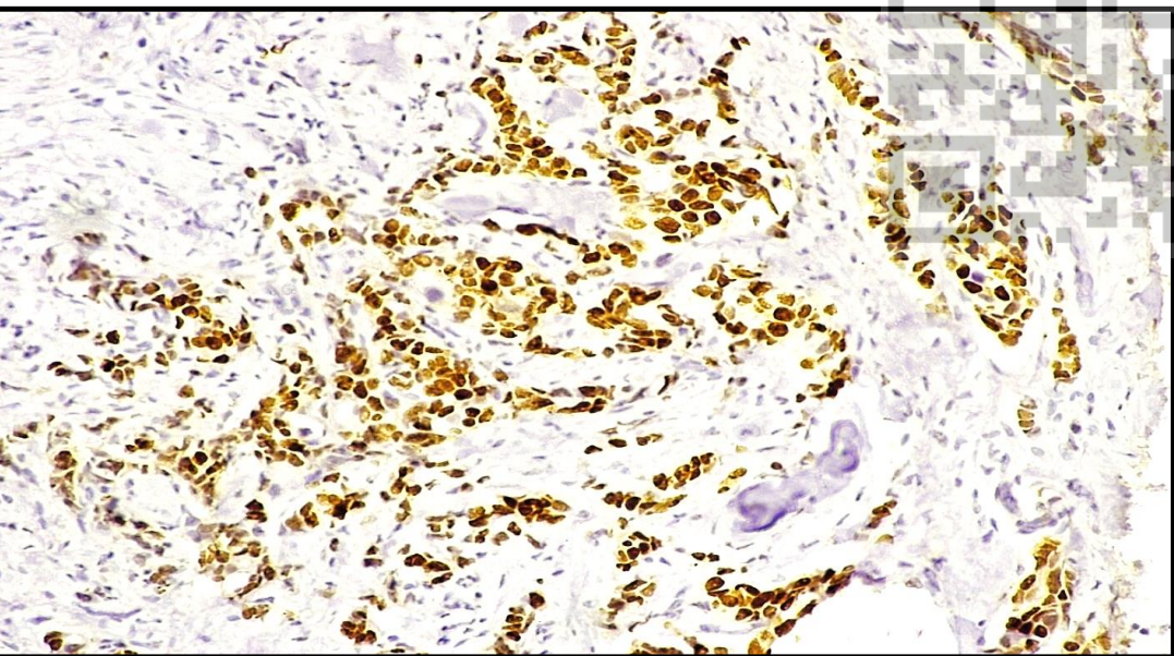
Immunohistochemical staining of Her2/neu.
- A case of invasive breast carcinoma showing Positive Membranous Her2/neu expression.
- This Her2/neu positivity indicates poor prognosis for this patient
Immunohistochemical staining of Estrogen Receptor (ER).
- A case of invasive breast carcinoma showing Positive Nuclear expression of ER.
- This ER positivity indicates good prognosis for this patient.
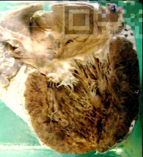
Metastasis of malignant melanoma of the heart
* Pathological process: Metastatic malignant tumor.
• A jar contains a heart showing multiple dark brownish nodules:
- Variable in size and shape.
- Seen on the pericardial and endocardial surfaces, in the myocardium and on the internal and external surfaces of the great vessels. - Represent metastatic malignant melanoma.
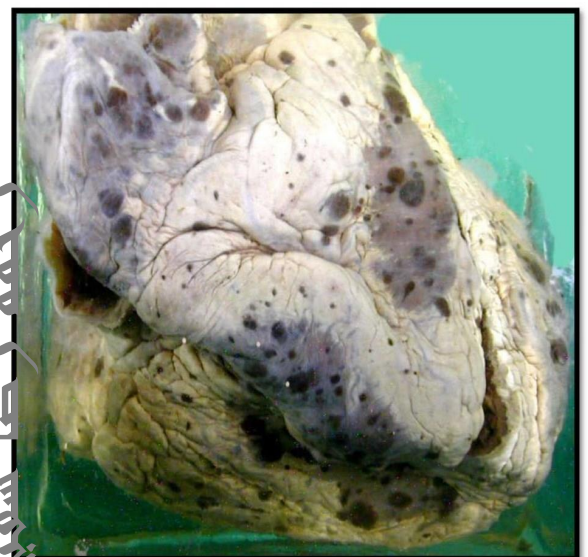
Metastasis of malignant melanoma of the heart
* Pathological process: Metastatic malignant tumor.
• A jar contains a heart showing multiple dark brownish nodules:
- Variable in size and shape.
- Seen on the pericardial and endocardial surfaces, in the myocardium and on the internal and external surfaces of the great vessels. - Represent metastatic malignant melanoma
Metastasis of malignant melanoma to the liver
* Pathological process: Metastatic malignant tumor.
A jar contains a piece of liver tissue
• The outer and cut surfaces of the liver show multiple blackish nodules that are:
- Well defined.
- Variable in size and shape.
- Some of them show umbilication (central necrosis and breakdown
Lymph nodes Secondaries of malignant melanoma
* Pathological process: Metastatic malignant tumor. •
• A jar contains two lymph nodes cut into two halves appear as:
• Large rounded black masses representing metastasis from malignant melanoma.
Melanoma, Skin
*Pathologic process: Malignant tumor.
Section in a tumour showing:
- A malignant tumour consisting of rounded or polyhedral cells and spindle shaped malignant cells arranged either in groups or nests or individually.
- The malignant cells are large, having hyperchromatic nuclei with prominent nucleoli and frequent mitoses.
- Their cytoplasm contains dark brown melanin pigment which can be seen extracellular as well. - The tumour is deeply infiltrative and shows areas of haemorrhage and necrosis
Cavernous Haemangioma
*Pathologic process: Benign mesenchymal tumor.
Section in a tumour showing:
• A hamartoma that is usually non- capsulated.
• It is formed of dilated vascular spaces which are:
- Variable in size and shape
- Lined by flat endothelial cells.
- Contain intact or hemolysed RBCs.
- Separated from each other by delicate connective tissue stroma
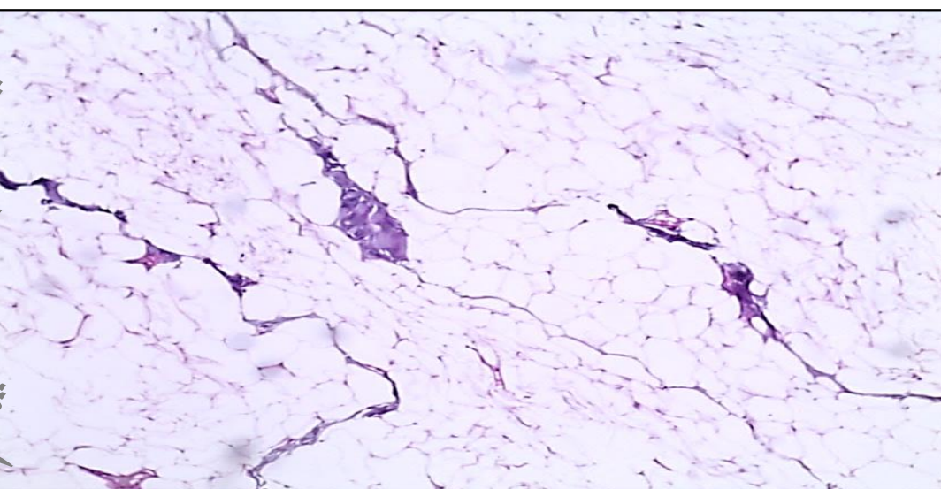
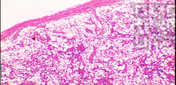
Subcutaneous Lipoma
*Pathologic process: Benign mesenchymal tumor.
Section in a tumor showing:
• A lobulated capsulated benign tumour composed of groups of mature fat cells separated by fibrous tissue septa
. • The mature fat cells:
- Large polygonal cells
- With peripheral compressed nuclei and clear cytoplasm (fat dissolves during preparation of paraffin sections giving the signet ring appearance).
Subcutaneous Lipoma
*Pathologic process: Benign mesenchymal tumor.
A jar contains well-defined benign subcutaneous tumor mass bulging above the surface of the skin😀
• Rounded or oval in shape.
• -It is well encapsulated.
-Color is yellowish & lobulated.
- Cut surface The fatty lobules are separated by strands of whitish fibrous tissue.
-Covering skin is thin & atrophic
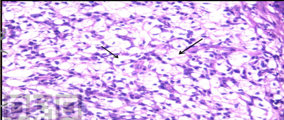
Osteoclastoma (Giant cell tumor) of the fibula.
*Pathologic process: Locally aggressive mesenchymal tumor.
Section in a tumour showing:
• Multinucleated osteoclastic giant cells:
- Large number
- Containing up to 100 rounded, small and vesicular nuclei scattered all over the cytoplasm or grouped in the centre.
• Mononuclear cells:
- Numerous - Oval and fusiform cells - Vesicular nuclei (the malignant component of the tumor). • The tumour may show areas of degeneration, necrosis and haemorrhage.
Osteoclastoma (Giant cell tumor) of the fibula.
Pathologic process: Locally aggressive mesenchymal tumor.
A jar contains a part of the fibula showing:
• Oval tumor mass located at the end of the fibula.
• This tumor causes expansion and thinning of the bone and the cortex is reduced to a thin shell
. • The cut surface shows areas of haemorrhage, necrosis, and cyst formation.
• No new bone formation.
Metastatic breast carcinoma to axillary lymph node
. *Pathologic process: Metastatic malignant epithelial tumor.
Section in a lymph node showing: • Replacement of the nodal tissue by nests and sheets of malignant epithelial cells surrounded by desmoplasia
• The primary of these cells is located in the breast.
The cells show all criteria of malignancy (Describe) .
Metastatic mucoid adenocarcinoma to the liver.
*Pathologic process: Metastatic malignant epithelial tumor.
• Section from the liver (Right) showing metastatic deposits from malignant epithelial glands with pools of mucin (Left) denoting metastatic mucoid adenocarcinoma to the liver.
• The epithelial glandular cells show all criteria of malignancy (Describe).
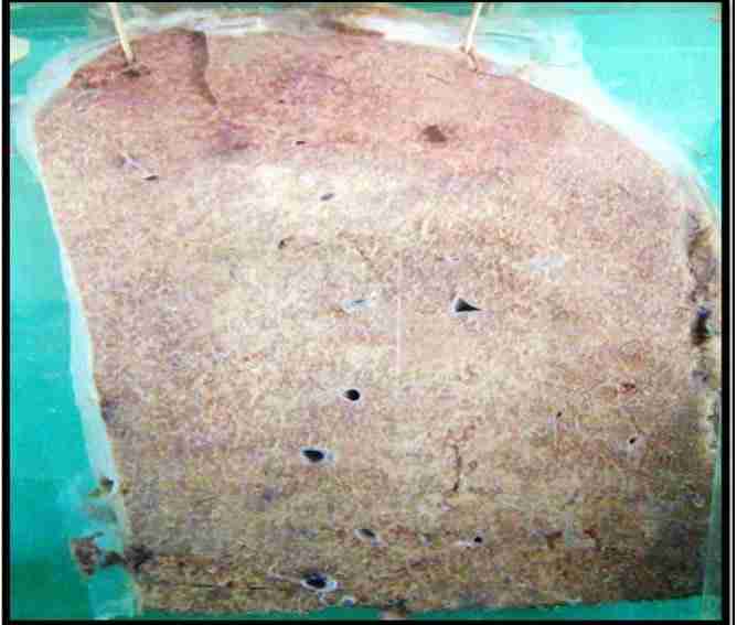
Multiple liver secondaries from carcinoma Pathologic process: Metastatic malignant epithelial tumor.
A Jar contains a slice of liver tissue showing multiple nodules:
• Roughly uniform in size and shape. • Distributed homogenously all over the liver substance.
• Yellowish in color.
• Some of them show umbilication (central ischemic breakdown).
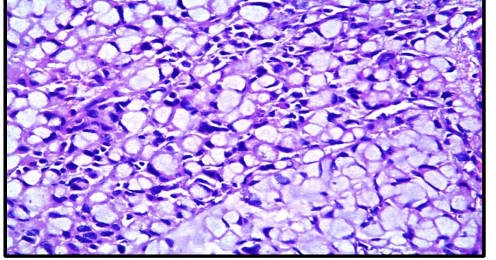
Signet ring cell carcinoma
• Pathologic process: • Malignant epithelial tumor .
• A slide showing signet ring cells
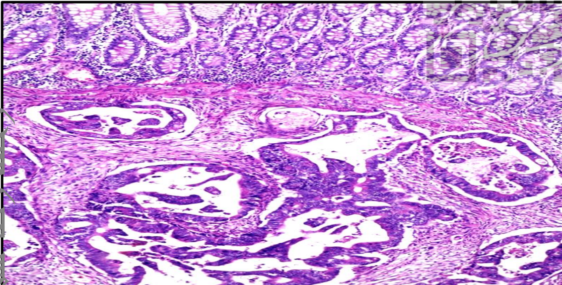
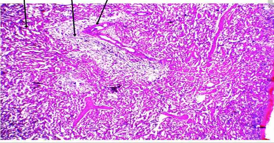
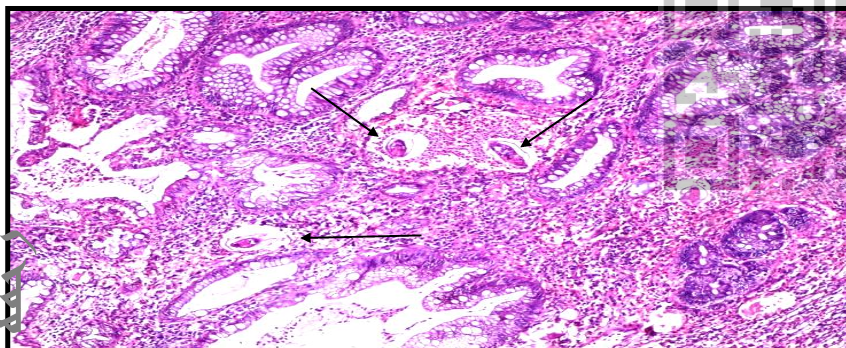
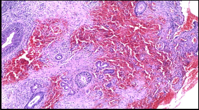
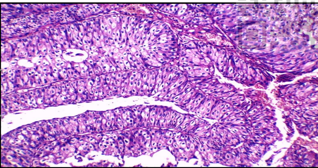
Adenocarcinoma, Colon
Pathologic process: Malignant epithelial tumor. - Normal mucosal covering formed of mucosal glands which are: ‘
• Regular sizes and shapes.
• Lined by one row of equal columnar cells with basal nuclei and apical mucin vacuoles. - Malignant glands are:
• Infiltrating the submucosa, muscle layer and serosa
• Irregular of variable sizes and shapes.
• Lined by one or more layers of malignant cells in irregular arrangement with loss of polarity. - The malignant cells show criteria of malignancy (Describe)
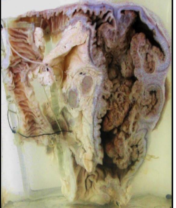
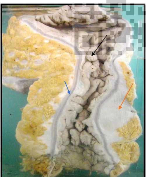
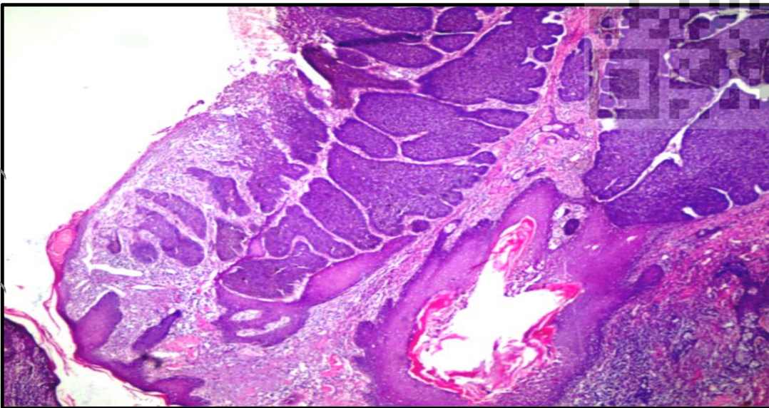
Adenocarcinoma of the large intestine with secondaries in the regional lymph nodes (fungating exophytic type):
Pathologic process: Malignant epithelial tumor
. A jar contains terminal ileum, caecum, appendix and a part of the ascending colon (i.e. right radical hemicolectomy specimen). -
There is a large fungating cauliflower mass:
• Projecting into the lumen of the caecum and infiltrating the colonic wall
. • Grayish brown in color
• The surface of tumor shows ulceration, hemorrhage and necrosis .
- Interruption of the muscle layer denoting infiltration to the underlying colonic wall.
- The pericolic lymph nodes showed metastatic grayish white tumor deposits
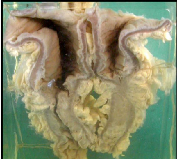
Adenocarcinoma of the large intestine (endophytic ulcerative type)
Pathologic process: Malignant epithelial tumor.
A jar contains a colonic segment (part of the recto sigmoid colon) showing malignant ulcer . -
The mucosa in the middle part is lost completely and replaced by an ulcerating malignant mass, with the following criteria:
1. Raised everted edges
2. Necrotic floor.
3. The base of the ulcer is fixed and indurated .
• The muscle layer is interrupted opposite the ulcerative mass, indicating invasion
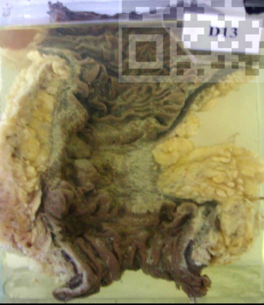
Stenosing Adenocarcinoma of the colon (endophytic stricture type)
Pathologic process: Malignant epithelial tumor. A jar contains a colonic segment cut longitudinally.
- The lumen is narrowed, even completely obliterated in some areas by grayish brown infiltrating tumor tissue.
- The muscle layer is interrupted opposite the stenosing mass, indicating invasion.
Papillary Transitional Cell Carcinoma (TCC). Pathologic process: Malignant epithelial tumor. Section in papillary tumour of the urinary bladder showing:
• Papillae with very thin fibrovascular core covered with malignant transitional epithelium (more than seven layers).
• The epithelial cells show all the criteria of malignanT
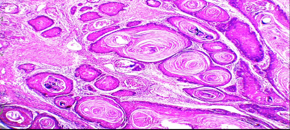
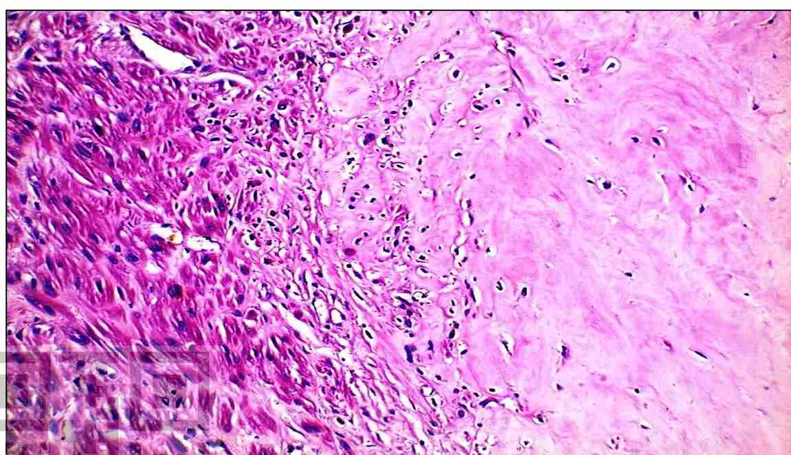
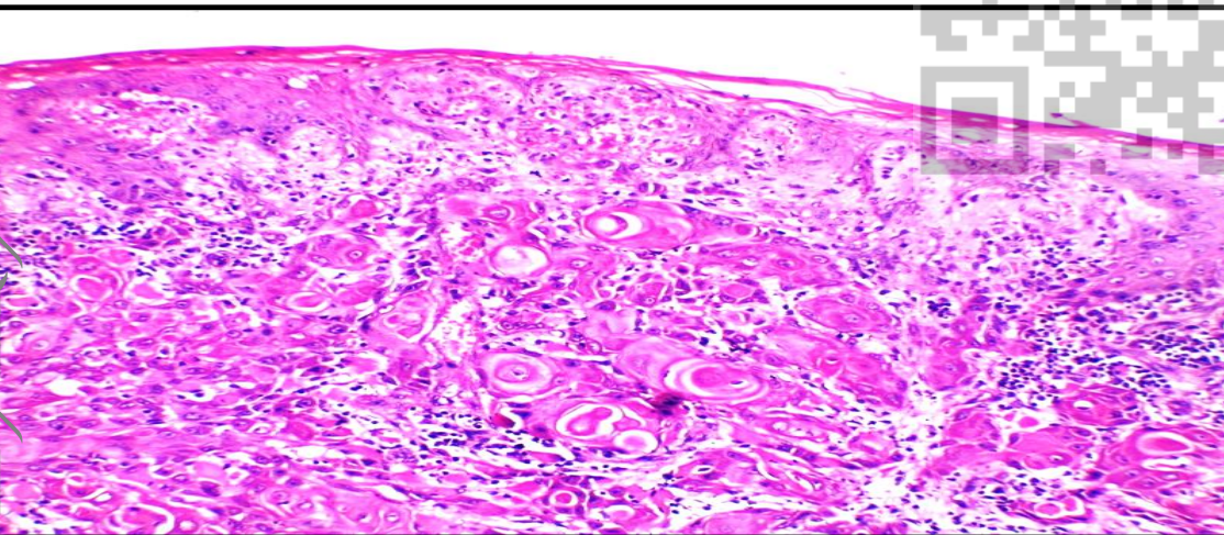
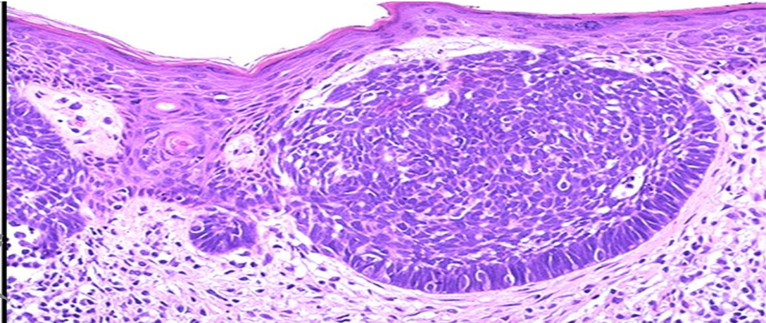
Squamous cell carcinoma, Skin
Pathologic process: Malignant epithelial tumor
. - A malignant tumor infiltrating the dermis and formed of masses of malignant epithelial cells forming cell nests of variable sizes and shapes.
- The cell nest is formed of:
• Peripheral layer of darkly stained small basal -like cells.
• Intermediate multiple layers of polyhedral cells resembling prickle cell layer
• Central cells show complete cornification forming dark red stained masses of keratin (Keratin pearls).
- The malignant cells show abundant eosinophilic cytoplasm with malignant characters (Describe)
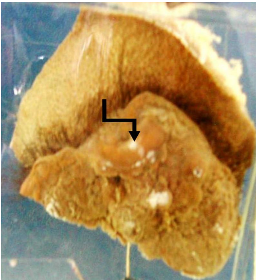
Squamous cell carcinoma of the scalp (malignant ulcer)
Pathologic process: malignant epithelial tumor.
A jar contains a piece of the skin of the scalp showing a large ulcerating mass:
- The ulcer shows the typical criteria of malignant ulcer which are:
• Number: single.
• Shape: irregular.
• Edge: raised everted
. • Floor: rough necrotic. -
The cut section shows invasion of the underlying tissue.
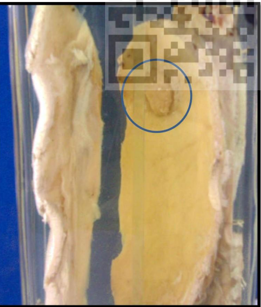
Squamous cell carcinoma of the scalp (nodular type)
Pathologic process: malignant epithelial tumor.
A jar contains 2 pieces of the scalp with the underlying bone of the skull showing big malignant tumor mass:
- Nodular
- Opaque grayish
- Arising from the skin of scalp
- Infiltrating the underlying tissues and skull bone to appear from its inner surface.
Basal Cell Carcinoma
Pathologic process: Locally aggressive epithelial tumor.
Section in a tumour showing 😀
- A locally aggressive tumour infiltrating the dermis formed of masses of malignant basophilic epithelial cells of variable sizes and shapes.
- The cell masses are formed of:
• Peripheral layer of columnar cells that are parallel to each other giving a palisading appearance of their nuclei.
• The central cells are polyhedral and rounded with no evidence of keratinization.
- The malignant cells show scanty and bluish cytoplasm with malignant criteria (Describe).
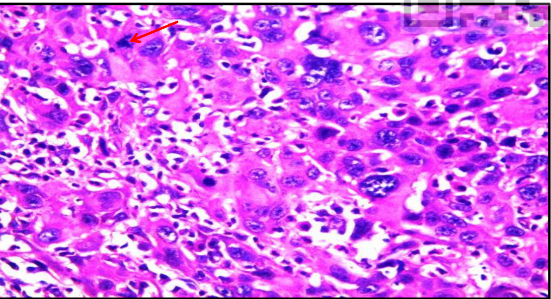
Malignant criteria
A section in malignant tumor showing malignant nuclear criteria in the form of: • Pleomorphism. • Hyperchromasia. • Increased N/C (Nucleo/Cytoplasmic) ratio. • Prominent nucleoli. • Abnormal mitosis (Red arrow).
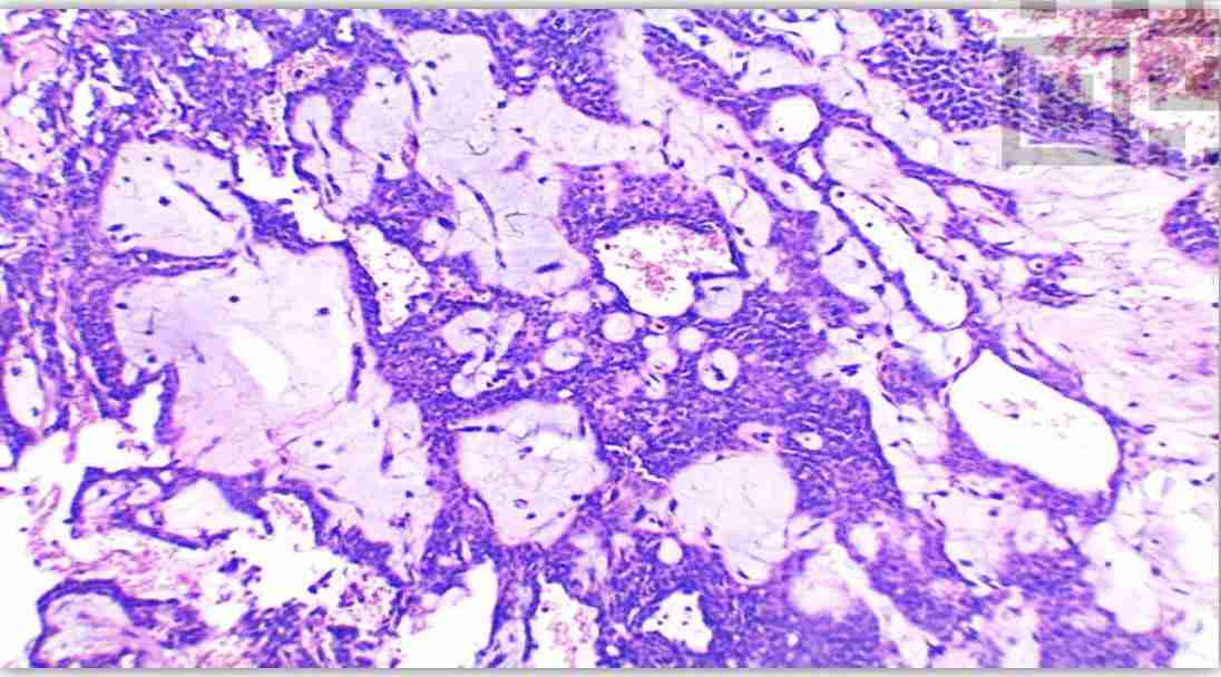
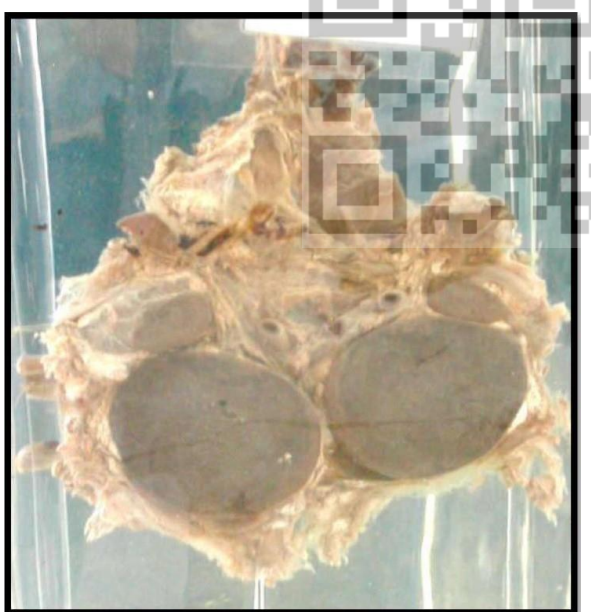
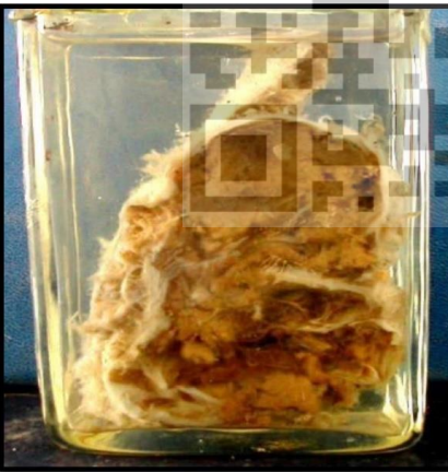
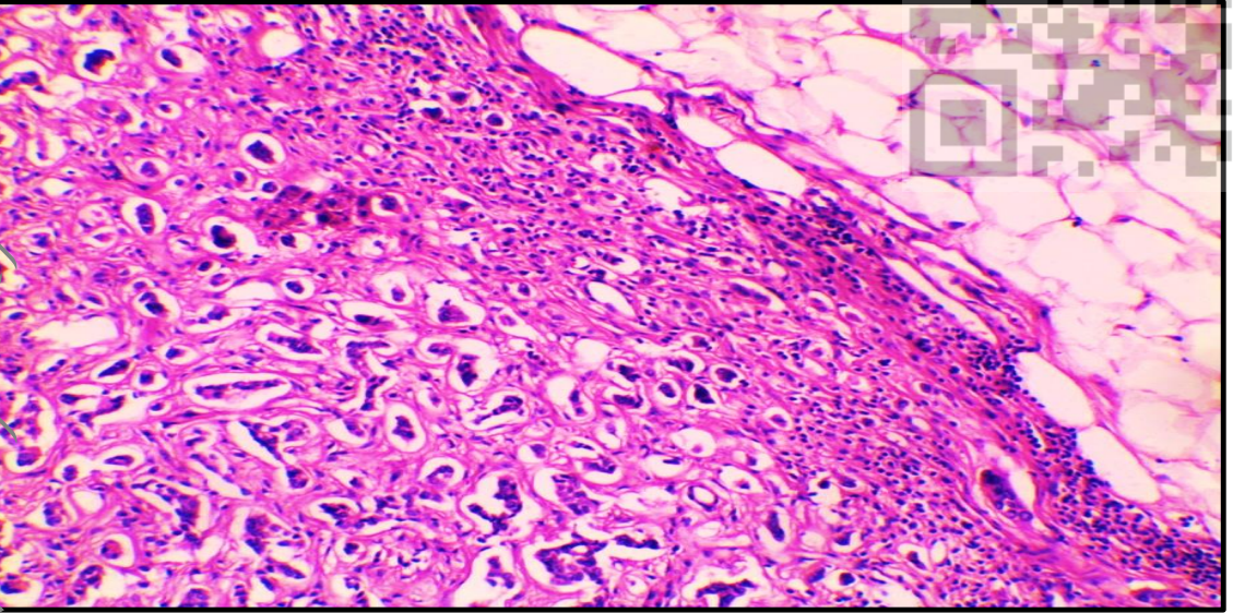
Mature cystic teratoma (Dermoid cyst) of the ovary
Pathologic process: Mature benign teratoma . Section in a benign ovarian cyst showing:
- Cyst wall is lined by stratified squamous epithelium.
- The cyst wall contains teratomatous elements: • Ectodermal elements such as sebaceous glands and hair follicles.
• Mesodermal elements in the form of cartilage and skeletal muscle.
Mature cystic teratoma (Dermoid cyst) of the ovary
Pathologic process: Mature benign teratoma .
A jar contains unilocular ovarian cyst cut into two halves showing:
- Multiple different structures projecting into the lumen from the wall such as tufts of hair, skin, and teeth.
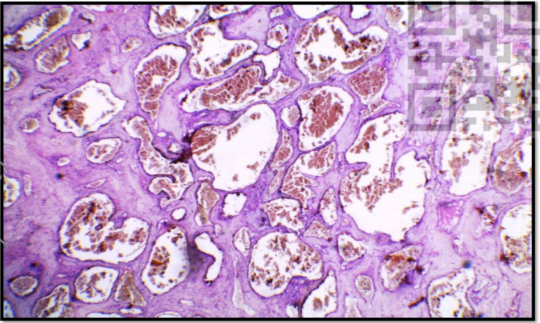
Thyroid adenoma
Pathologic process: Benign epithelial tumor. Section in a tumour showing:
• A well - encapsulated benign tumour tissue
. • Inside the capsule: - Closely packed follicles forming solid masses. - Some follicles contain colloid.
• Outside the capsule: - Normal thyroid follicles, regular uniform and filled with colloid.
- The follicles appear totally different from the neoplastic tissue inside the capsule
. • Within the capsule: There are compressed follicles. • No capsular or vascular invasion by the tumor cells
Thyroid Adenoma (solitary thyroid nodule) Pathologic process: Benign epithelial tumor.
A jar contains a solitary (single) thyroid nodule cut into two halves:
The nodule is: - Well encapsulated by fibrous tissue capsule. - Brownish in color due to gelatinous brownish colloid
Mucinous cystadenoma
Pathologic process: Benign epithelial tumor.
A section in cystic tumor showing:
A cyst wall lined by a single layer of mucinous epithelium
. - The lining epithelial cells:
• Columnar in shape • Nuclei are basally located.
• Cytoplasm contains mucin.
• Lack any atypia or mitosis.
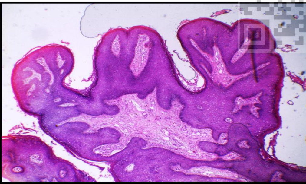
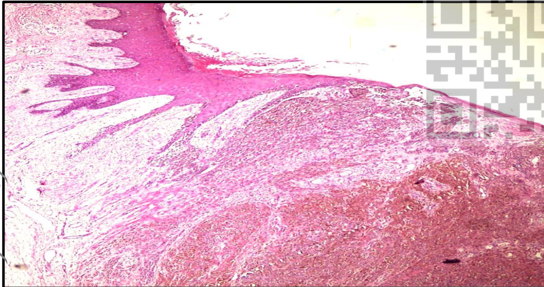
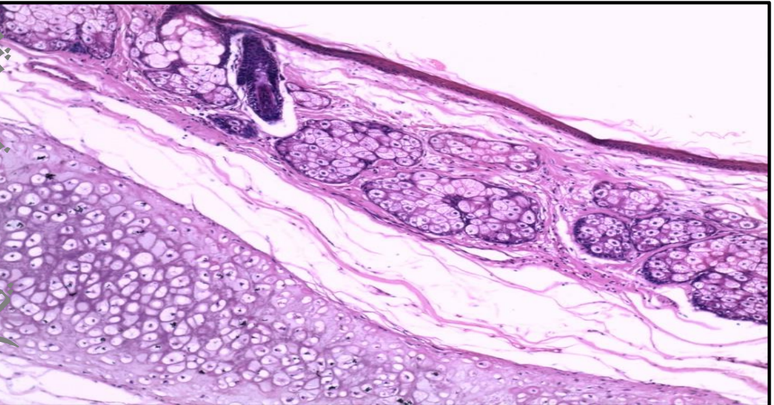
Squamous cell papilloma of skin
: Pathologic process: Benign epithelial tumor
• A branching central core of connective tissue covered with a thick layer of stratified squamous epithelium.
• The covering epithelium shows: -Hyperkeratosis (thick keratin layer) -Acanthosis (thick prickle cell layer) -Parakeratosis (retained nuclei of the keratin layer). -Basal cell layer hyperplasia
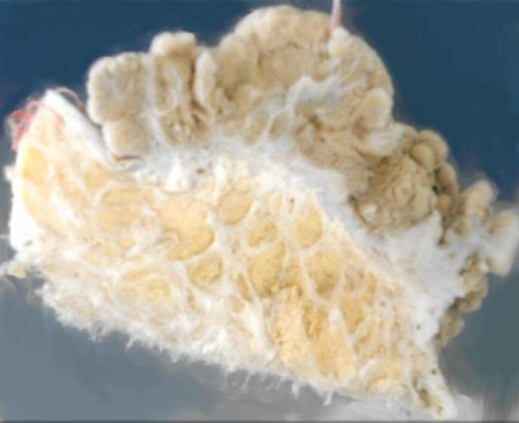
Squamous cell papilloma of skin Pathologic process:
Benign epithelial tumor A jar contains piece of the skin and subcutaneous tissue showing
: A large cauliflower mass arising from the skin surface:
➢ The mass is formed of: - Multiple compound papillary projections - Connected to the skin surface by a pedicle.
➢ No invasion of the subcutaneous tissue (line of demarcation between tumor and underlying subcutaneous
no surface ulceration
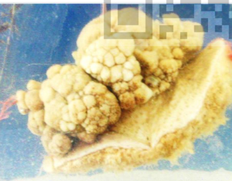