final exam - anatomy 2 lab
1/118
There's no tags or description
Looks like no tags are added yet.
Name | Mastery | Learn | Test | Matching | Spaced | Call with Kai |
|---|
No analytics yet
Send a link to your students to track their progress
119 Terms
inspiration
air moving from atmosphere into lungs
expiration
air expelled from respiratory tree into atmosphere
types of pressure gradients
atmospheric pressure, intrapulmonic pressure, intrapleural pressure
atmospheric pressure
pressure exerted by atmosphere on body
intrapulmonary pressure
pressure within the lungs
intrapleural pressure
pressure within pleural cavity
diaphragm during inspiration
contracted and flattened
diaphragm during expiration
relaxed and concave
during inspiration
thoracic volume increases, pressure decreases
during expiration
thoracic volume decreases, pressure increases
inspiratory muscles
external intercostals, diaphragm (prime mover)
forced expiratory muscles
internal intercostals, rectus abdominis, external abdominal obliques
tidal volume
amount of air inhaled or exhaled in one breath
inspiratory reserve volume (IRV)
amount of air that can be forcefully inhaled
expiratory reserve volume (ERV)
amount of air that can be forcefully exhaled
vital capacity (VC)
amount of air that can be inhaled and then exhaled with maximum effort; deepest possible breath
inspiratory capacity (IC)
maximum amount of air that can be inhaled after a normal tidal expiration
functional residual capacity
amount of air remaining in the lungs after a normal tidal expiration; can never be emptied from lungs
total lung capacity (TLC)
maximum amount of air the lungs contain
vital capacity formula
VC = expiratory reserve volume (ERV) + tidal volume + inspiratory reserve volume (IRV)
inspiratory capacity formula
IC = tidal volume + inspiratory reserve volume (IRV)
functional residual capacity
FRC = residual volume (RV) + expiratory reserve volume (ERV)
total lung capacity formula
TLC = residual volume (RV) + vital capacity (VC)
hypoventilation (slow, shallow breathing)
raises CO2, lowers blood pH
more H+ means
lower pH
hyperventilation (rapid, deep breathing)
losing large quantities of CO2, little change in O2, higher blood pH
obstructive diseases
asthma, COPD, chronic bronchitis
restrictive disease
pulmonary fibrosis
digestion
process of breaking down molecules small enough to be absorbed across the cell membrane
five basic processes of digestion
ingestion, movement of food, digestion, absorption, defecation
organs of digestive system
mouth, esophagus, stomach, small intestine, large intestine
acessory organs of digestive system
teeth, tongue, salivary glands, pancreas, liver, gall bladder
salivary glands
parotid glands, sublingual glands, submandibular glands
four major areas of stomach
cardia, fundus, corpus, pylorus
fundus produces
hydrochloric acid
pyloric glands secrete
mucus
small intestine does
90% of absorption of nutrients
small intestine parts
duodenum, jejunum, ileum
ileum
ileocecal sphincter at distal most portion
large intestine divisions
cecum, ascending colon, transverse colon, descending colon, sigmoid colon, rectum
liver
upper right quadrant, under diaphragm; two lobes; produces bile
bile
detergent, not an enzyme; emulsify fat
gall bladder
pear shaped sac in depression of liver; stores bile and concentrates it
pancreas
produce digestive enzymes
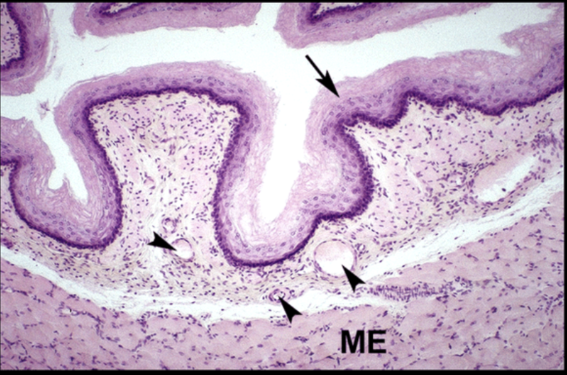
esophagus histology
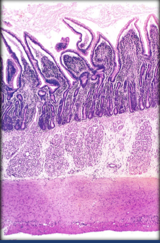
stomach histology
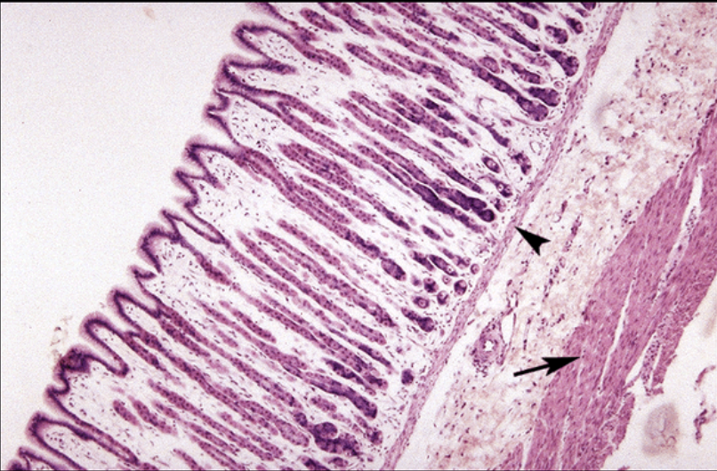
small intestine histology
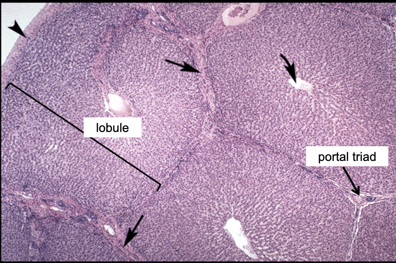
liver histology
portal triad histology
portal triad consists of
hepatic artery, portal vein, bile duct
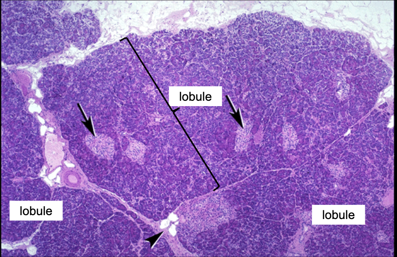
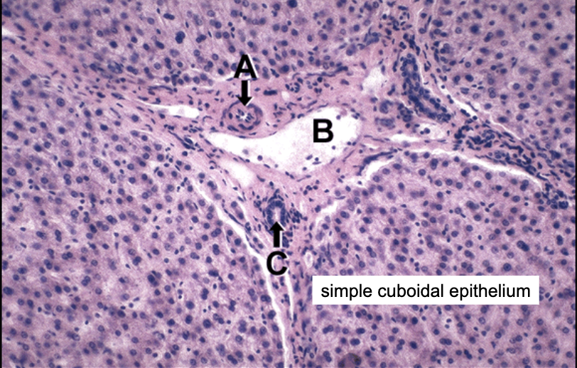
pancreas histology
components of urinary system
kidney, ureter, bladder, urethra
function of urinary system
removing and restoring water and solutes from blood, excreting urine
urine consists of
water, nitrogenous wastes, toxins, H+, ions
kidney external anatomy
hilus, renal artery, renal vein, ureter
renal hilus
entrance/exit for ureter, renal artery, and vein, nerve, lymphatics; entrance to renal sinus
renal fascia
renal capsule (collagen and elastin); adipose capsule, protects and anchors kidney

diagram of renal capsule
renal capsule
kidney internal anatomy (layers)
renal cortex, renal medulla
renal cortex consists of
renal columns
renal medulla consists of
renal pyramids, papilla, minor and major calyx, and renal pelvis

diagram of renal cortex
renal cortex
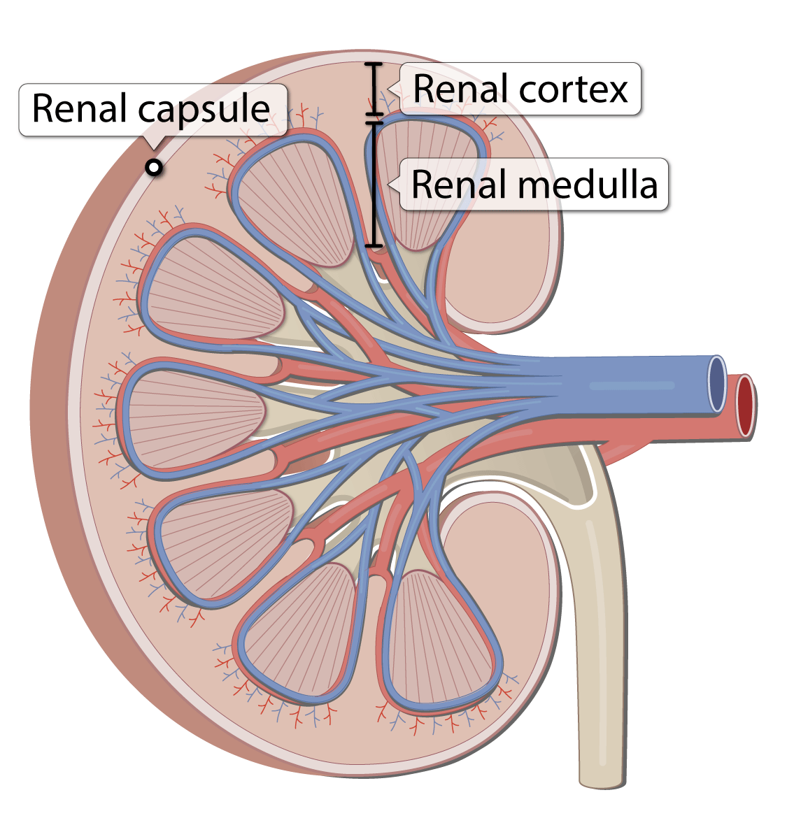
diagram of renal medulla
renal medulla

diagram of renal pyramid
renal pyramid
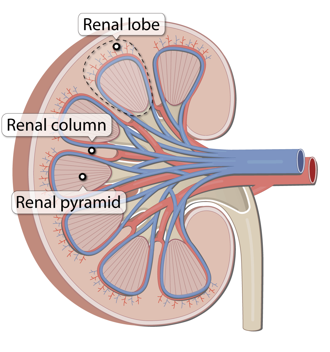
diagram of renal column
renal column
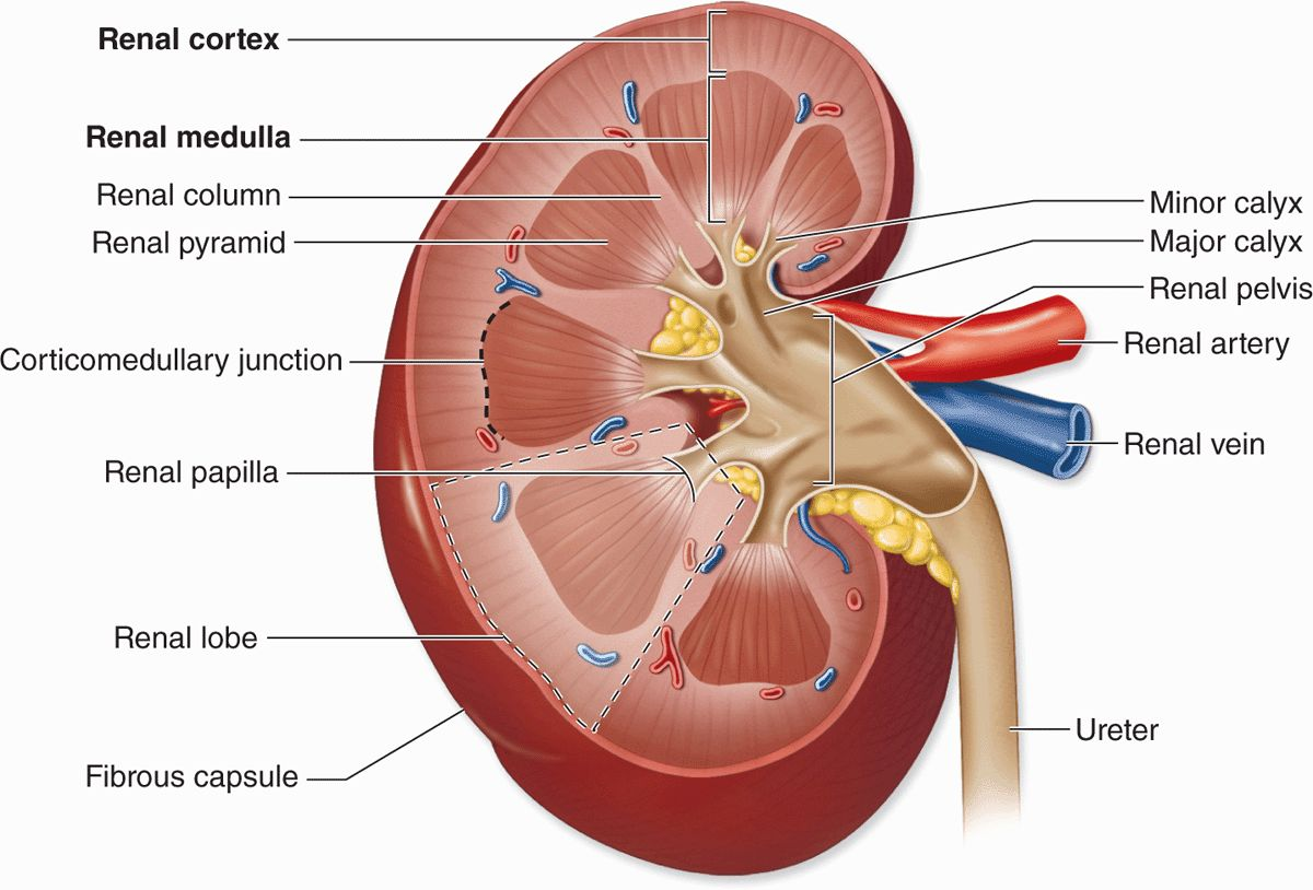
diagram of renal papilla
renal papilla
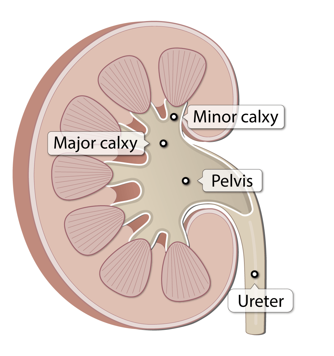
diagram of calyces
minor and major calyx

diagram of renal pelvis
renal pelvis
function of nephron
filtration, reabsorption, secretion
filtration
pressure across endothelium
reabsorption
diffusion and active transport
secretion
tubule cells secrete additional materials directly into filtrate
nephron is located in
renal pyramid
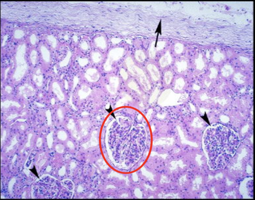
renal corpuscle consists of
glomerulus, bowman’s capsule (parietal, visceral, capsular space)
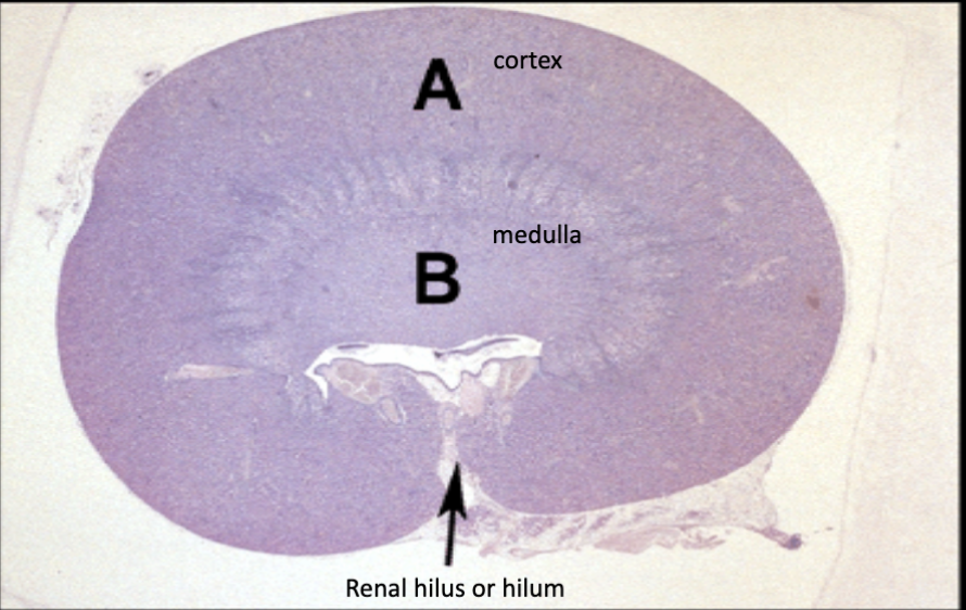
kidney histology
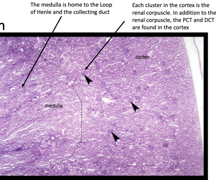
nephron histology
renal corpuscle histology
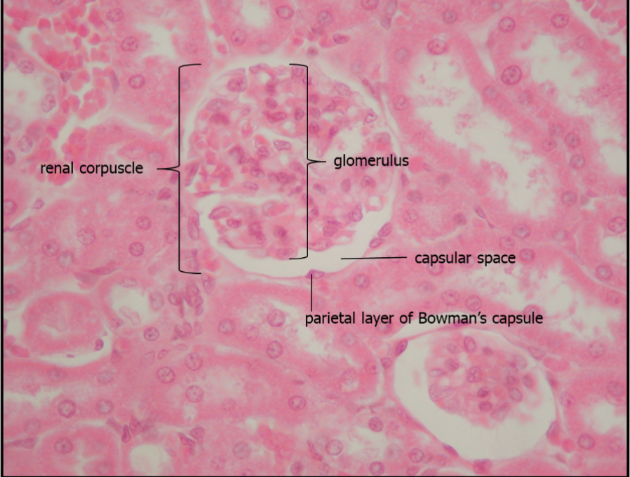
glomerulus histology
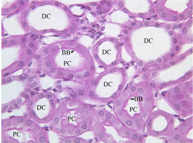
pct - more abundant (longer than the dct); brush border present, dct - less abundant; no brush border
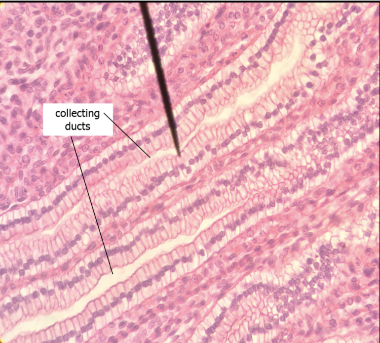
collecting duct histology
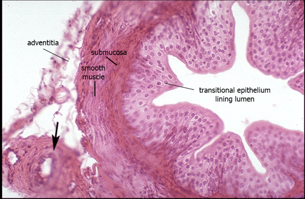
ureter histology
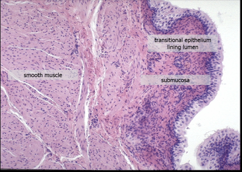
urinary bladder histology

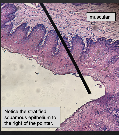
male urethra histology
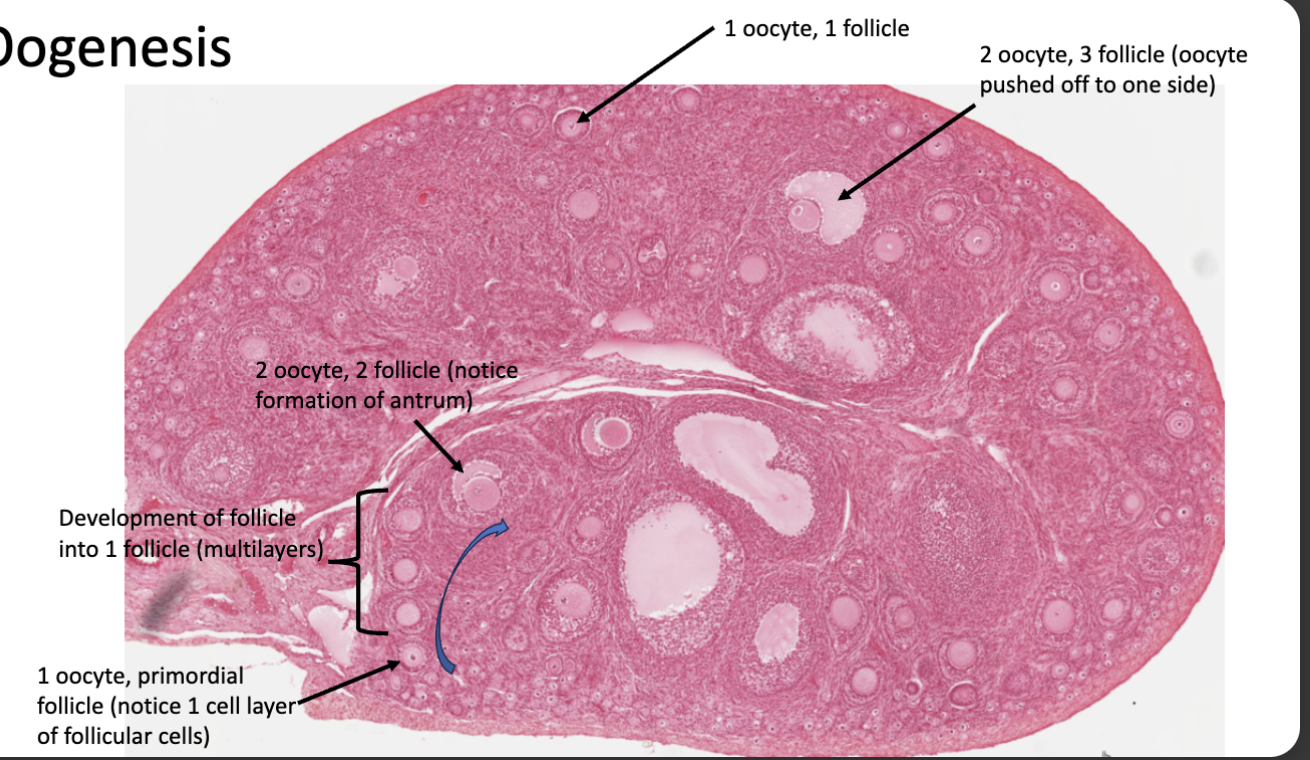
oogenesis histology
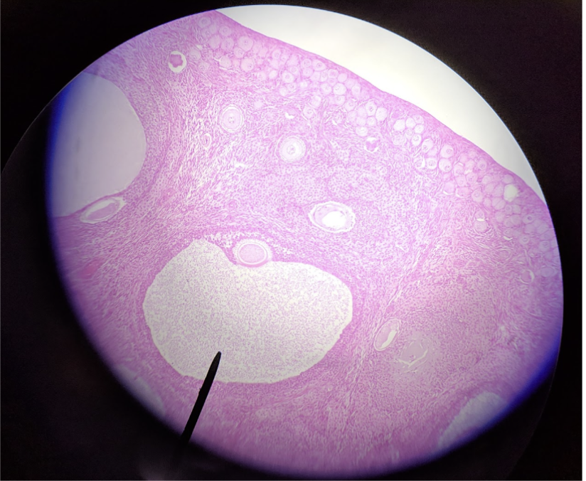
secondary oocyte in tertiary follicle
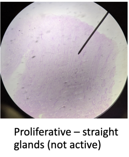
proliferative phase - growing back of the stratus functionalis; long, straight uterine glands
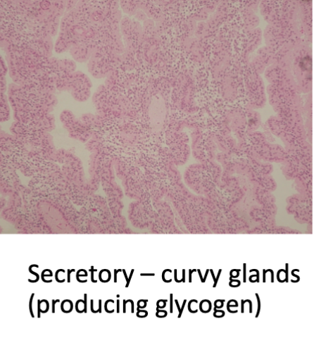
secretory phase - signals for water retention, softening of uterine lining, glands (convoluted) provide glycogen, substantial blood supply, awaiting fertilized egg implantation
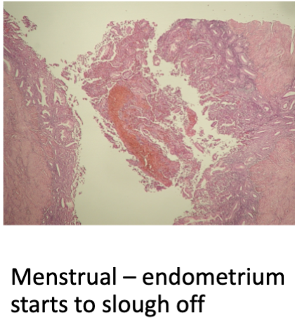
menstrual phase - if NO fertilization, uterine lining shed (endometrium thinner, in disarray)
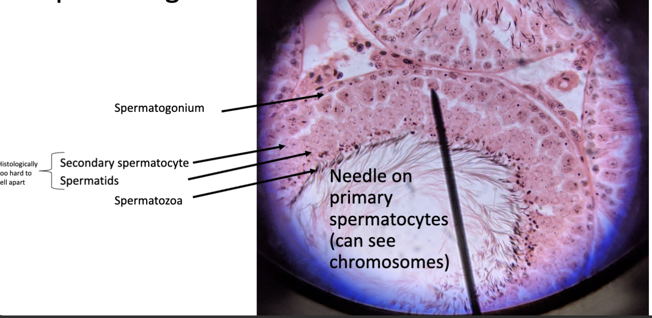
spermatogenesis
most reabsorption takes place in the
proximal convoluted tubule; fine tuning after in loop of henle, distal convoluted tubule, and collecting duct
mechanisms for movement
active transport, diffusion, facilitated diffusion, secondary active transport, osmosis
ureter
union with bladder, no valve, prevention of reflux of urine into ureters, pressure on bladder wall; function to transport urine, peristaltic waves
urinary bladder
hollow muscular organ, lies retroperitoneally in pelvic cavity, rises in abdominal cavity as fills
urethra
final passageway for urine from body, sphincters (external and internal), trigone zone (2 ureteral openings and urethral opening)

female urethra (stained blue)
female urethra
flow of urine
proximal convoluted tubule, loop of henle, distal convoluted tubule, collecting duct, renal papillae, minor calyx, major calyx, renal pelvis, ureter, urinary bladder, urethra
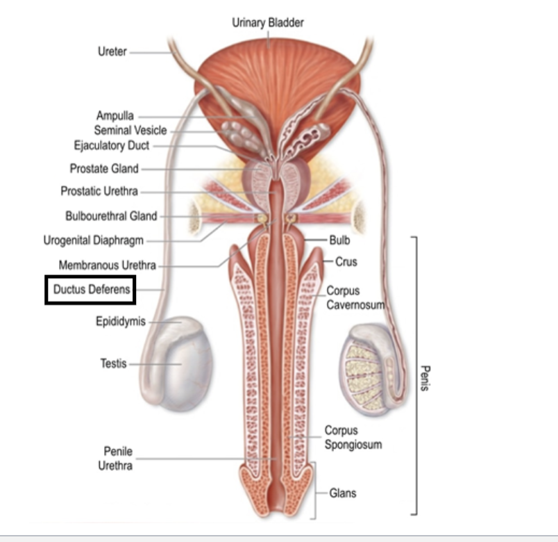
male reproductive structures
spermatogenesis occurs in
seminiferous tubules
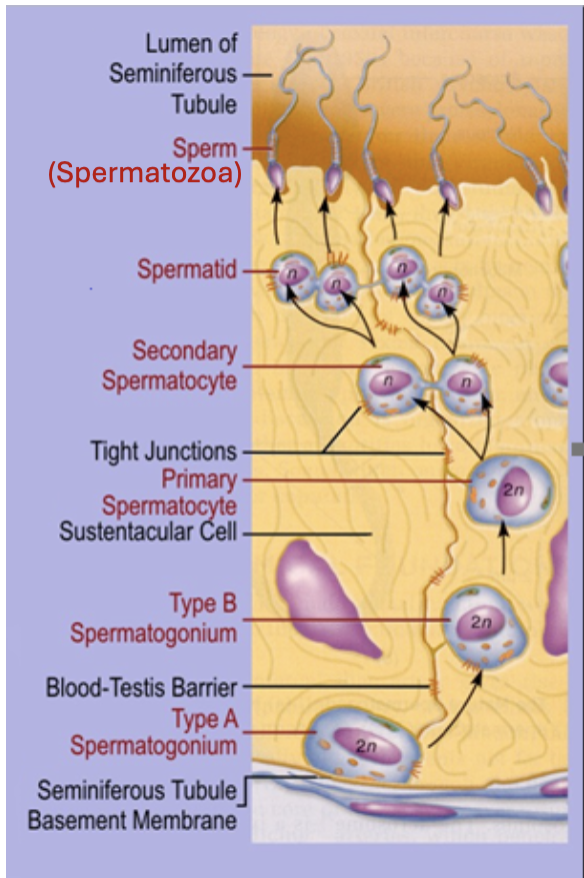
process of spermatogenesis