biology 30-ib: cell division and molecular genetics
 Call Kai
 Learn
 Practice Test
 Spaced Repetition
 Match
 Flashcards
 Knowt Play
1/74

Earn XP
Description and Tags
homie chromies are homologous chromosomes
75 Terms
griffith experiment and griffith’s transforming principle
trials done on mice with pathogenic and non-pathogenic bacteria. mice with pathogenic bacteria died while non-pathogenic and heat-treated pathogenic lived. when a mix of heat-treated pathogenic and non-pathogenic were injected, the mouse died.
griffith's transforming principle: the heat treated bacteria 'transferred' something to the harmless bacteria to transform it.
hershey and chase experiment
aimed to find the genetic material in the cell. people thought it was either protein or DNA that was the inheritable unit.
two groups of bacteriophage: radioactive sulfur-35 (found in protein only) and radioactive phosphorus-32 (found in DNA only).
grew viruses in radioactive medium
let viruses reproduce in vitro
centrifuge to separate
check pellet and supernatant for radioactivity
supernatant: contains viruses, liquid portion of solution
pellet: contains bacteria, solid portion of solution
%reactivity in supernatant of the phosphorus-32 was lower than the sulfur-35, meaning DNA was injected into the bacteria.
watson and crick
determined the structure of DNA from x-ray crystallography images (performed by rosalind franklin)
structural features of DNA
polymer of nucleotides
2 twisting strands that form a helix, both are complementary and antiparallel
A-T and C-G bonds are of equal length (helix stability)
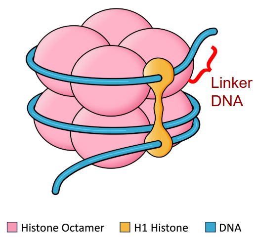
DNA is wrapped around histones forming an organised package called a nucleosome
“backbone” of DNA are repeating unites of deoxyribose sugar and phosphate
“rungs” are nitrogenous bases
sketch DNA
label:
deoxyribose
phosphates
nitrogenous bases + type (ATGC)
direction of strands (5’ and 3’)
strands pointing in opposite directions (tip of deoxyribose pointing towards 5’ end)

nucleotides
phosphate group, deoxyribose sugar, and nitrogen base
4 DNA nucleotides which split into two groups
purines: adenine and guanine
pyrimidines: cytosine and thymine
bases can only hydrogen bond to their complementary pair (i.e. A-T and G-C)
tetranucleotide hypothesis
early proposals suggested a square shape containing all four nucleotides.
erwin chargaff falsifies this by showing that all four nucleotides were not found in equal amounts.
chargaff’s rules
number of purines = number of pyrimidines, or A+G=C+T
in other words, A=T;G=C
5’ and 3’ ends
five prime and three primes are used to indicate the direction of individual strands of DNA. deoxyribose has polarity so carbons are counted.
because of this, DNA is “anti-parallel” and “complementary”.
purine-pyrimidine stability
A-T and G-C pairs have equal length, i.e. regardless of the location, DNA will have the same overall width which makes it more stable.
nucleosome
made of 8 core histone proteins. 1 additional histone protein forms the linker DNA.

DNA replication
replication starts at a specific sequence, called the replication origin.
step 1: initiation
DNA helicase separates the strands of DNA forming a replication fork
step 2: RNA priming
RNA primase will make a short RNA piece (primer)
step 3: synthesis
DNA polymerase III reads the original DNA and makes a complementary copy
nucleotides are extended on the 3’ end
when mistakes occur, DNA pol III reverses direction and removes it
problem: DNA pol III can only make DNA in the 3’ direction, which causes the two strands to replicate differently
leading strand: DNA pol III extends until replication is finished
lagging strand: DNA pol III extends in short okazaki fragments
step 4: replacing the primer
DNA polymerase I removes RNA primers and synthesises DNA in its place
step 5: fixing the backbone
DNA ligase will bond gaps in backbone through dehydration synthesis to complete the covalent bonds between 5’ and 3’ ends
ending result: each piece of DNA contains one old and one new strand of DNA, called semi-conservative replication
semi-conservative replication
after DNA replication, each piece of DNA contains one old and one new strand of DNA
protein
composed of 20 unique amino acids with many different functions.
monomer: amino acid
polymer: polypeptide, protein
protein synthesis transcription
uses the gene to create an mRNA copy of the protein to be made
transcription factors bind promoter on the DNA
RNA polymerase finds the promoter and unzips DNA
RNA pol makes an mRNA strand with nucleotides complementary to the DNA code (always extending the 3’ end of RNA)
completed mRNA is moved out of nucleus
RNA pol falls off and DNA strands rejoin
translation occurs after this.

post-transcription modification
in eukaryotes, mRNA is usually modified before it is translated
5’ cap and polyA (adenine) tail stabilise ends of mRNA to prevent digestion by nucleases
non-coding regions of mRNA called introns are removed
protein synthesis translation
makes proteins based on the mRNA code
codons: groups of three nucleotides that code for one amino acid
i.e. there are 1/3rd the amount of amino acids compared to DNA/mRNA
universal for all life!
occurs after transcription.
initiation:
ribosome attaches to mRNA from 5’ end
a tRNA binds to AUG start codon to start synthesis
elongation: EPA sites
anticodon for AUG is bound to P site
second tRNA pairs with the next codon in A site
condensation reaction attaches the two amino acids
P site tRNA has no amino acid and is moved to the E site when the ribosome shifts to the next codon
termination:
ribosome reaches a stop codon on mRNA and falls off
stop codons do not add an amino acid!
some polypeptides require additional modifications before being functional
mRNA
messenger ribonucleic acid
usually found as a single strand
contains adenine, cytosine, guanine, and uracil instead of thymine
proteins are made outside the nucleus and DNA is too large and important to be moved, so mRNA is small, mobile, and temporary
there are also rRNA: ribosomal RNA, and tRNA: carries amino acids to the ribosome
sketch deoxyribose vs ribose

non-coding DNA
do not code for proteins but have other functions:
code for tRNA and rRNA
enhancers and silencers (affect rate of transcription)
telomeres: form the ends of chromosomes
introns: sequences that are edited out of mRNA
promoters: binding site for RNA polymerase, typically requires a transcription factor
alternative splicing
some exons may be removed or added to modify and specialise proteins for different purposes
e.g. adding or removing a membrane anchor to determine if the protein is membrane-bound or free moving
recycling amino acids
proteins can be targeted for recycling by adding a ubiquitin tag
ubiquinated proteins will be brought to a proteasome for dismantling
genome
complete set of genetic information in an organism/species
transcriptome
complete set of RNAs currently being transcribed in a cell
proteome
complete set of proteins currently being transcribed in a cell
mutations
a change in DNA sequence.
can have beneficial or detrimental effects.
germ-line/gametic mutations: occur in gametes and can be passed on to future zygotes.
somatic mutations: occur in body cells, (not passed on) can lead to cancer.
cause of mutations
can be spontaneous or induced:
spontaneous: mistakes during proofreading in DNA rep.
induced: caused by mutagenic substances.
mutagens
any agent that causes changes in DNA.
physical mutagens: ionizing radiation (UV, x-ray, gamma rays), asbestos, etc.
chemical mutagens: carcinogens such as nitrites, gas fumes, cigarette smoke
cannot occur naturally with the intent of creating a deliberate change (lamarckian)
degeneracy/wobble hypothesis
multiple codons code to the same amino acid
wobble pairing: the last letter in a codon is usually the least important, so it can change without changing the entire protein
this DNA is considered a degenerate code
point mutations
changes one nucleotide
silent mutations
has no effect on the protein sequence, degeneracy (sometimes a wobble pairing if it is the last letter in the codon)
e.g. GTA → GTT, both are valine
missense mutations
changes a single amino acid
e.g. CCC → ACC, proline → threonine
nonsense mutations
substitutes an amino acid to create a stop codon
e.g. TAC → TAG, tyrosine → stop
frameshift mutation
insertion or deletion of a single nucleotide shifts the entire amino acid code.
e.g. huntington’s disease: insertion of multiple CAG nucleotides in HTT gene on chromosome 4, mutated protein has an adverse effect on brain cells
e.g. cystic fibrosis: deletion of the 7th, 8th, and 9th nitrogen bases, inability to produce a protein that regulates Cl- channels, lung secretions are thick and block airways.
single-nucleotide polymorphisms
occur in both coding and non-coding regions, presence is associated with certain diseases
can look for SNPs to determine predisposition to develop a disease in the future.
DNA sequencing
method to record the DNA sequence of any piece of DNA.
mtDNA
circular mitochondrial DNA passed down from mother to child.
replicates independently from the nucleus DNA.
can help trace human migration histories.
polymerase chain reaction
scientific process to create copies of DNA through multiple cycles of:
heating: distrupts hydrogen bonds between DNA strands
annealing: attachment of designed primers
extension: DNA replication by the temperature resistant taq polymerase: heat resistant form of DNA pol I, to make copies of DNA
gel electrophoresis
separates pieces of DNA based on size
DNA sits in an electrolyte bath, current passes through and DNA fragments move through a gel
DNA is negative due to phosphate groups so they are attracted to positive cathodes
small pieces move faster than larger ones
DNA fingerprinting
gel electrophoresis is performed on randomly cut genetic material
each individual has their own unique size of cut genetic material, matches can be used for relation between DNA samples
e.g. criminal cases, determining paternity
process for creating mutants
restriction endonucleases (DNA scissors) cuts palindromic sequence into the circular plasmid DNA, creating sticky ends
plasmids can replicate on their own without the main chromosome
technologists design genes to have the complementary sticky ends to help the mutated gene insert
DNA ligase joins a mutant gene to the plasmid DNA
plasmid is introduced into bacterial cell
chromosomes
DNA is bundled in the cells in thread-like structures called chromosomes
chromosomes contain one long double strand of DNA
DNA is packaged with histones and folded into a chromatin fibre
chromosomes can be:
condensed or uncondensed (i.e. tightly coiled or loose)
duplicated or unduplicated (i.e. is it past the s-phase of mitosis?)
interphase
the process of cell activity between cell divisions.
G1: gap/growth 1, rapid cell growth
S: synthesis, duplication of chromosomes
G2: gap/growth 2, cell growth and organelles increase in number, preparation for mitosis
mitosis occurs after G2
G0: cells that go not go beyond G1 stay here, may be permanent or temporary
e.g. nerve, heart, and red blood cells
mitosis
asexual cell division that maintains the normal number of chromosomes after cell division.
46 chromosomes in humans.
prophase:
chromatin condenses, chromatids are held together by the centromere
centrioles duplicate in animal cells
spindle fibres (or microtubule) extend from the centrioles and attach to the centromeres
nuclear membrane begins to break
metaphase:
chromosomes moved to equator of the cell by spindle fibres
centrioles migrate to the poles of the cells
anaphase:
spindle fibres shorten and rip sister chromatids to opposite poles of the cell
sister chromatids are now referred to as chromosomes
telophase:
chromosomes reach the opposite poles
cell membrane begins to pinch in animal cells (cell furrow)
nuclear membrane begins to reform
chromosomes start to uncondense
spindle fibres are broken up
cytokinesis:
cell furrow continues until two separate cells are created
cell wall created in plant cells
new cells enter G1 phase of interphase
in some cases, daughter cells will not have equal amounts of cytoplasm (oogenesis/yeast)
meiosis
sexual cell division that produces cells with half the number of chromosomes after cell division.
23 chromosomes in humans.
mitosis is done twice but there is no s-phase between the two
meiosis I: involves all mitosis stages (PMATC)
prophase I: chromosomes find their homologous pair to form a tetrad, homie chromies exchange portions of their chromatids in synapsis
metaphase I: tetrads line up along the equatorial plate
anaphase I: homologous chromosomes are separated (not chromatids!)
telophase I and cytokinesis I: nuclear membrane reforms and the cells completely separate
cells are not identical to original cell due to synapsis and reduced number of chromatids
meiosis II: identical to mitosis!
sister chromatids are split into each new cell (gametes)
in humans, females make one ovum and three polar bodies while males make four sperm cells
somatic cells
cells that have two copies of each unique chromosome
2 copies x 23 unique chromosomes = 46 chromosomes total
gamete cells
specialised cells that have 23 unique chromosomes.
sperm and egg cells in humans
ploidy
the number of complete chromosomes in a cell.
haploid: contains a single set of chromosomes, found in gametes
n = # of chromosomes
diploid: contains two sets of chromosomes, found in somatic cells
2n = total # of chromosomes
homologous chromosomes
a matched pair of chromosomes from each parent that share the same length, centromere position, and gene expression
autosomes
any chromosome that is not a sex chromosome, 22 pairs in a human
sex chromosomes
chromosomes that determine an individual’s sex; larger chromosome is X and smaller is Y
supercoiling
chromosomes that are condensed by supercoiling DNA around histones until a tight, short, and wide package is created.
chromatid
a replicated chromosome, each copy is known as a chromatid.
sister chromatids are two identical copies.
held together by the centromere.
telomeres
non-coding DNA caps at the end of eukaryotic chromosomes
reduce in length each time a cell goes through the cell cycle and divides
may have a role in cell aging and the behaviour of cancer cells
the enzyme telomerase maintains the length of the telomere and slows cell death
cell proliferation
creation of new identical cells in mitosis to replace old or damaged cells
cyclins
control the cell cycle
when cyclins reach a threshold concentration, they activate other proteins needed to move to the next phase through phosphorylation
they form part of the checkpoints of the cell cycle
if a checkpoint is not passed, programmed cell death is possible
stem cells
cells that can develop into many different specialised cell types.
totipotent: able to become any cell in the body
pluripotent: able to become almost any cell in the body
multipotent: able to become only a few cells in the body
twins
there are two types of twins:
identical twins, or monozygotic: form when a cell breaks free from the undifferentiated embryo and develops on its own, one sperm and one egg
fraternal twins, or dizygotic: form when two eggs are fertilized and implant at the same time, two sperms and two eggs
cloning
occurs when identical organisms are produced from the DNA of a single totipotent cell
cancer
a state of constant mitosis that can suction energy from other parts of the body to grow these cells
oncogenes
genes that may cause cancer, mutate from proto-oncogenes (which normally help increase cell growth and continuation of the cell cycle) and cause cells to grow and divide uncontrollably.
cancerous mutations
there are two types of mutations:
mutations in proto-oncogenes: turns them into oncogenes which then undergo constant mitosis, like too much gas in a car
mutations in tumour suppressor genes: stop triggering apoptosis, like no brakes in a car
mutations in either sets of genes can lead to cancers
tumour
a mass of cancerous cells within an otherwise normal tissue
benign tumours: do not usually cause serious problems and can be removed through surgery
malignant tumours: impair functions of one or more organs
carcinomas: malignant tumours caused by carcinogens
unlike normal cells, cancer cells do not stick together and therefore can move throughout the body from the blood stream (metastasis)
types of malignant tumours
primary: where the cancer has originated
secondary: when metastasis has occured
mitotic index
the ratio of the number of cells undergoing mitosis to the total number of cells
cells in mitosis ÷ total number of cells = mitotic index
nondisjunction
when anaphase does not occur properly in meiosis
this will result in some gametes having more chromosomes and others having less
in most cases, this makes gametes inviable (i.e. they cannot develop into offspring)
nondisjunction types that create 45 or 47 chromosomes can develop into surviving babies
cells with older telomeres are more likely to go through nondisjunction
nondisjunction in anaphase I
homologous chromosomes do not separate
results in two gametes with the ploidy n+1 and two with n-1
nondisjunction in anaphase II
sister chromatids do not separate
results in one gamete with the ploidy n+1, one with n-1, and two with n
karyotype
a visual representation of an individual’s chromosomes
chromosomes are identified by length, banding, and centromere position

trisomy
a nondisjunction disorder where there are three of a homologous chromosome
ploidy is 2n+1=47
trisomy 21 (down syndrome): three copies of the 21st set of chromosomes
disomy
a nondisjunction disorder where two copies of the same sex chromosome are passed on from one parent.
XXY (klinefelter syndrome): three sex chromosomes instead of two, one extra X chromosome.
monosomy
a nondisjunction disorder where there is one copy of a homologous chromosome.
ploidy is 2n-1=45
monosomy X: only one X chromosome instead of two.
genetic testing for chromosome abnormalities
amniocentesis: a needle is used to extract a sample of the amniotic fluid that surrounds the fetus.
chorionic villus sampling: cells are extracting from the chorion of the developing embryo.
alternation of generations
when an organism switches between a diploid stage (sporophyte) and a haploid stage (gametophyte) during their life cycle.
diploid stage is the zygote which goes through meiosis immediately.
this creates somatic haploid cells which go through mitosis to create gametes.
there are two main examples of this:
mosses: exist mostly in haploid form (gametophyte)
sporophyte will go through meiosis to make more gametophyte spores
ferns: exist mostly in diploid form (sporophyte)
sporophyte will go through meiosis to create haploid cells which will be fertilized and create the sporophyte gametes
rules for changes in ploidy
2n → 2n or n → n means mitosis has occured
2n → n means meiosis has occured
n + n → 2n is fertilization