Anatomy test 3
 Call Kai
 Learn
Learn Practice Test
 Spaced Repetition
 Match
Match Flashcards
 Knowt Play
1/212

There's no tags or description
Looks like no tags are added yet.
Name | Mastery | Learn | Test | Matching | Spaced | Call with Kai |
|---|
No analytics yet
Send a link to your students to track their progress
213 Terms
Vertebral column
Contains 7 cervical, 12 thoracic, 5 lumbar, 5 sacral and 4 coccygeal bones
Functions of the vertebral column
Support body weight, transmit forces through pelvis to lower limbs, carry and position head, brace and help manoeuvre the upper limbs and protect spinal cord
Cervical curve
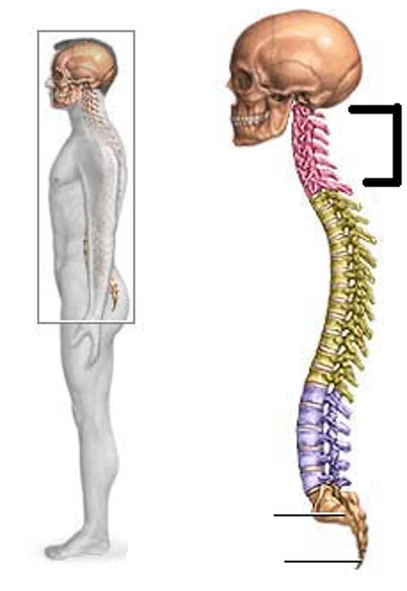
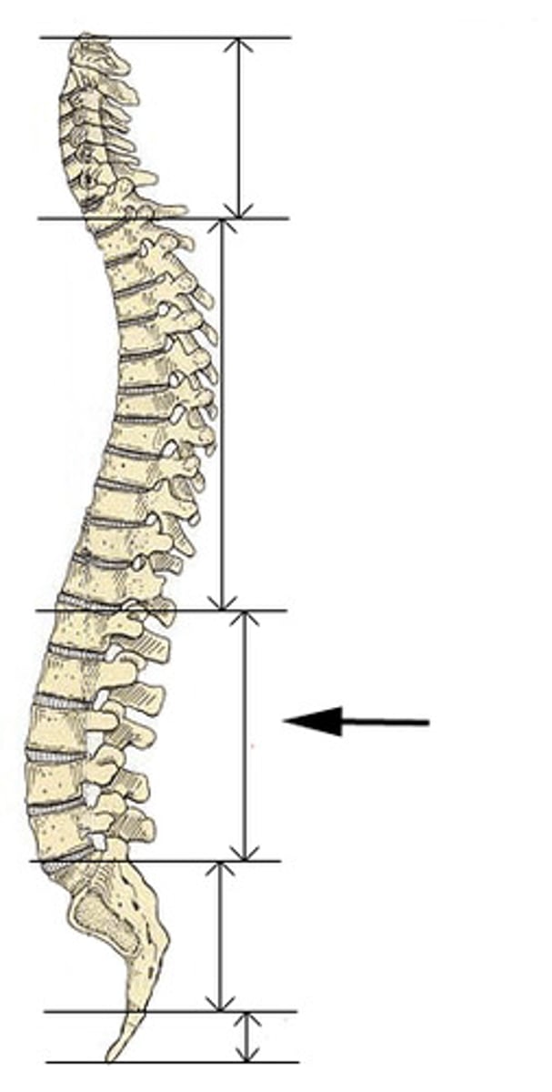
Lumbar curve
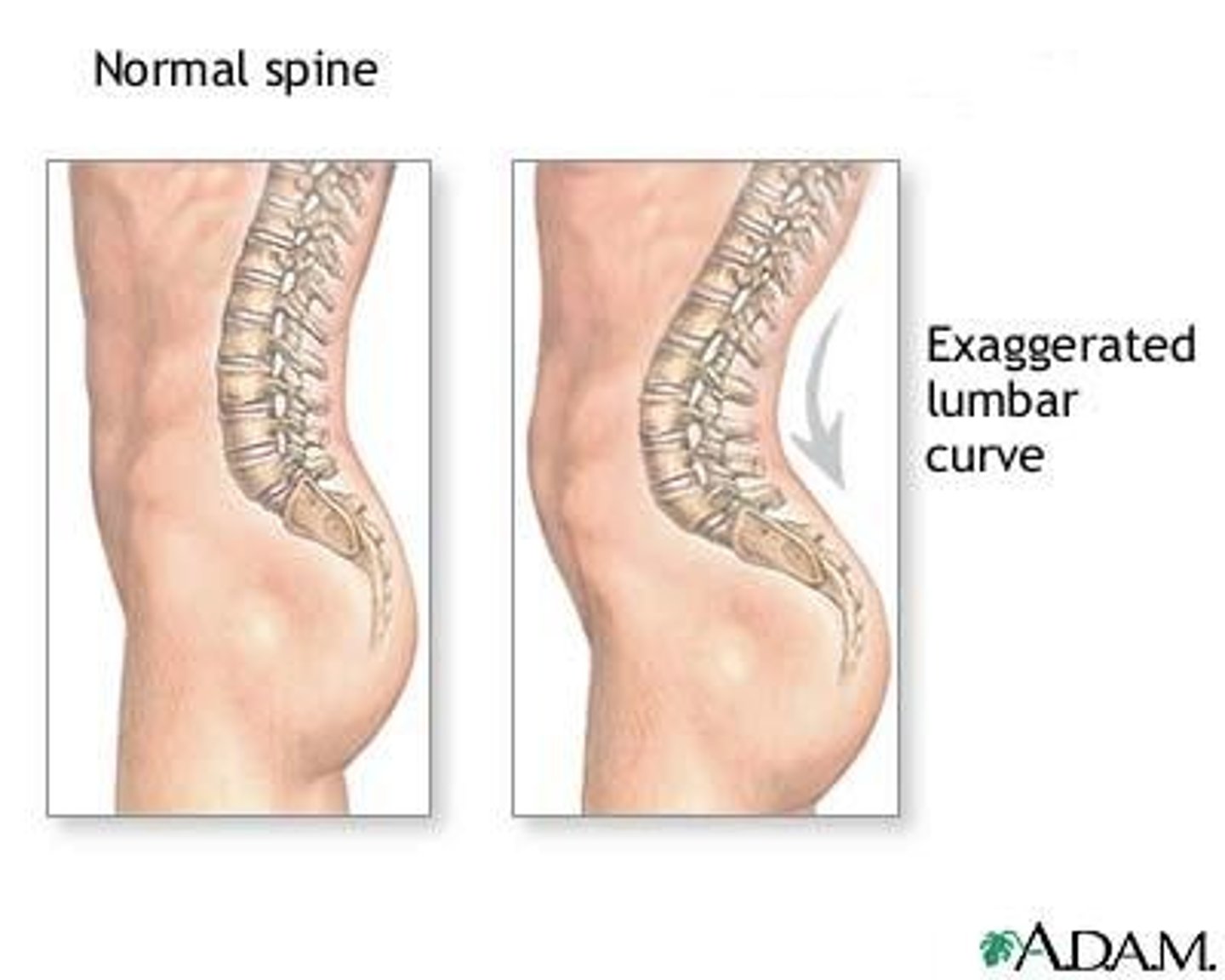
Lordosis
Kyphosis
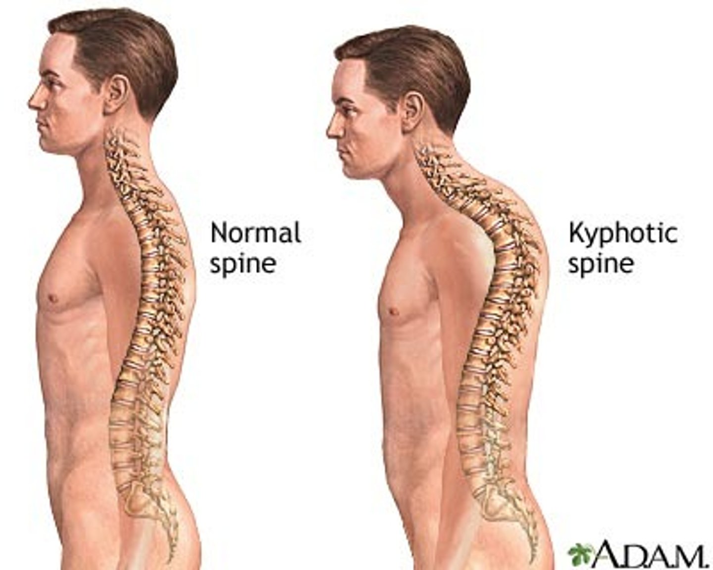
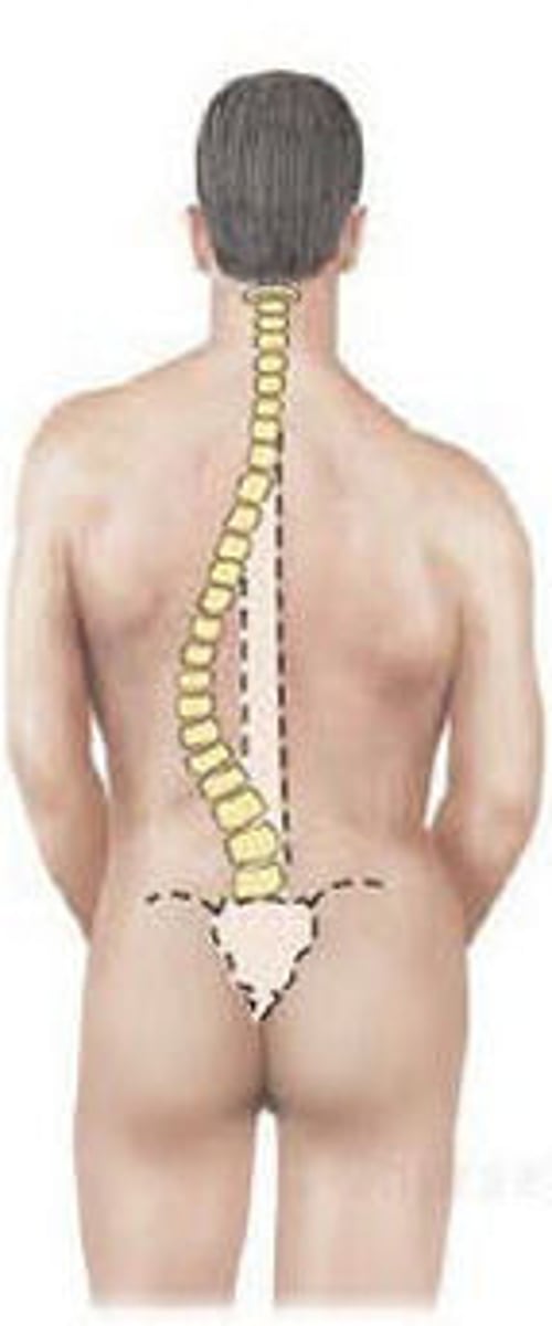
Scoliosis
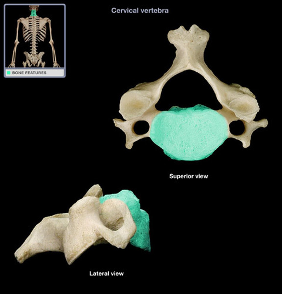
Body of vertebrae
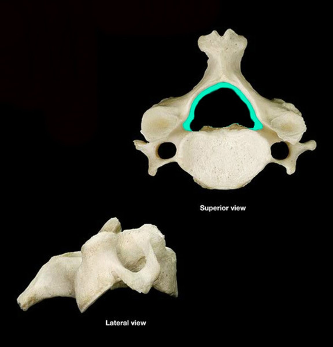
Vertebral arch
Pedicle
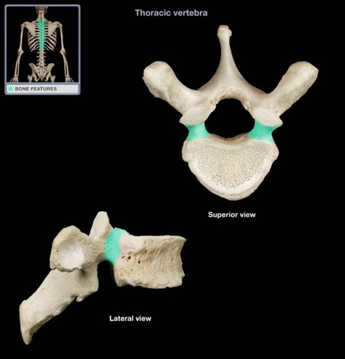
Lamina
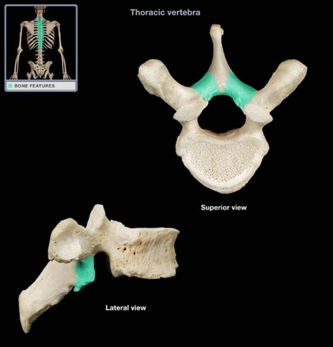
Vertebral foramen
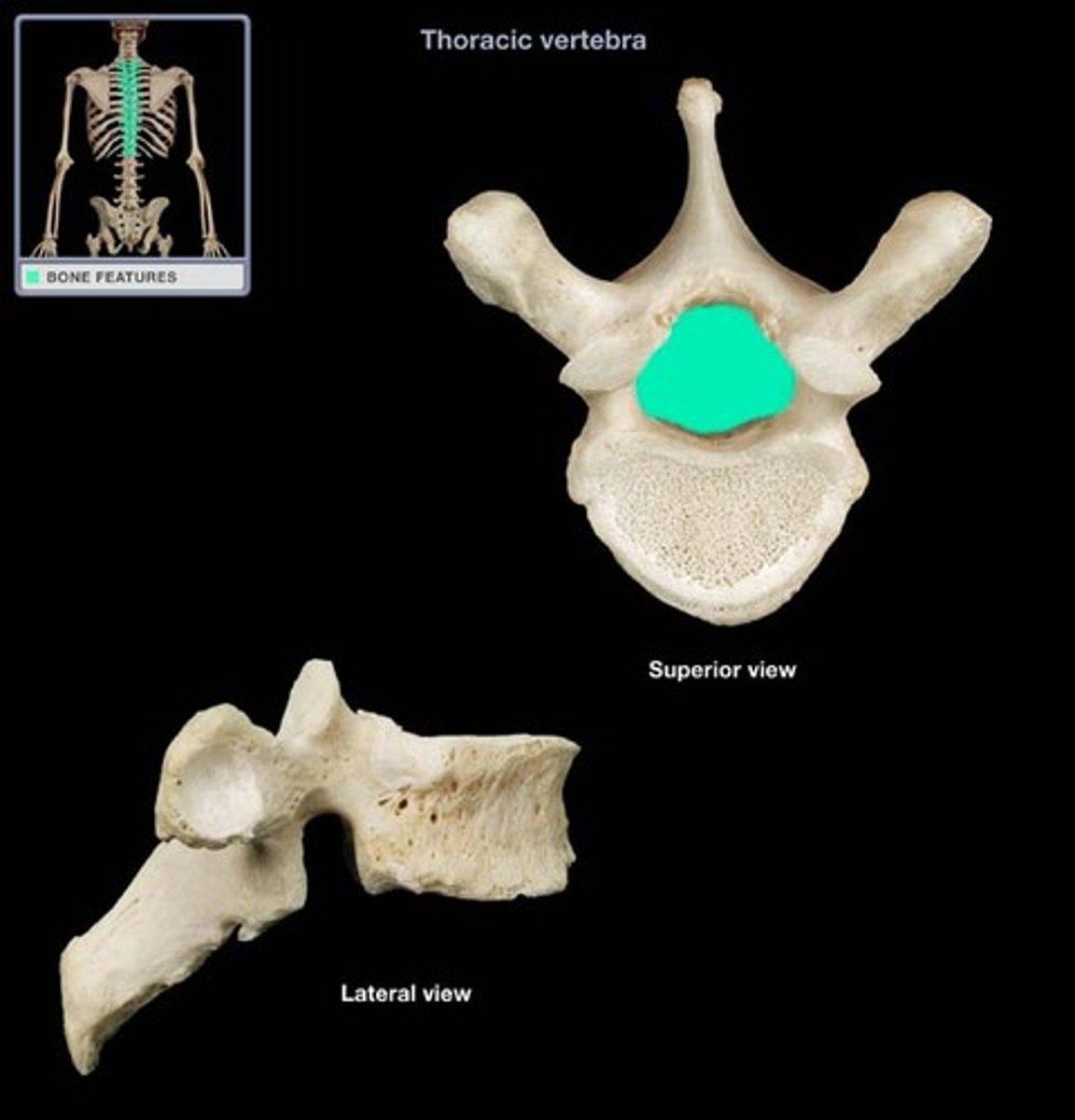
Transverse processes
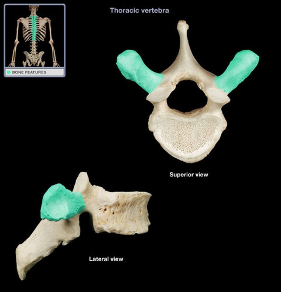
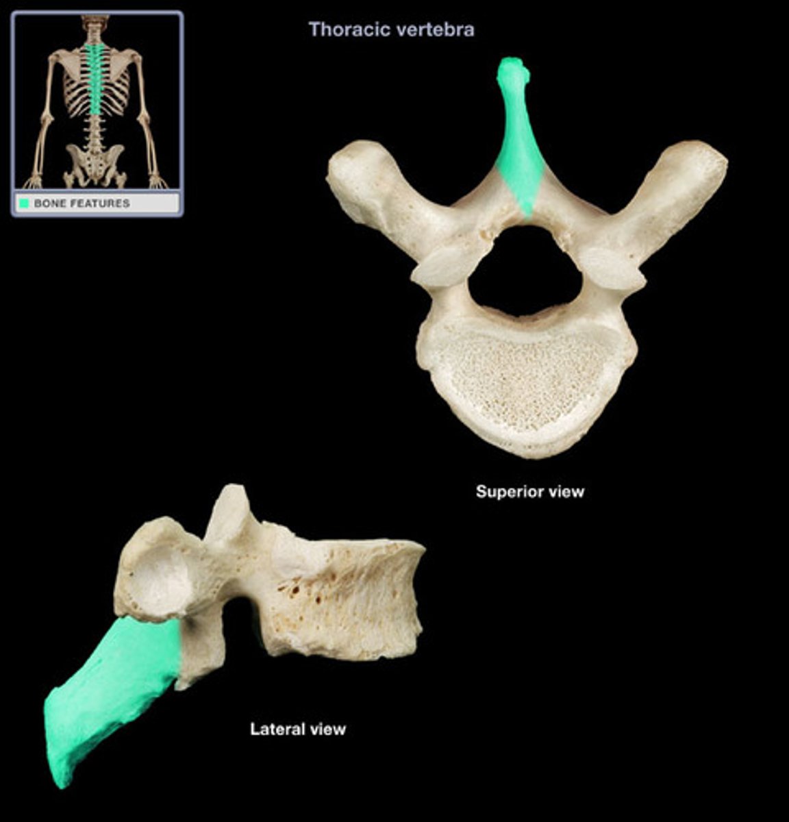
Spinous process
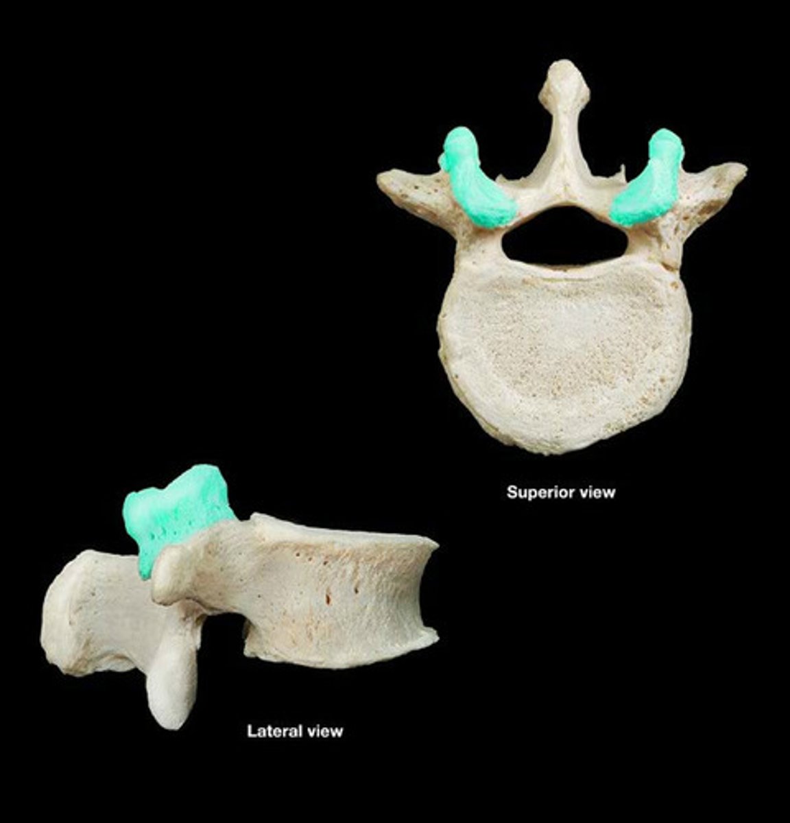
Articular facet
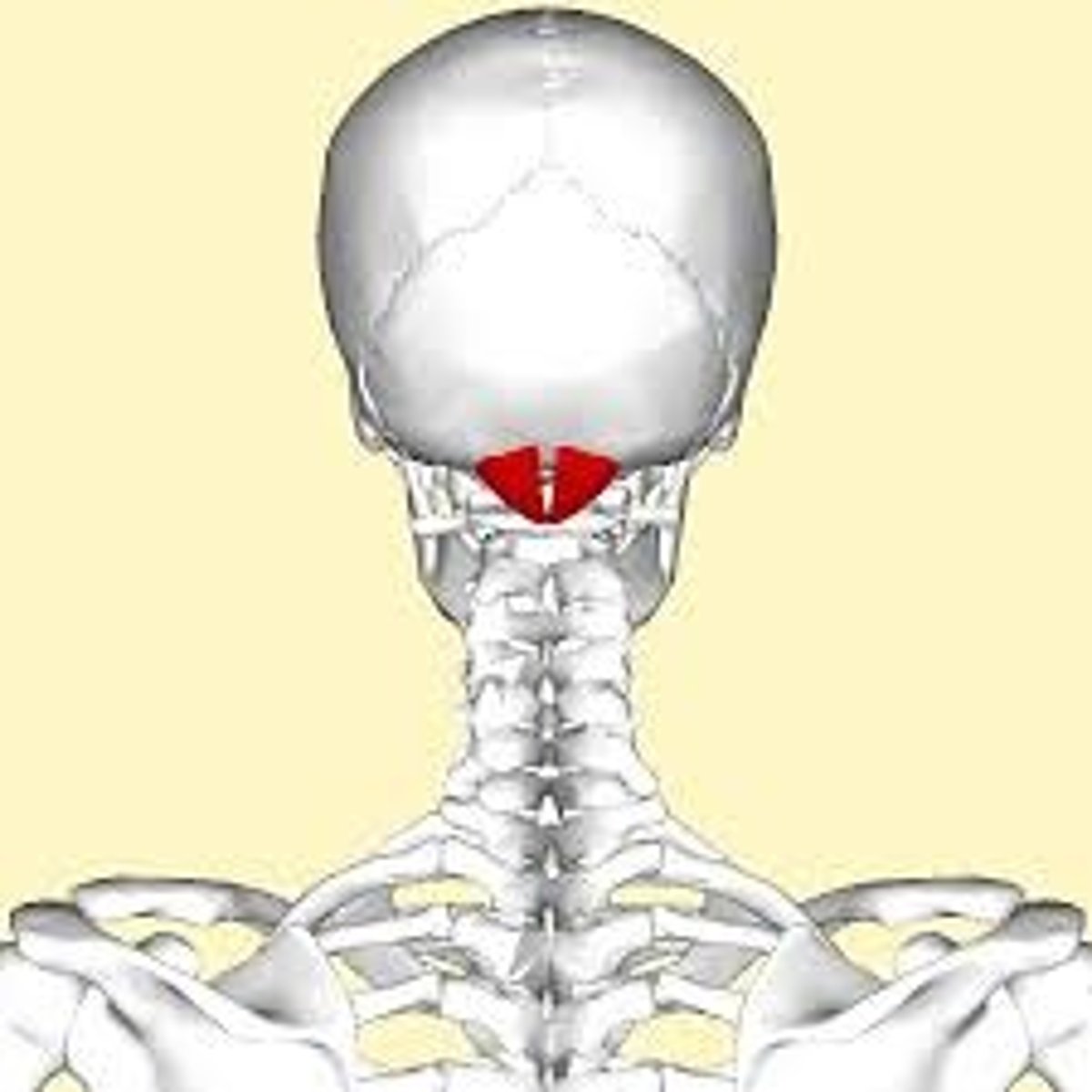
Atlanto-occipital joint
Creates a 'yes' motion, between C1 and skull
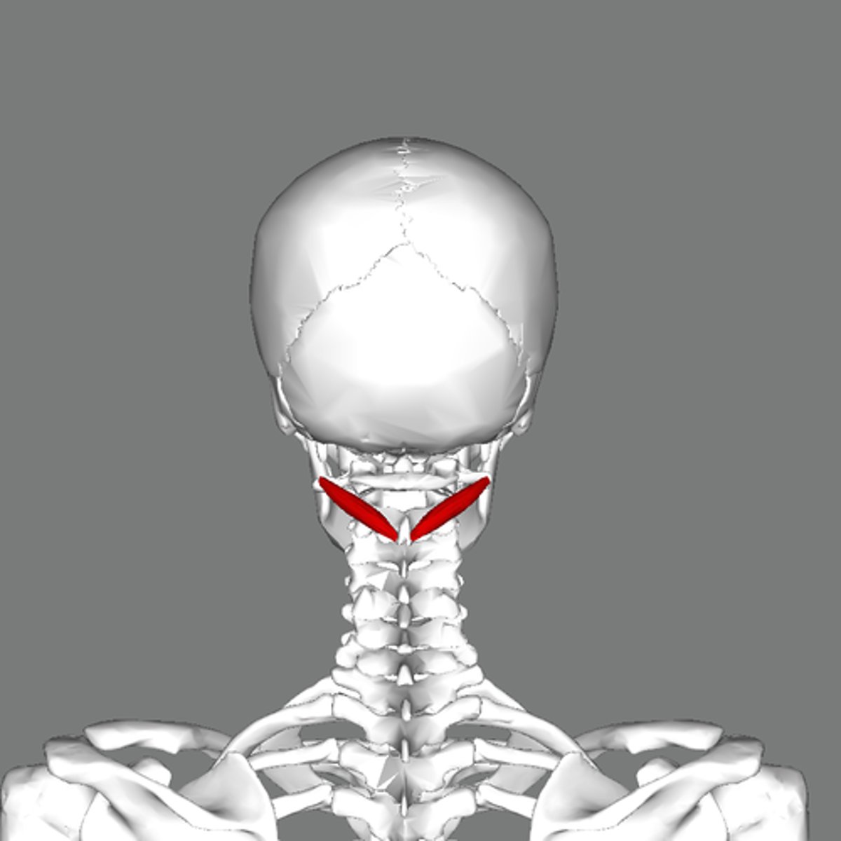
Atlanto-axis joint
Creates a 'no' motion, between C1 and C2
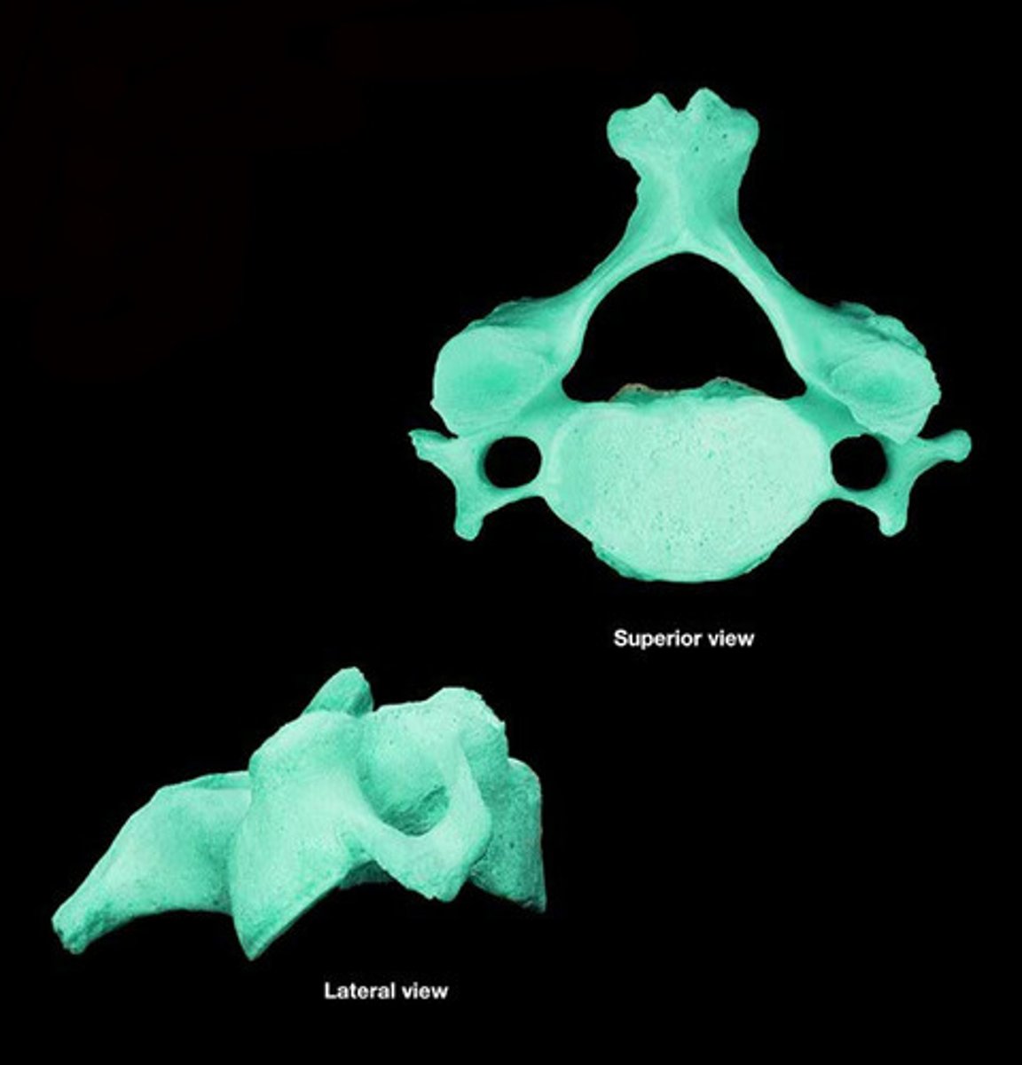
Cervical
Neck, 7 vertebrae, small vertebral body, transverse foramen
Thoracic
Chest/upper back, vertebrocostal articulations, long spinous process facing down
Lumbar
Lower back, large bean shaped body, transverse process is long, spinous process faces back
Sacral and coccygeus
5 fused sacral vertebrae and 4 fused coccygeal vertebrae
Typical vertebrae have how many joints?
6 joints, 4 synovial, 2 symphyses
Zygapophyseal joints
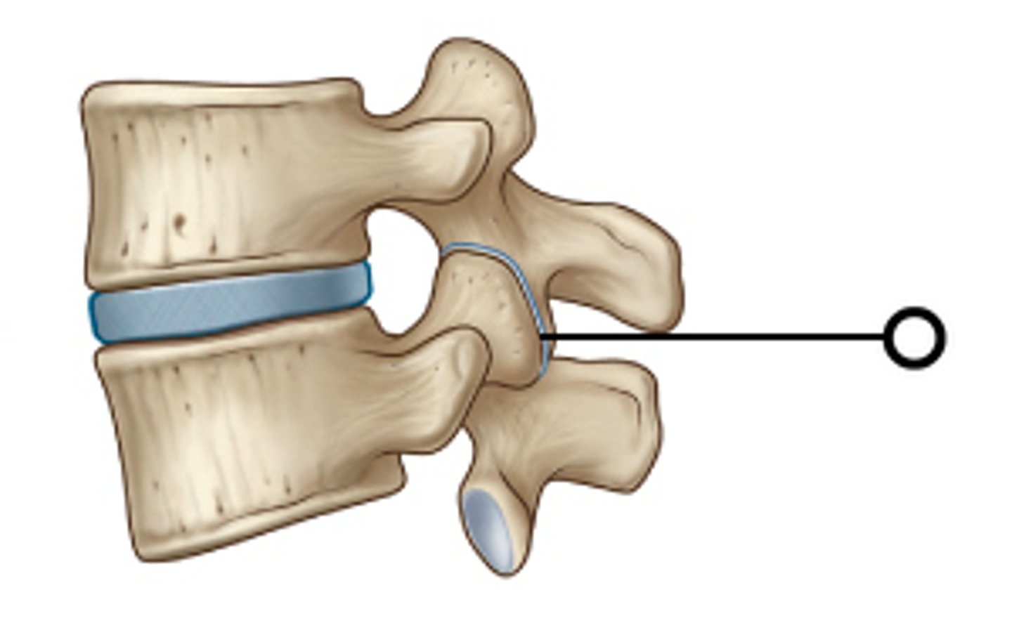
Cervical zygapophyseal joints
Allows flexion and extension
Thoracic zygapophyseal joints
Allows rotation
Lumbar zygapophyseal joints
Allows flexion/extension
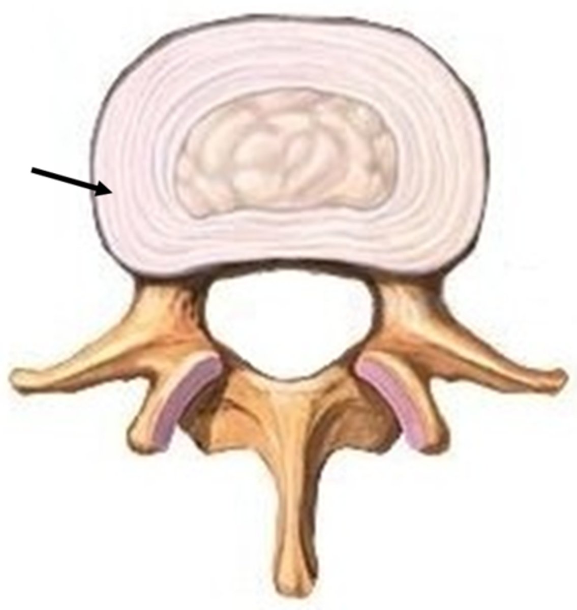
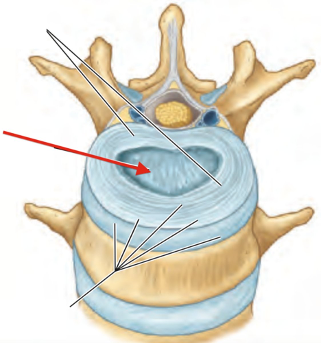
Intervertebral discs
Fibrocartilage pads that separate and cushion the vertebrae
Annulus fibrous of intervertebral disc
Nucleus pulposus of intervertebral disc
Afferent
Sensory
Efferent
Motor
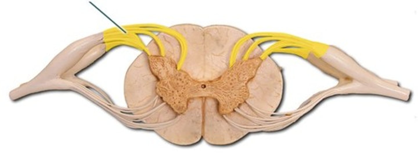
Posterior roots
Fibres carry sensory information
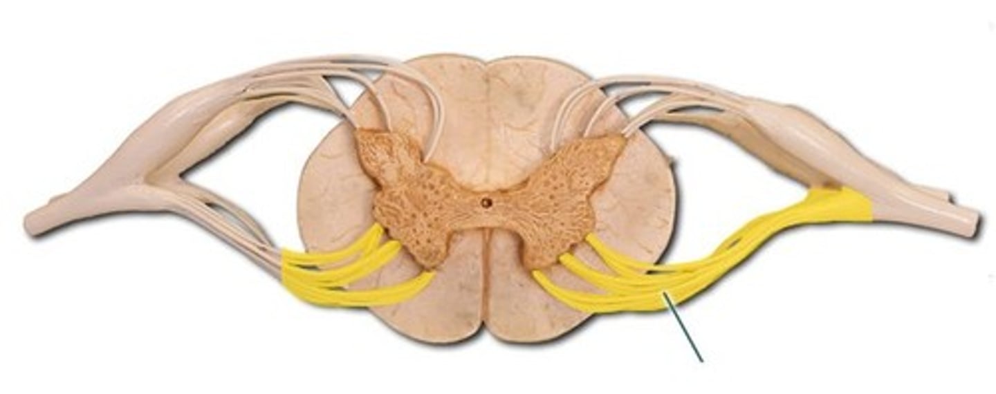
Anterior roots
Fibres carry motor information
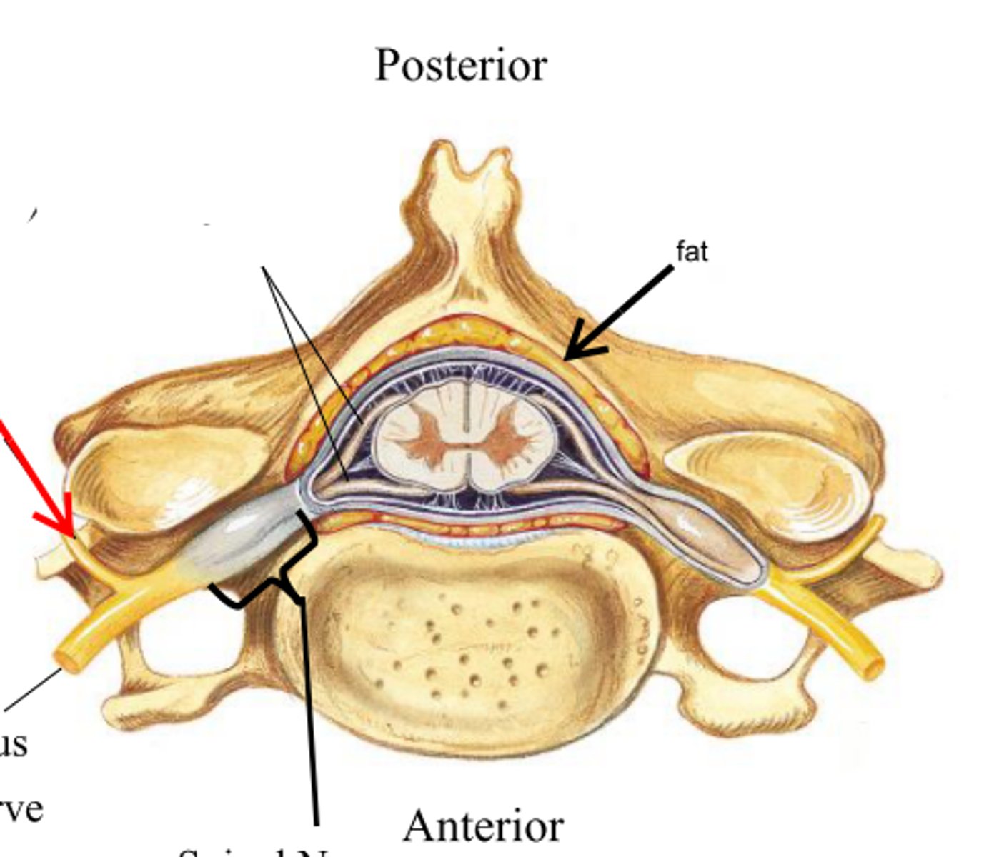
Posterior ramus
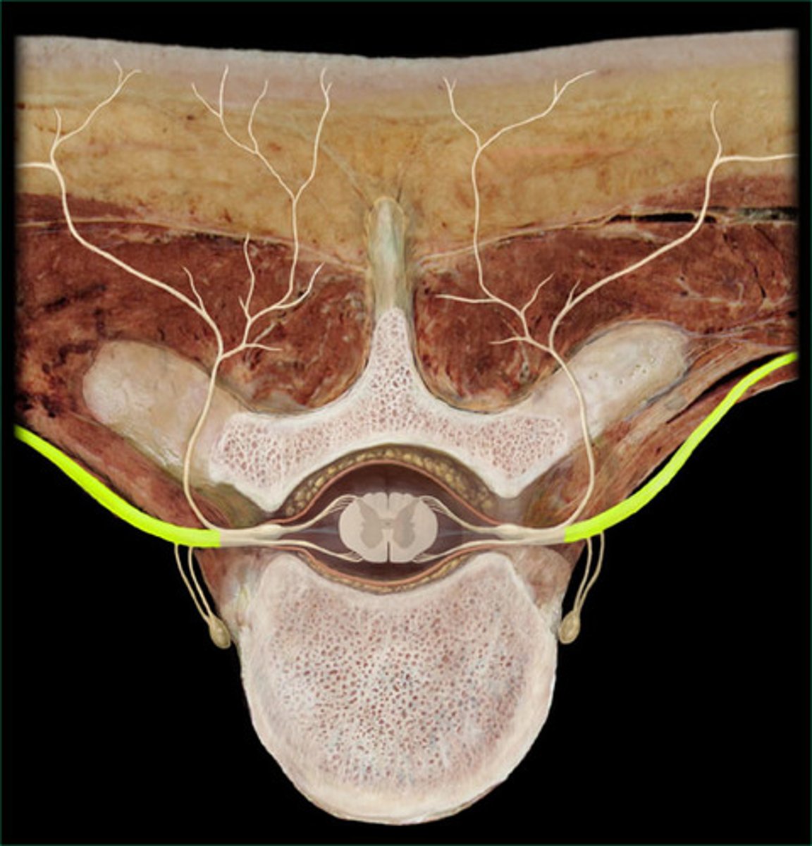
Anterior ramus
Spinal meninges
3 protective sheaths around spinal cord and brain
Pia mater
Innermost layer of the meninges
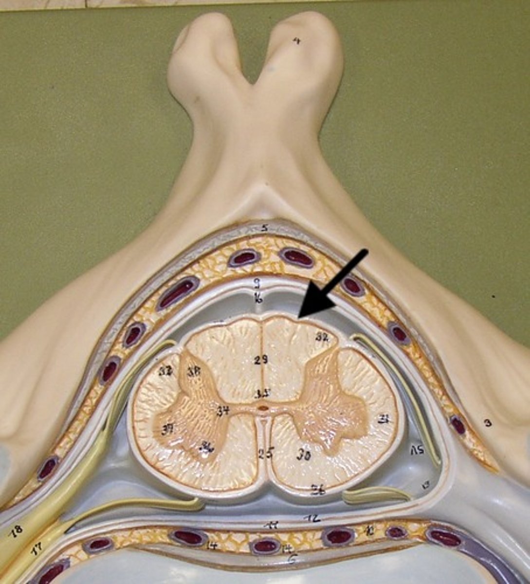
Arachnoid mater
Middle layer of the meninges
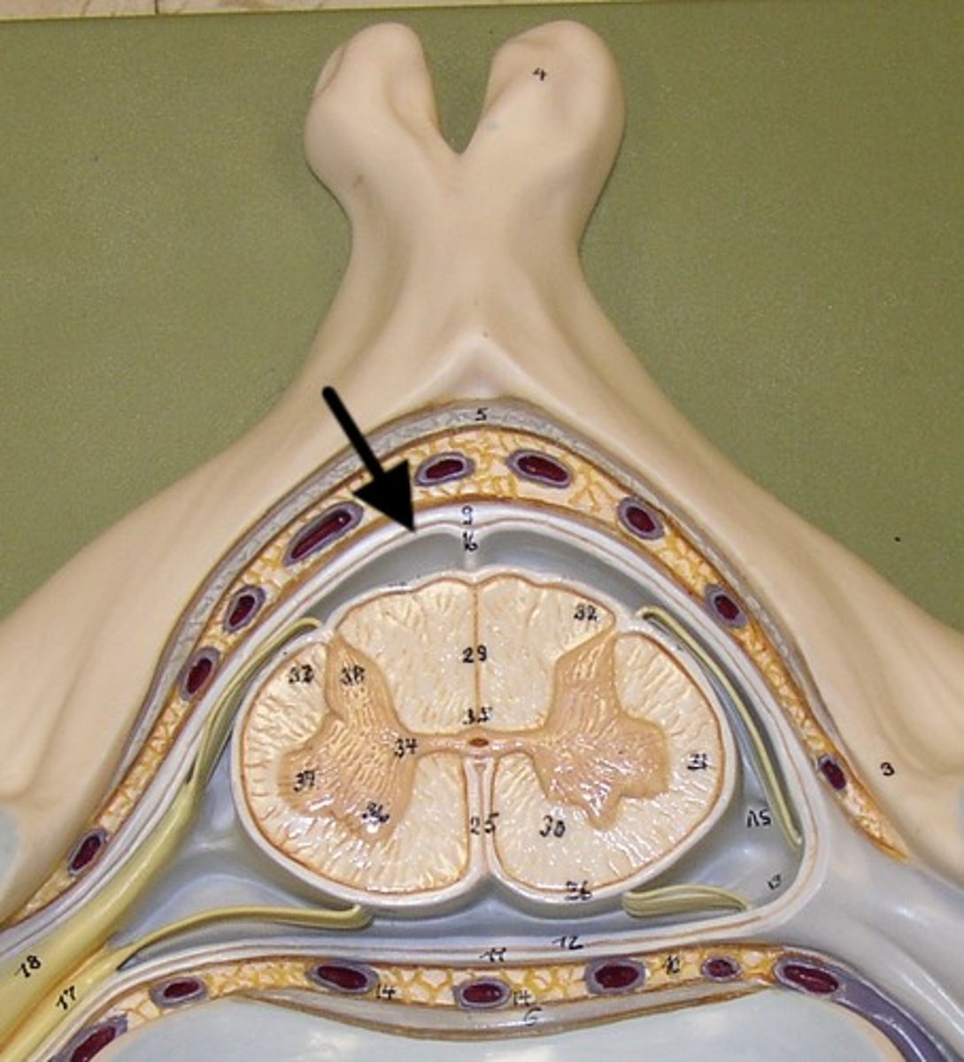
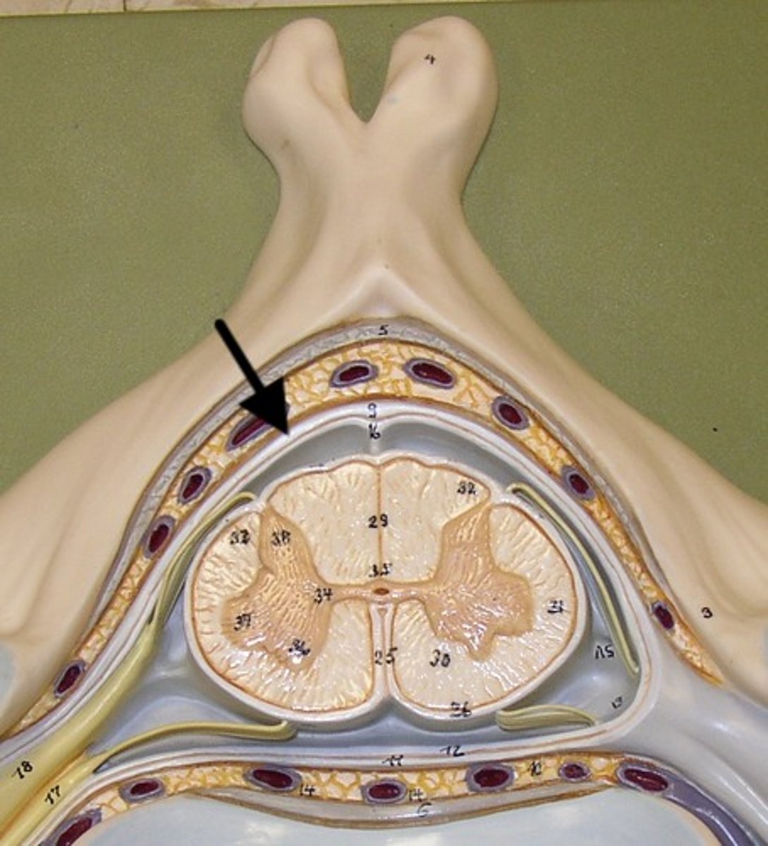
Dura mater
Outermost layer of the meninges
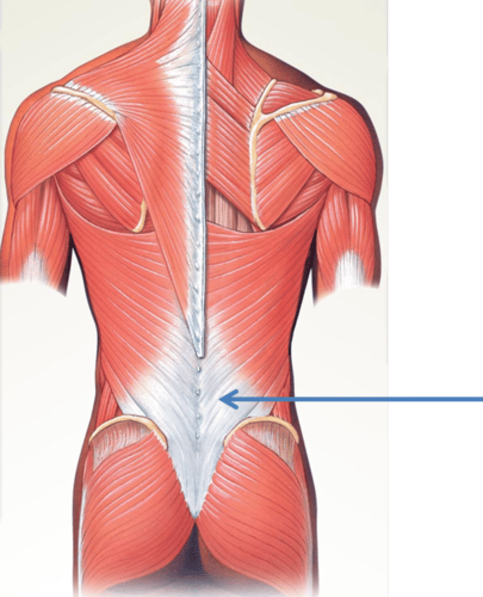
Thoracolumbar fascia
Sheet of connective tissue on lower portion of the back
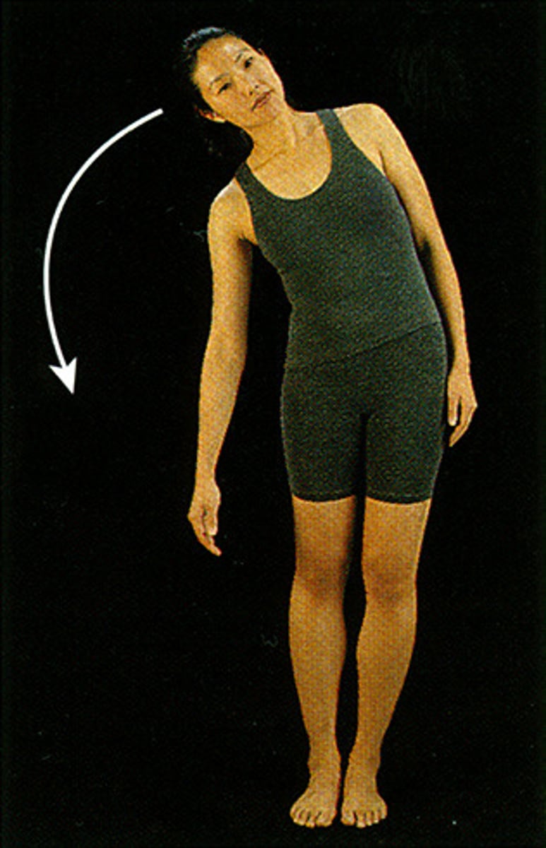
Lateral flexion of the spine
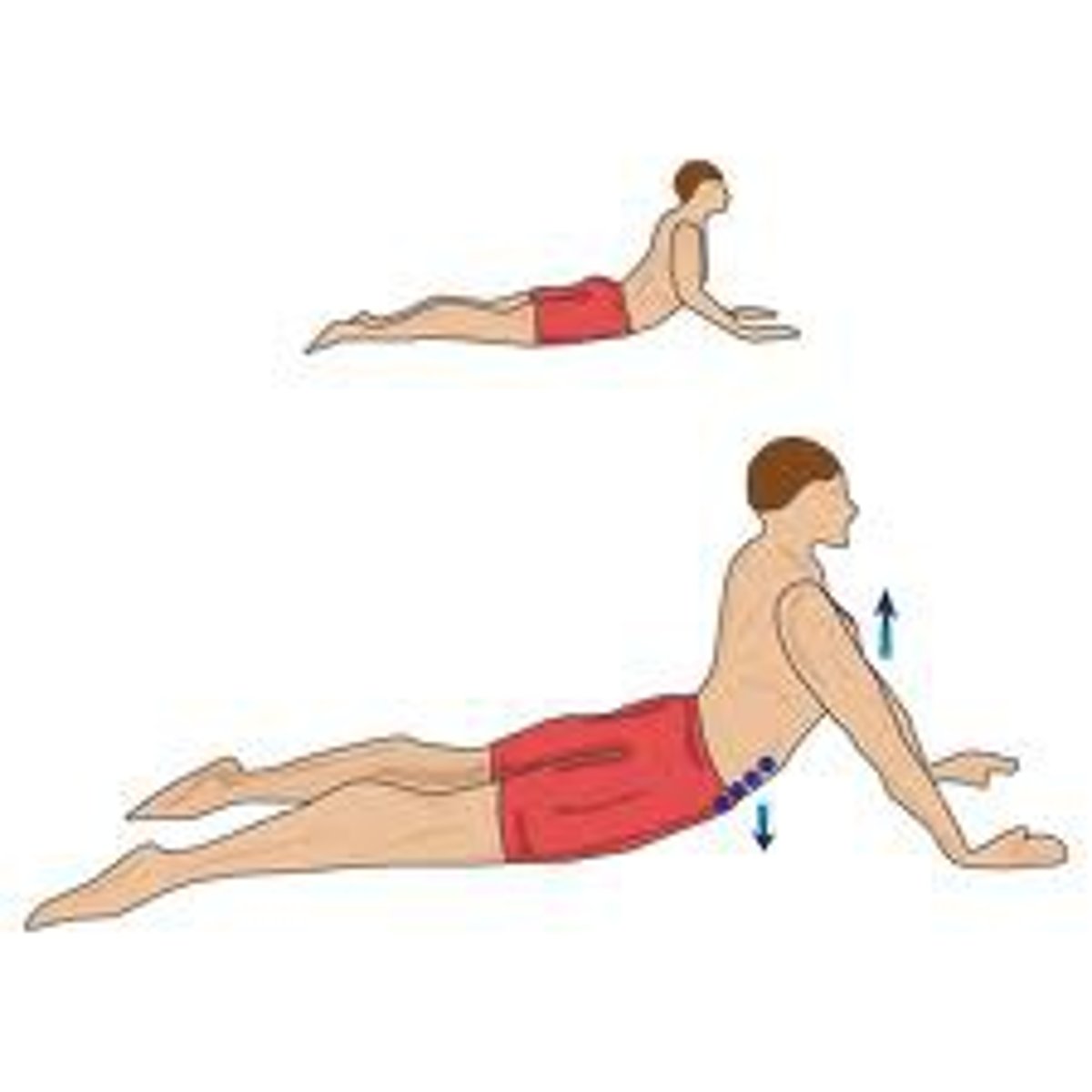
Extension of the spine
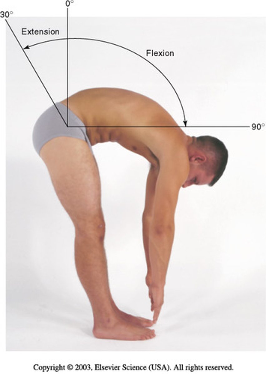
Flexion of the spine
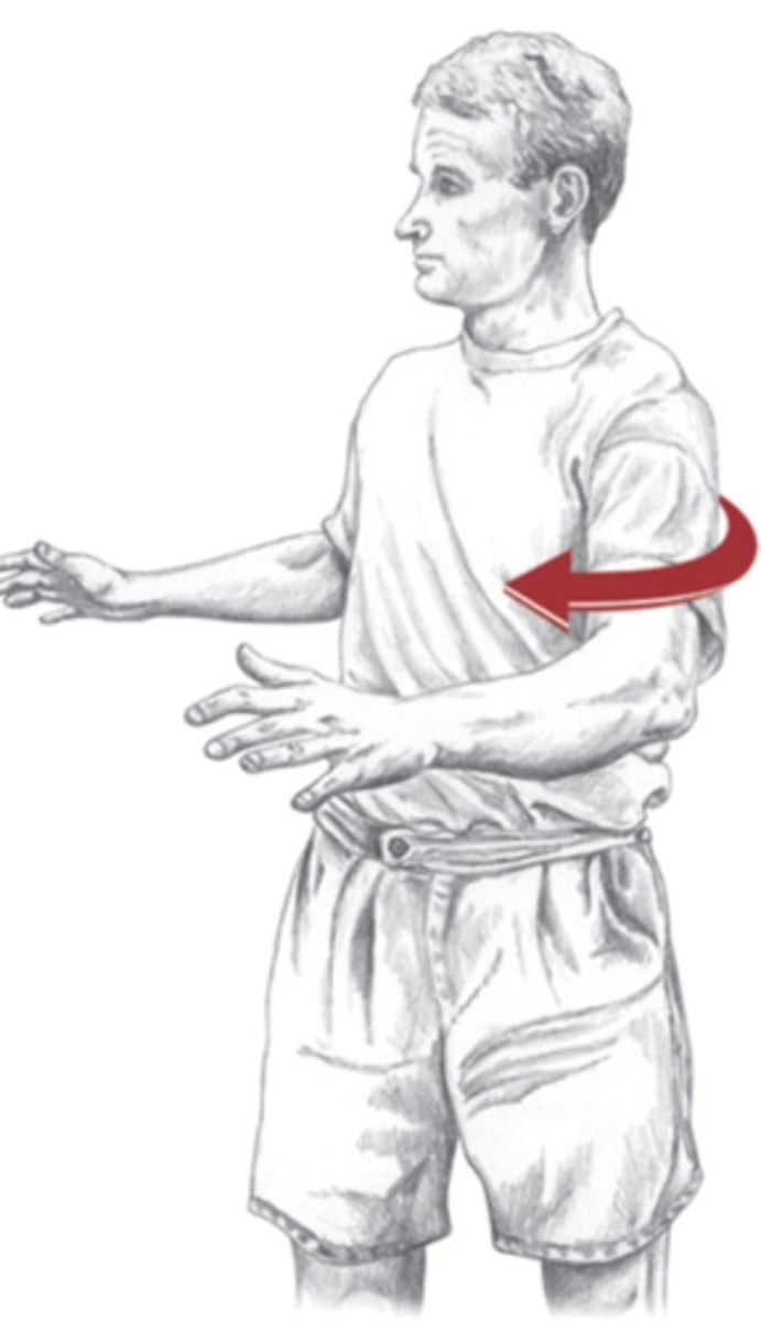
Rotation of the spine
Erector spinae
Extends vertebral column
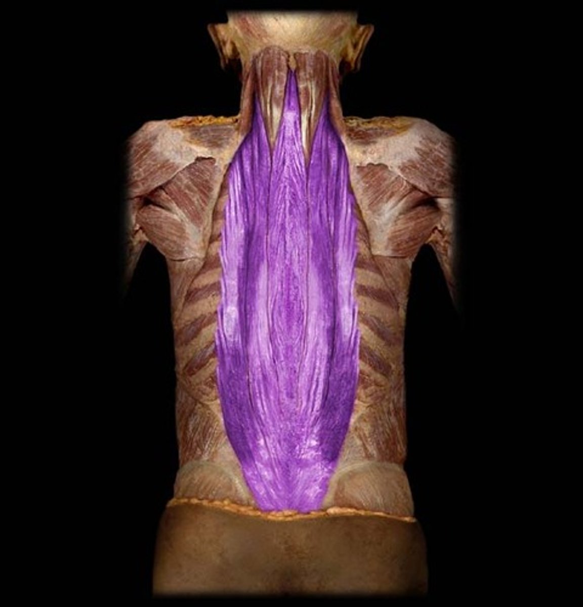
Muscles of the erector spinae
Iliocostalis, longissimus, spinalis
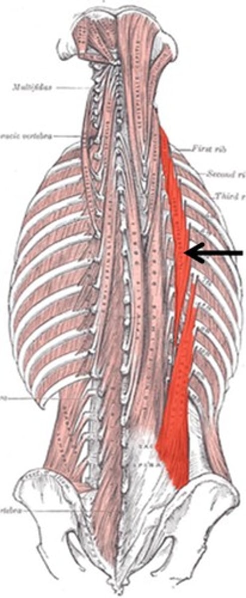
Ilicostalis
Spine extension and lateral flexion (unilateral)
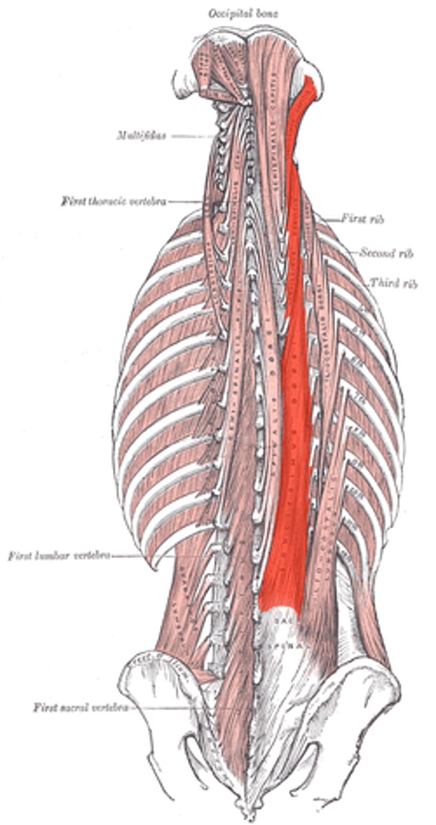
Longissimus
Extends spine
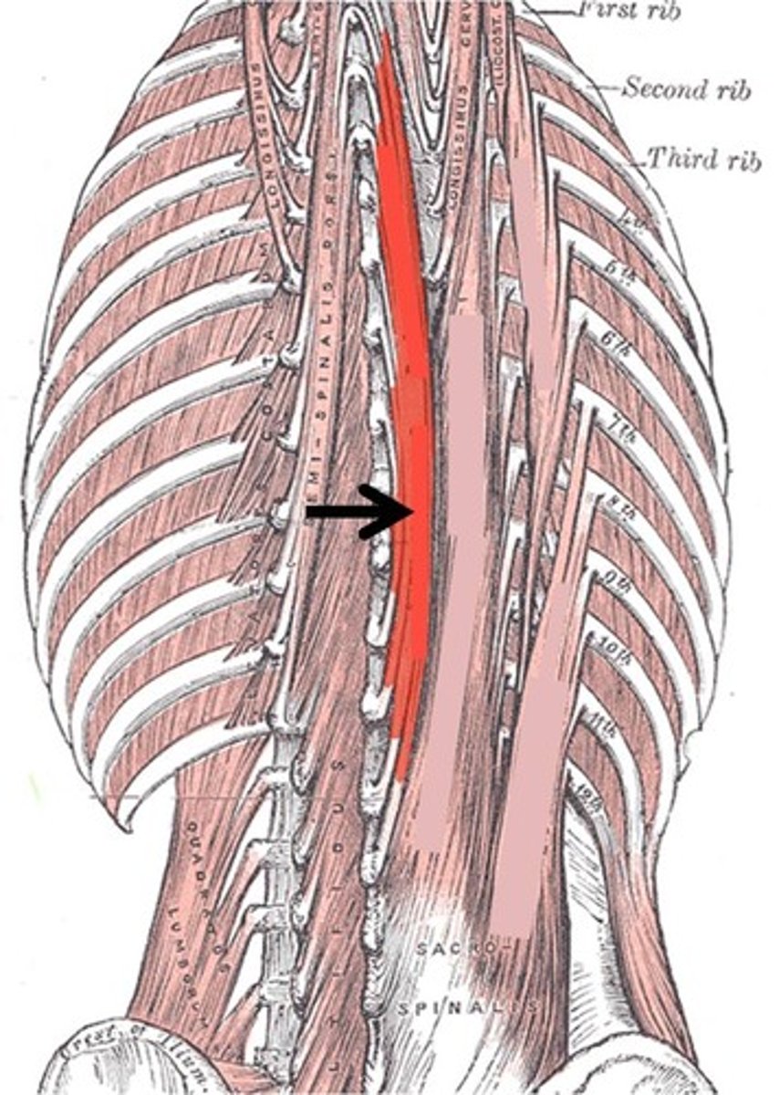
Spinalis
Extends vertebral column and allows lateral flexion (unilateral)
Muscles that lie anterior to the spine
Diaphragm, anterior lumbar vertebrae, quadratis lumborum, tsoas major and minor, iliocostalis
Abdominal muscles
Rectus abdominis, transverse abdominis, external oblique, internal oblique
Transversus abdominus
Compress abdomen
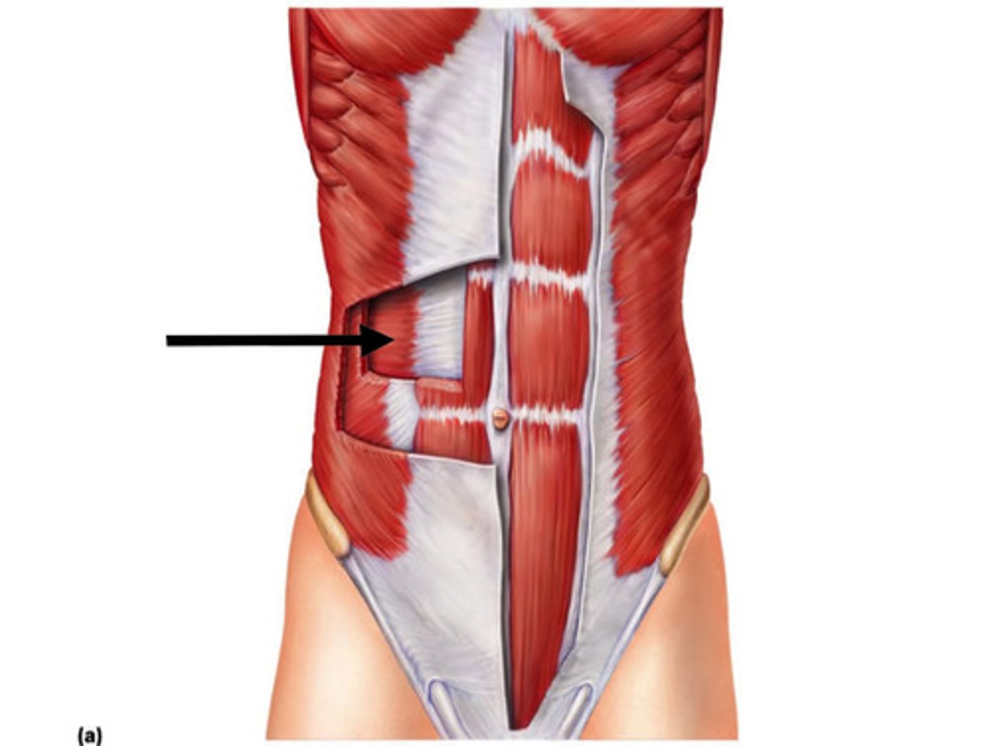
Internal oblique
Compresses abdomen, flexes trunk, rotates trunk (unilateral)
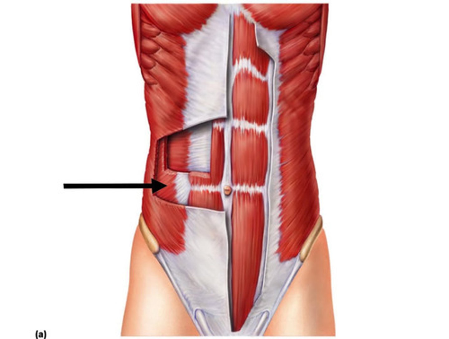
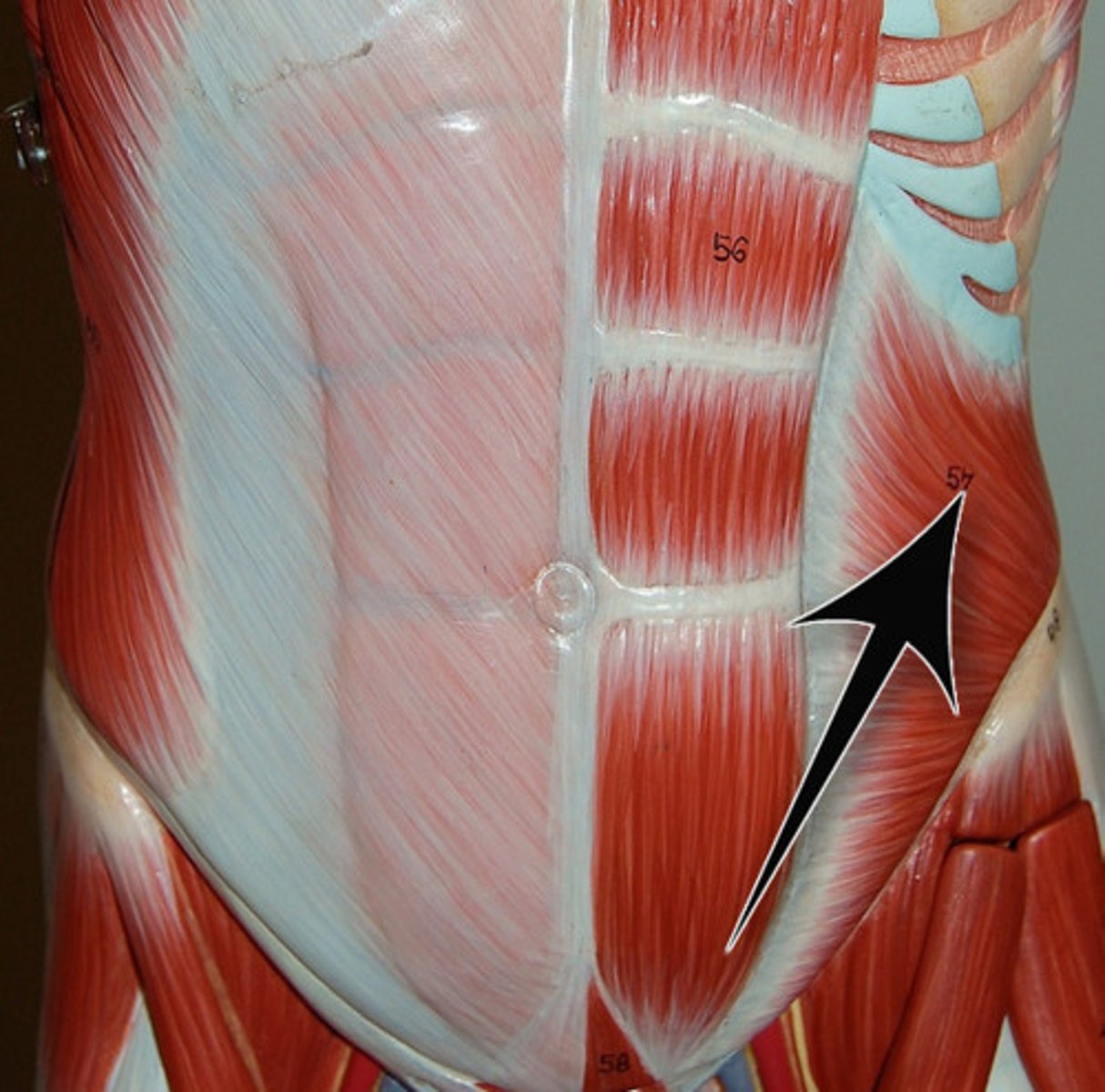
External oblique
Compresses abdomen, flexes trunk, rotates trunk (unilateral)
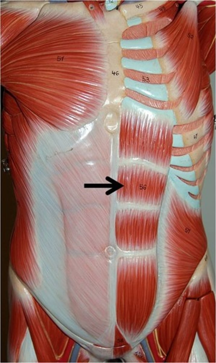
Rectus abdominus
Flexes trunk
Cervical vertebrae
Thoracic vertebrae
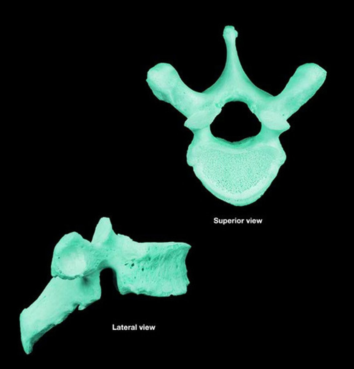
Lumbar vertebrae
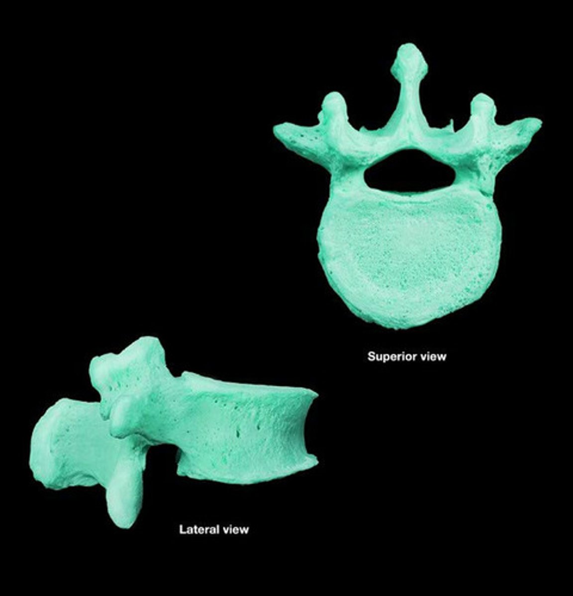
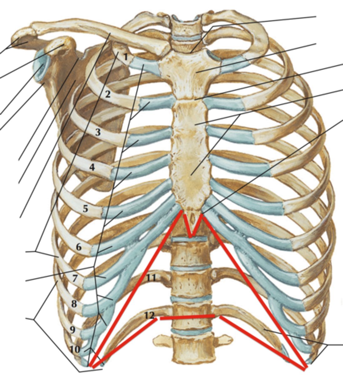
Thorax
Protects vital organs, resists intrathoracic pressure during respiration, attachment for upper limbs and support weight of upper limb, attachment for muscles of thorax and arm
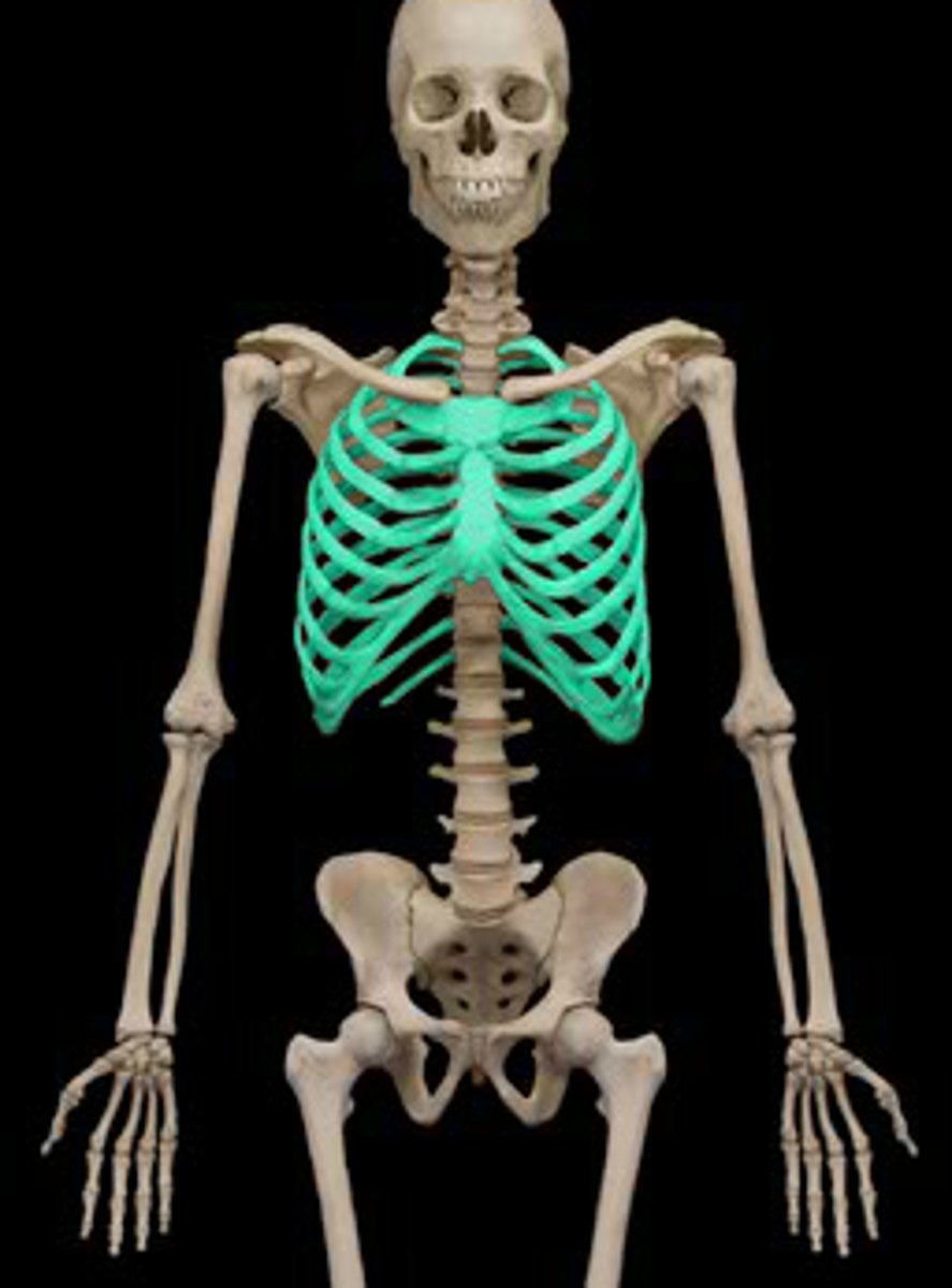
What do the superior 7 ribs articulate with?
Sternum
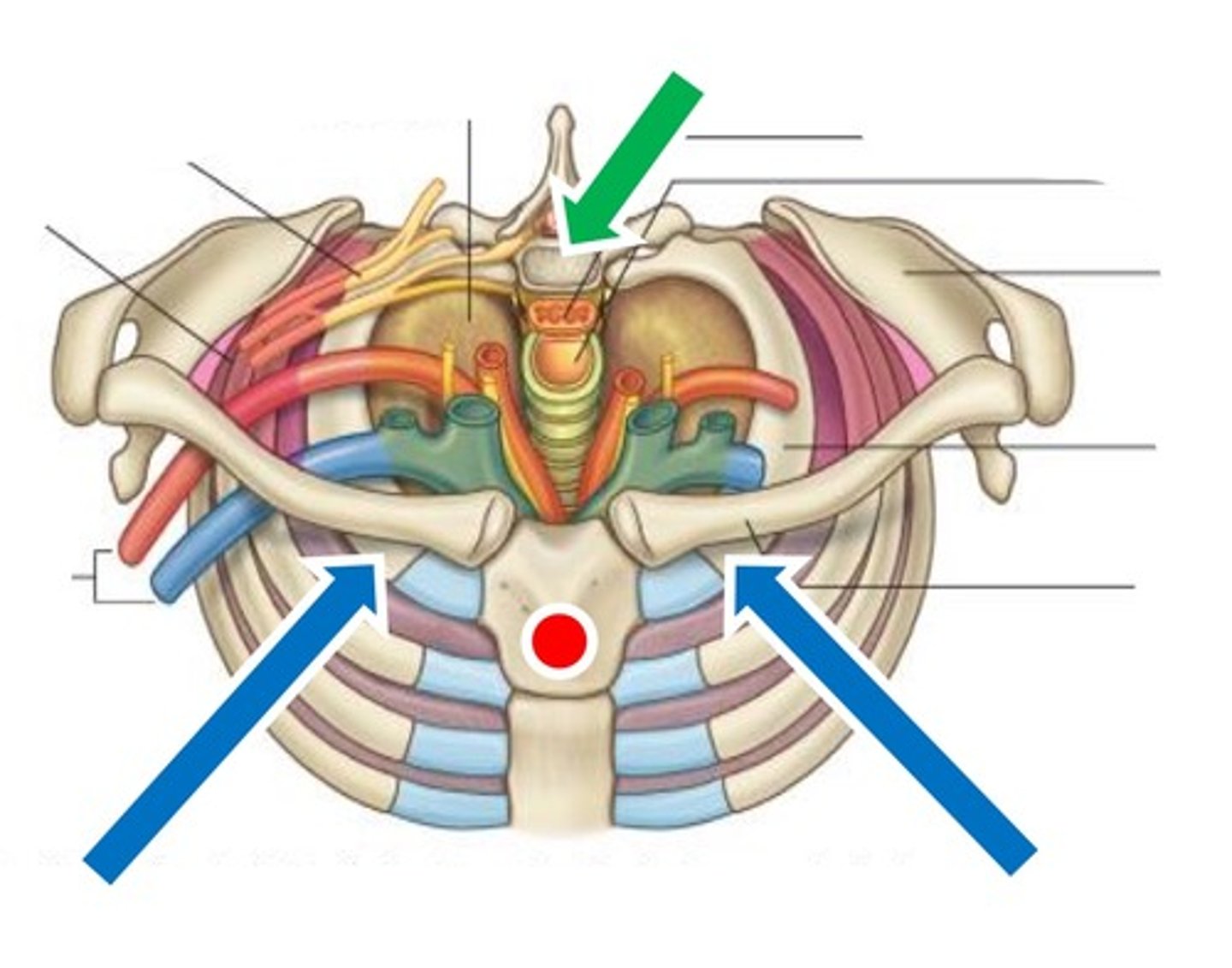
Superior thoracic aperture
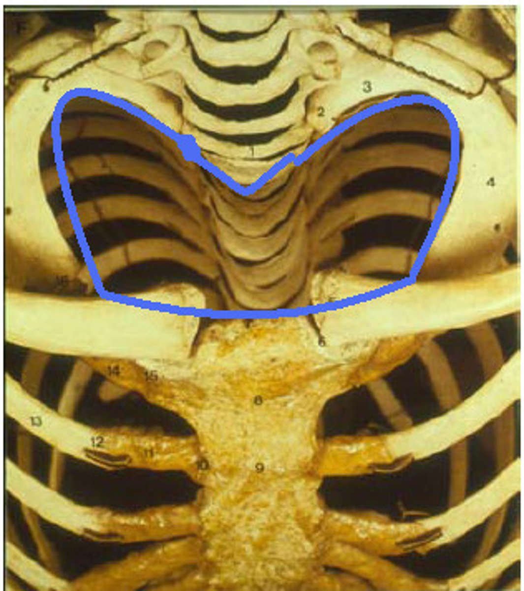
Inferior thoracic aperture
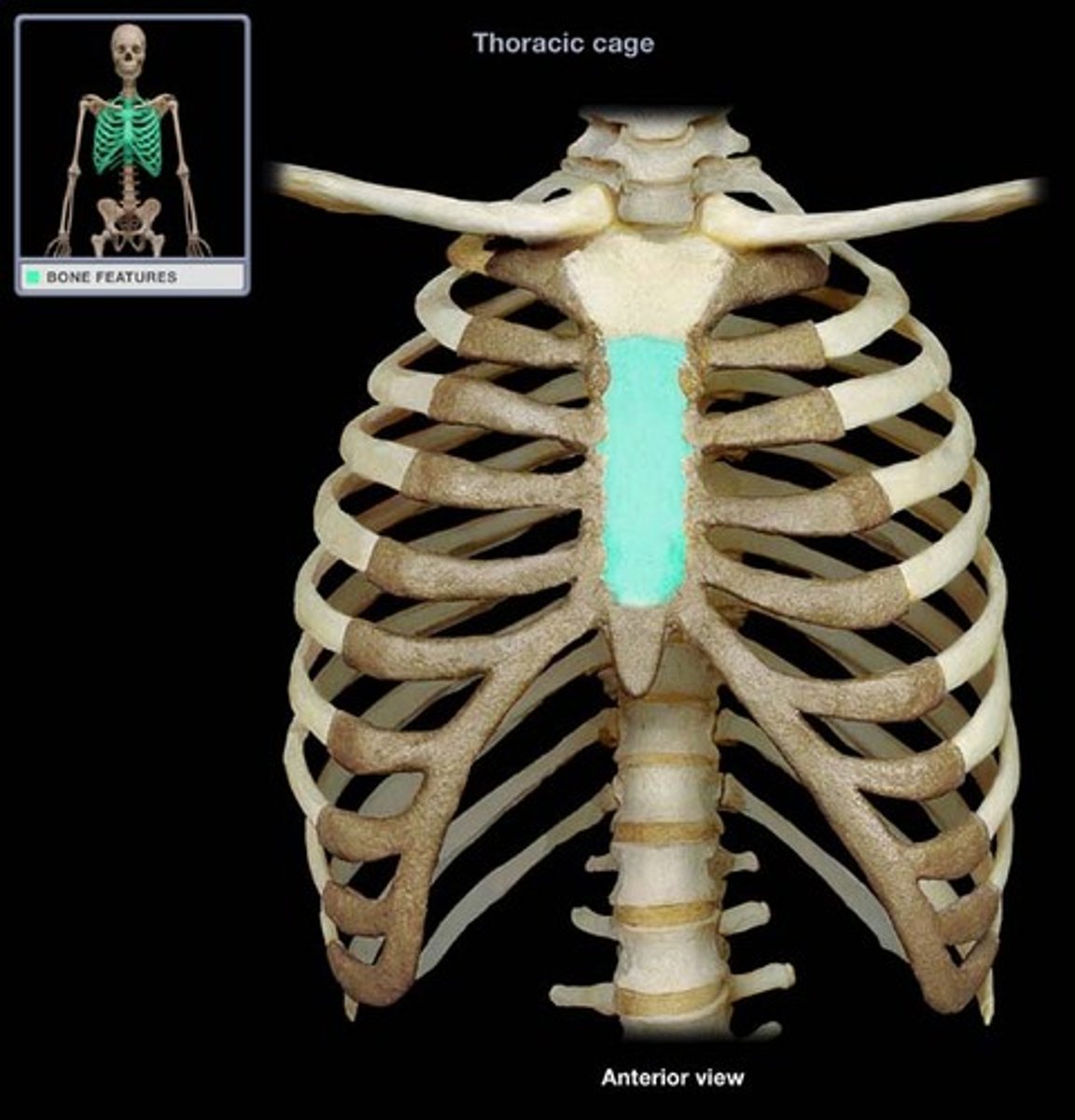
Sternum
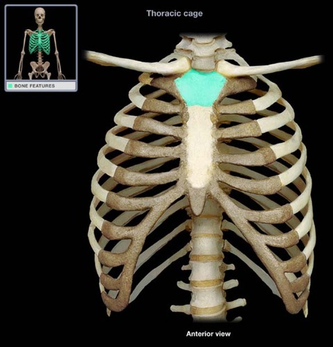
Manubrium
Clavicle and 1st ribs attach
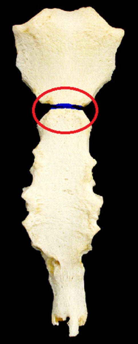
Sternal angle
Ridge between manubrium and body at second rib
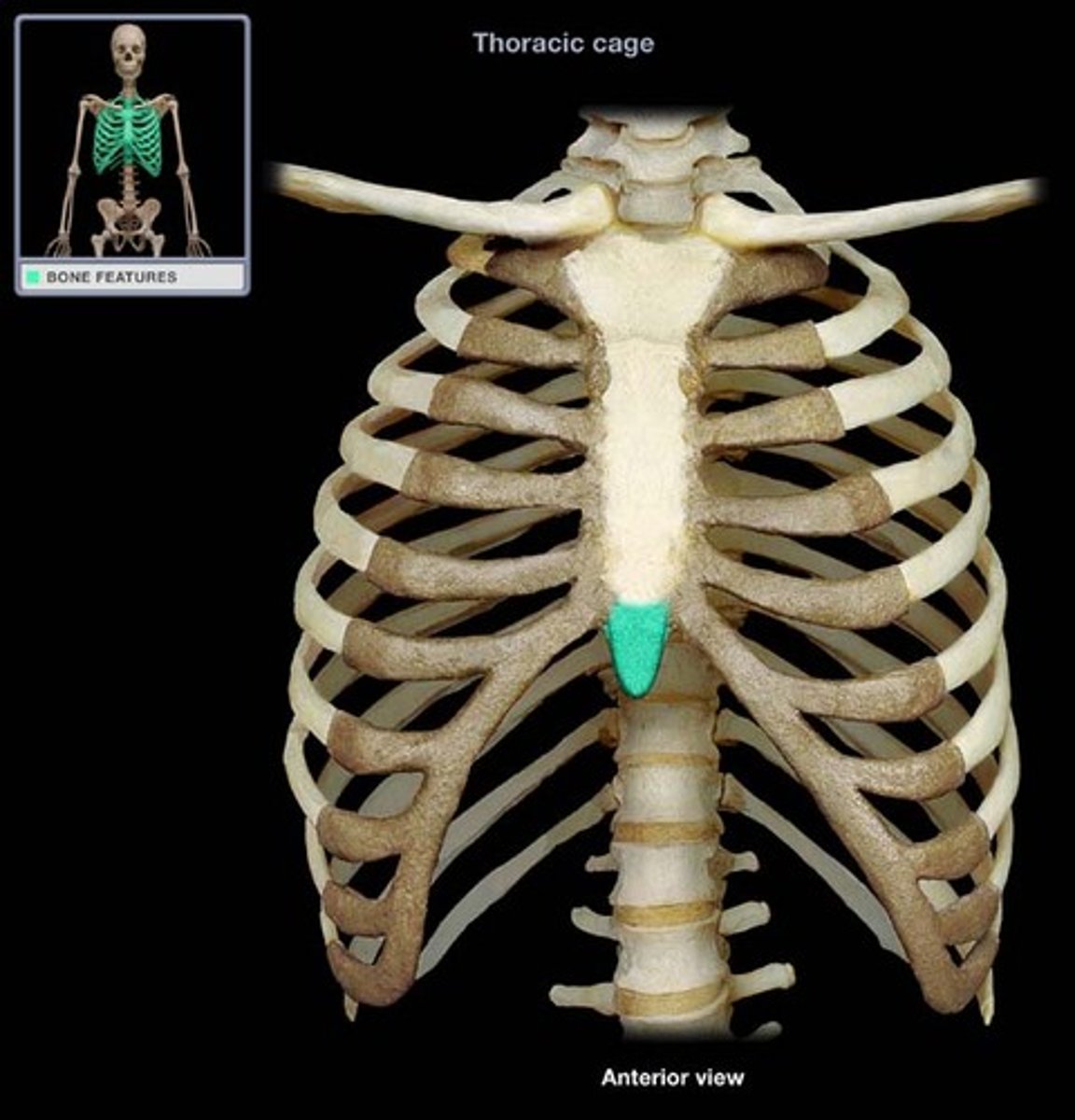
Xiphoid process
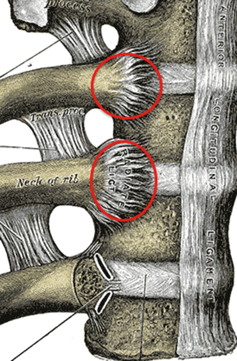
Costovertebral joint
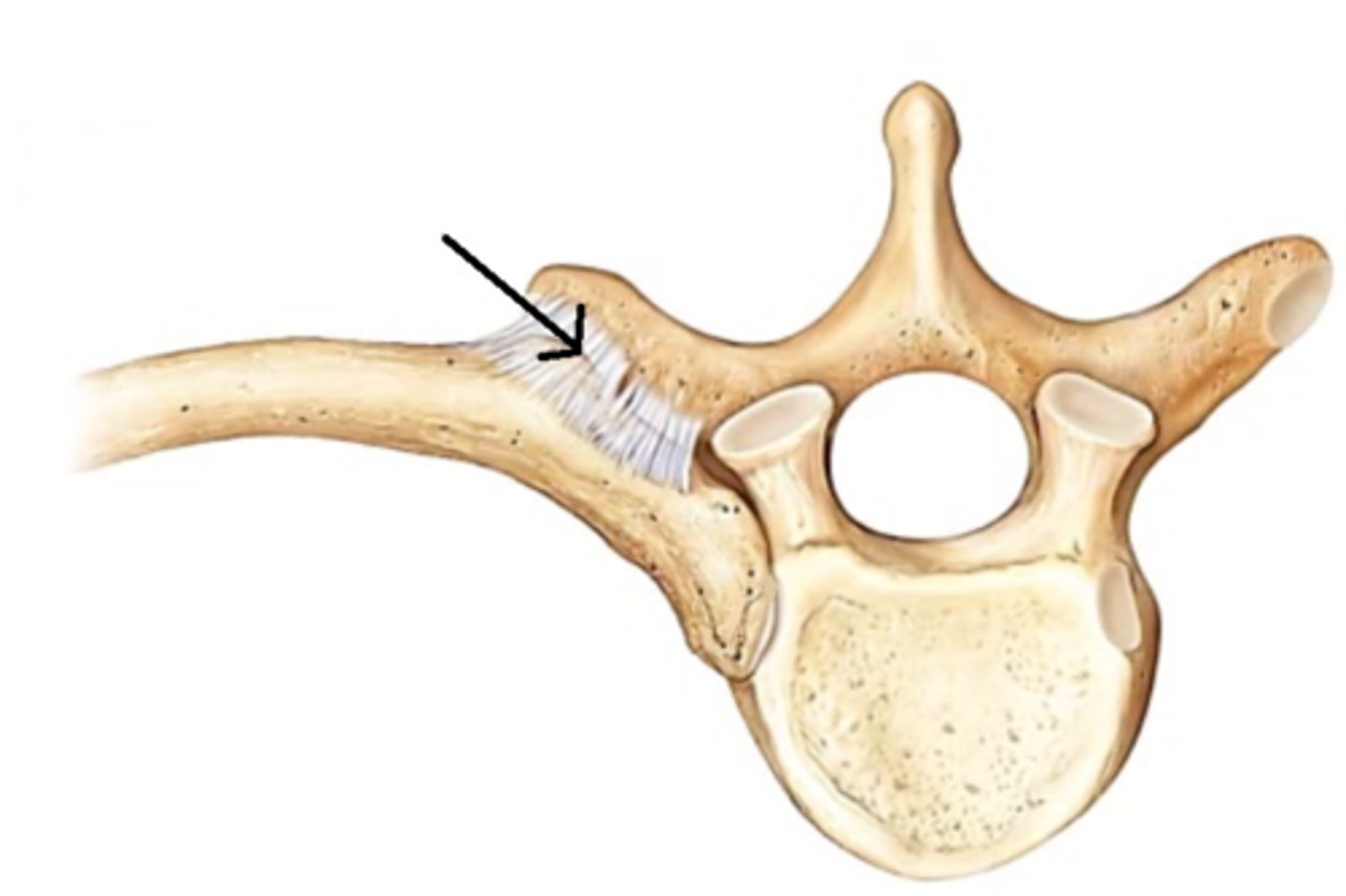
Costotransverse joint
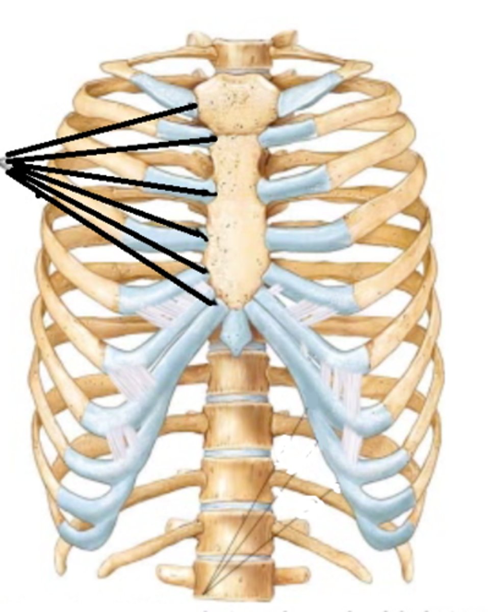
Sternocostal joint
Costochondral joint
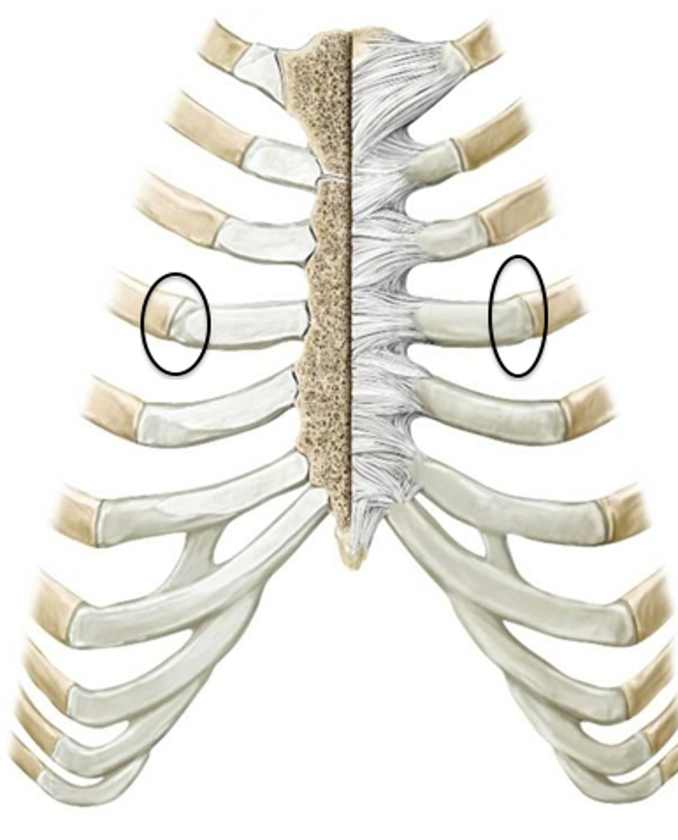
Costal cartilage of 1st ribs
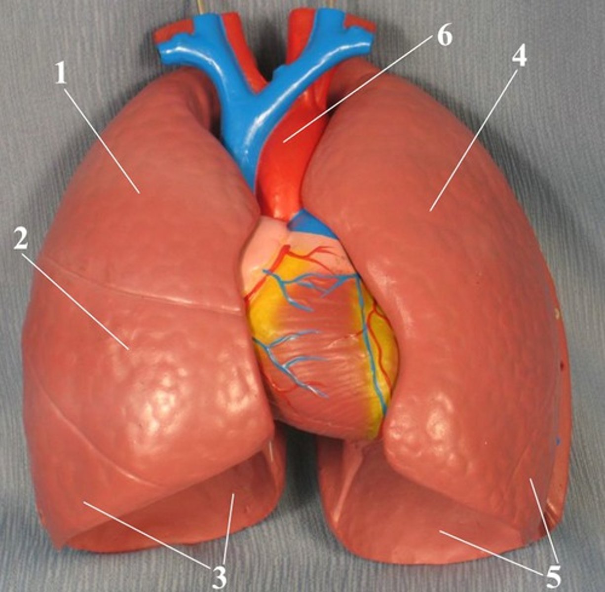
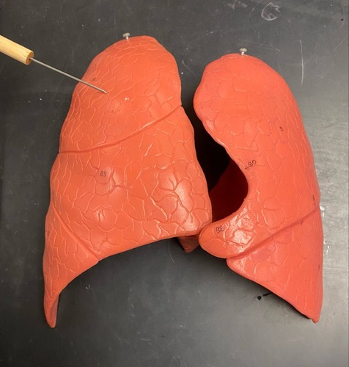
Superior lobe of lungs
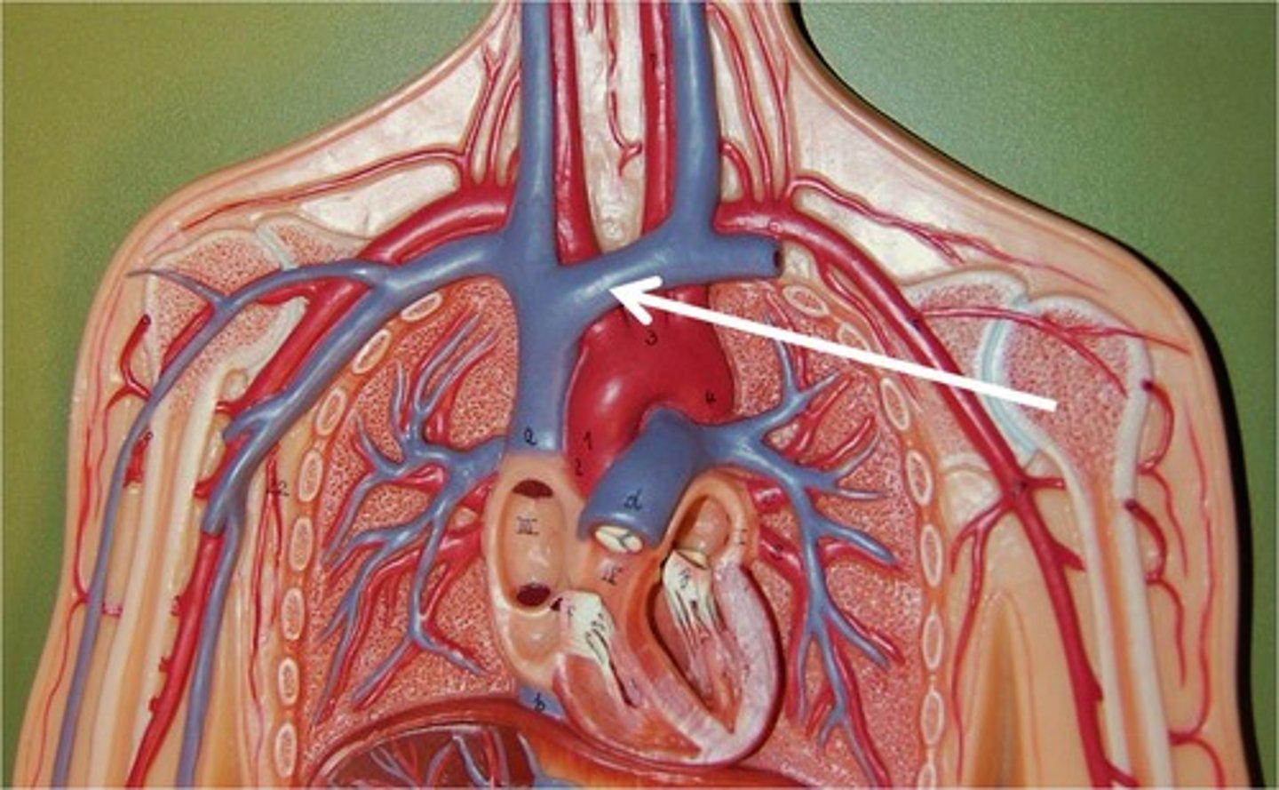
Brachiocephalic veins
Carotid artery
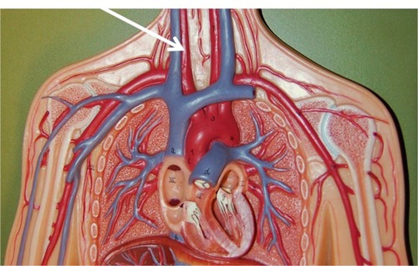
Subclavian artery
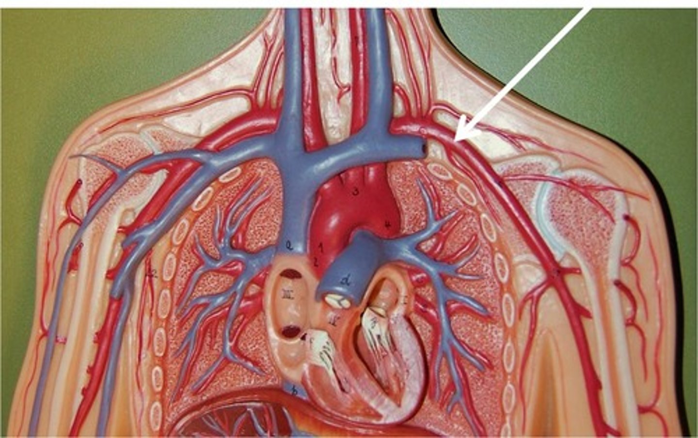
Trachea
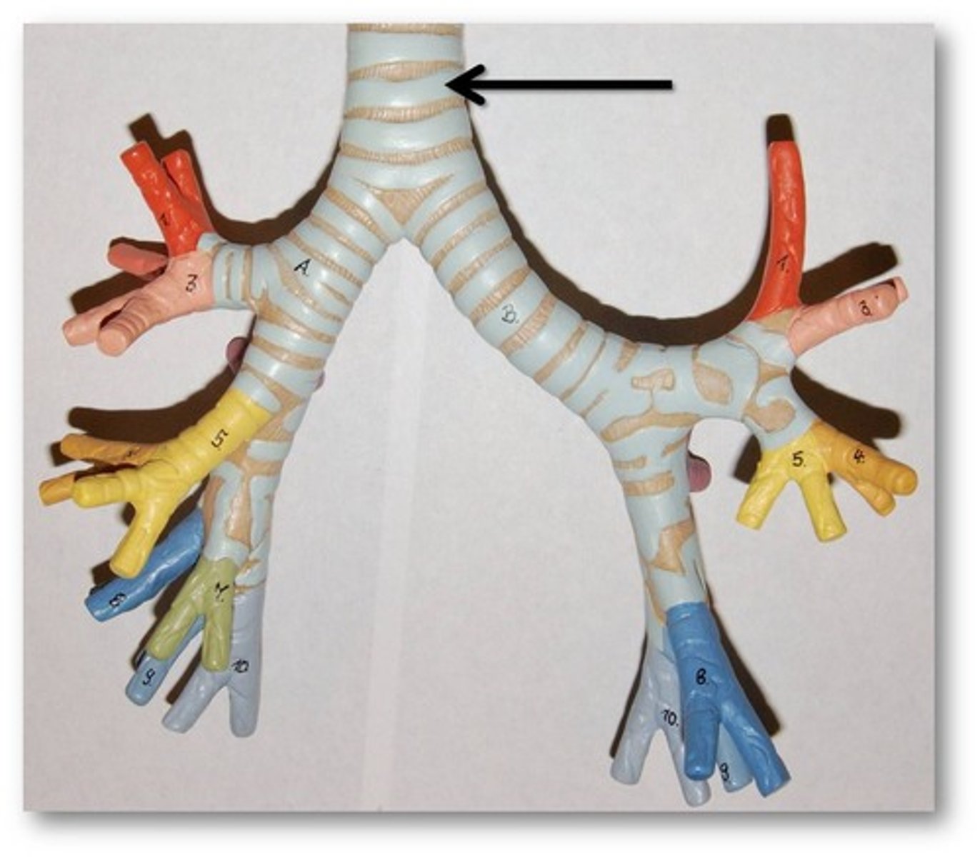
Esophagus
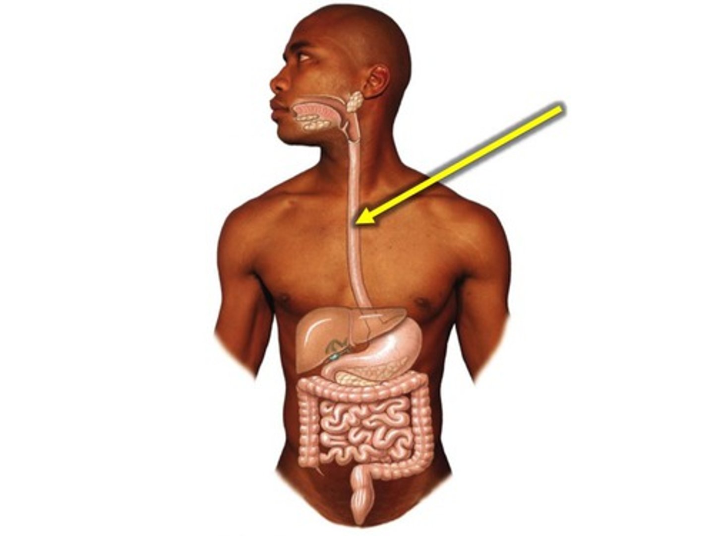
Sympathetic trunks
Paired bundles of nerves and ganglia that run parallel, length of the spine
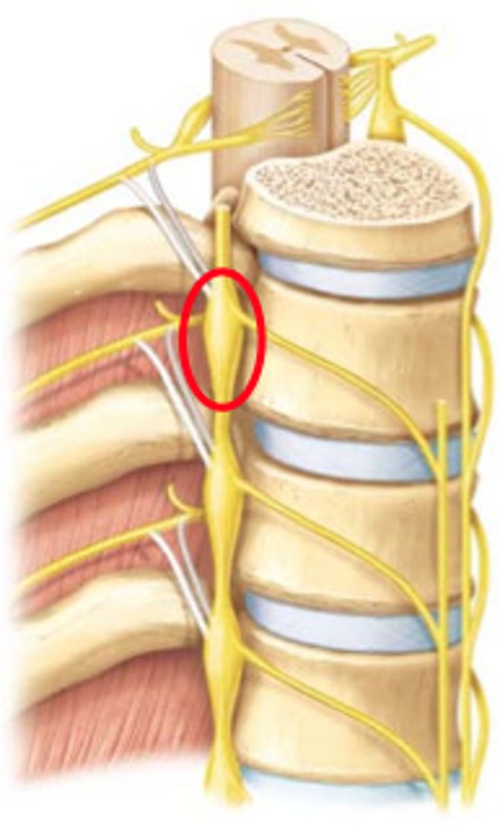
Phrenic nerve C3, 4 and 5
Motor, sensory, sympathetic nerves
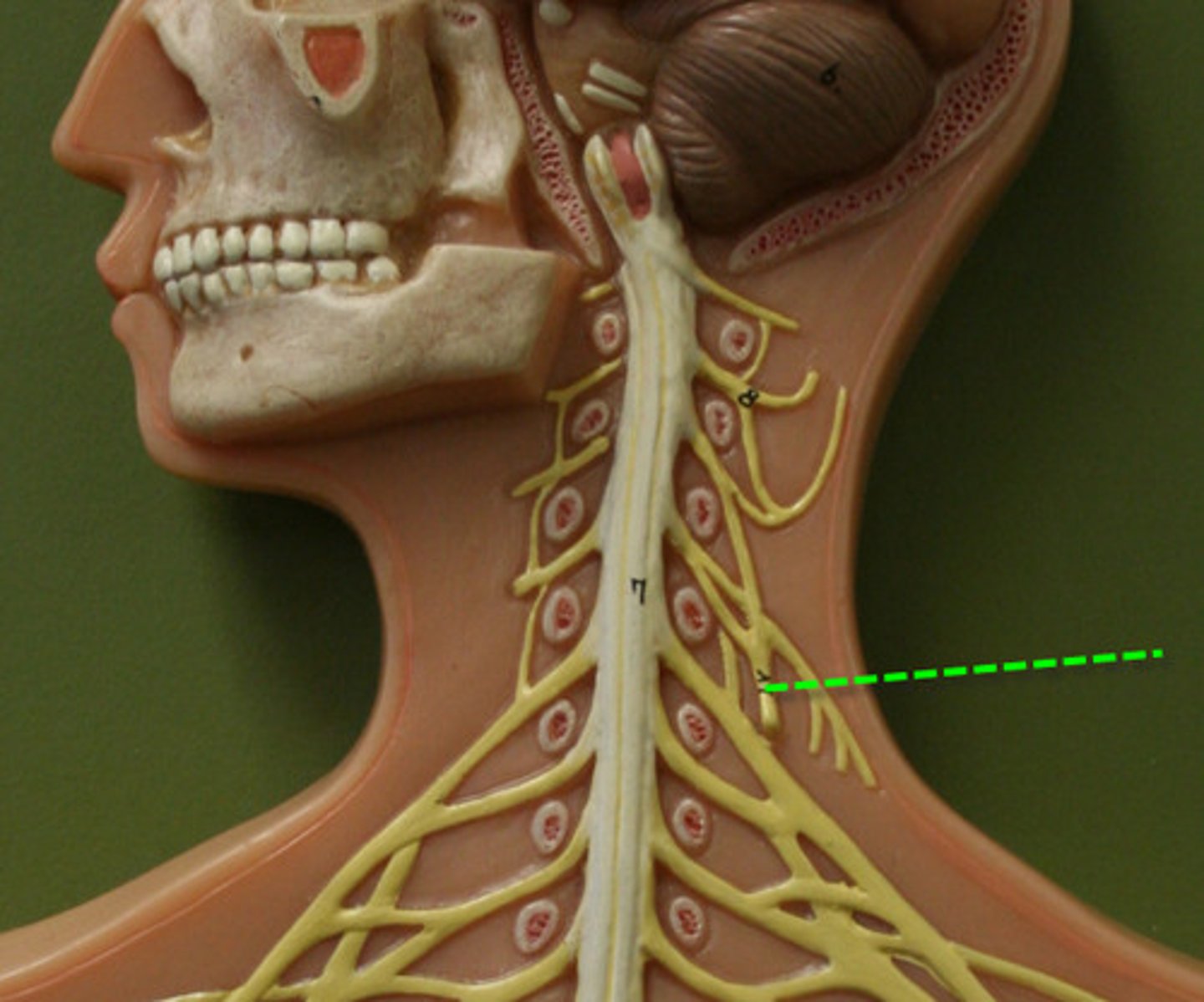
Vagus CX
Parasympathetic to organs
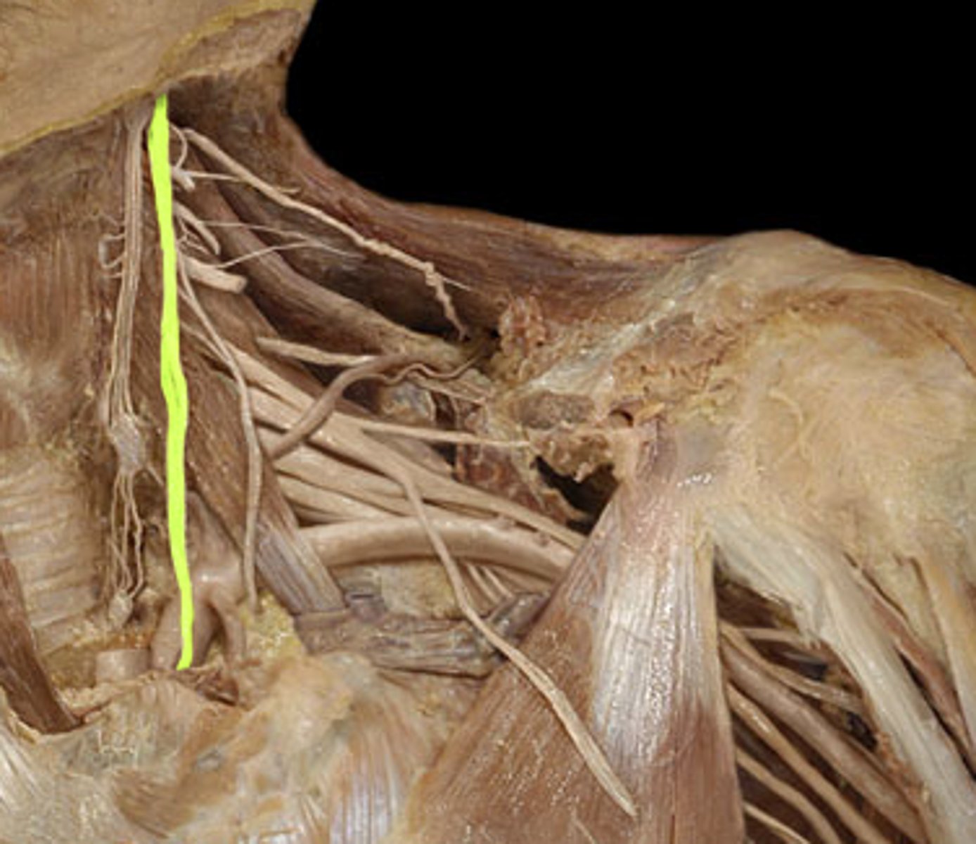
Thoracic duct
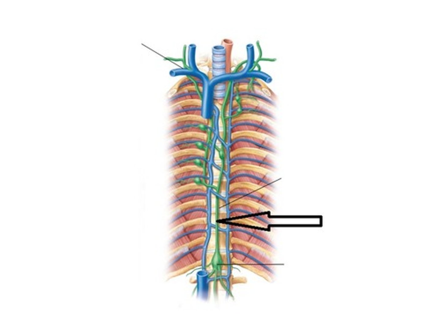
Structures that run through the superior thoracic aperture
Sympathetic trunks, phrenic nerve C3, 4 and 5, vagus CX, thoracic duct
Thoraco-abdominal diaphragm
Draws central tendon down (inspiration)
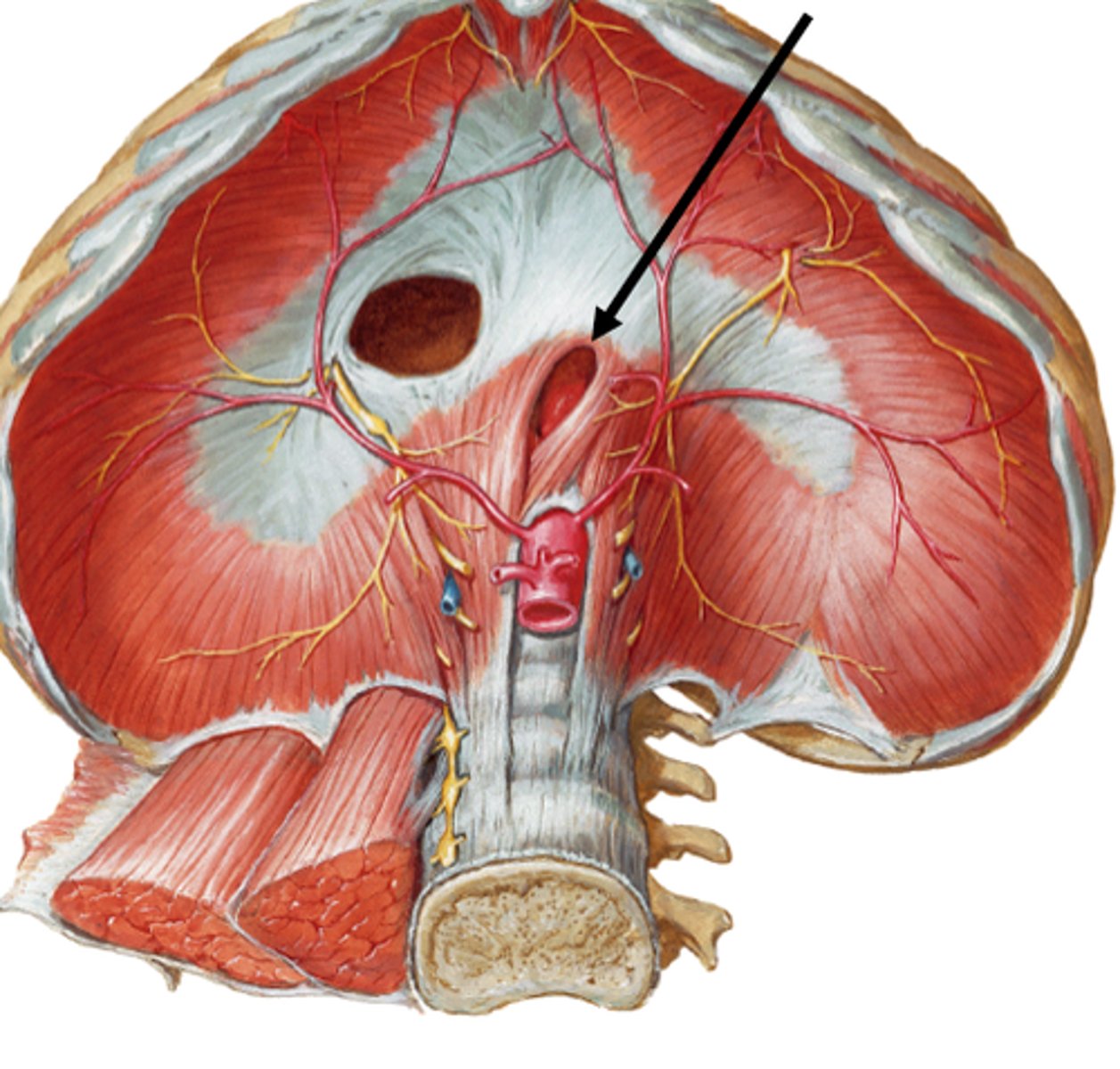
Structures that pass through the diaphragm
Inferior vena cava, esophagus, descending aorta, thoracic duct, azygusvein, sympathetic chains
External intercostals
Aids inspiration, elevates ribs
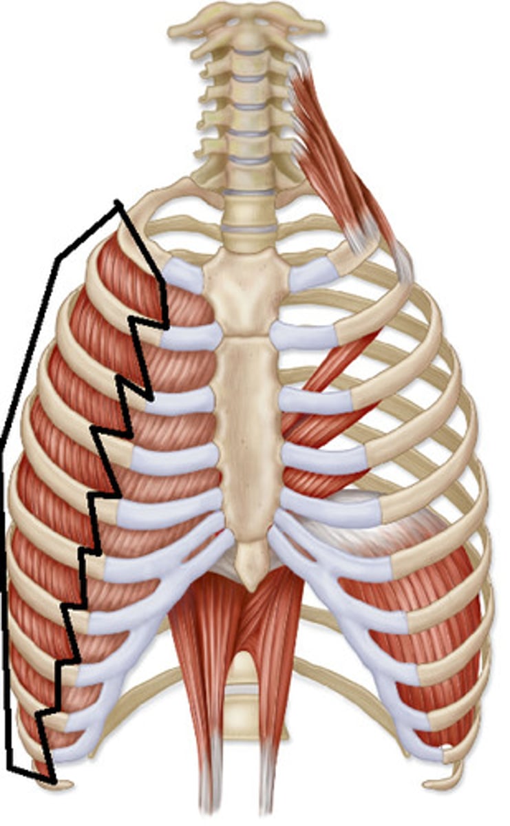
Internal intercostals
Aid forced expiration, depress ribs
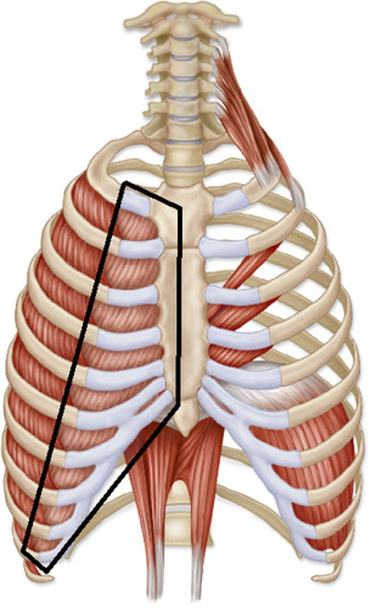
Sternocleidomastoid
Rotate head, flex neck, elevates ribcage (inhalation)
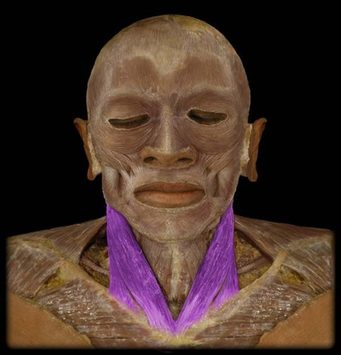
Scalene muscles
Inhalation
Abdominal muscles
Expiration
Upper respiratory system
The nose is lined with mucous membranes and hair. Allows the air to become moist, warm and clean. The medial walls undulate and cause air to whirl as it passes
Pharynx
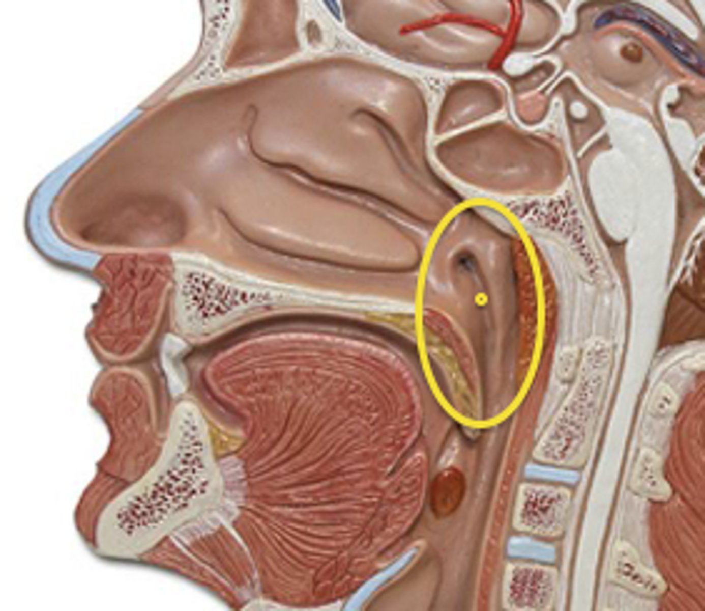
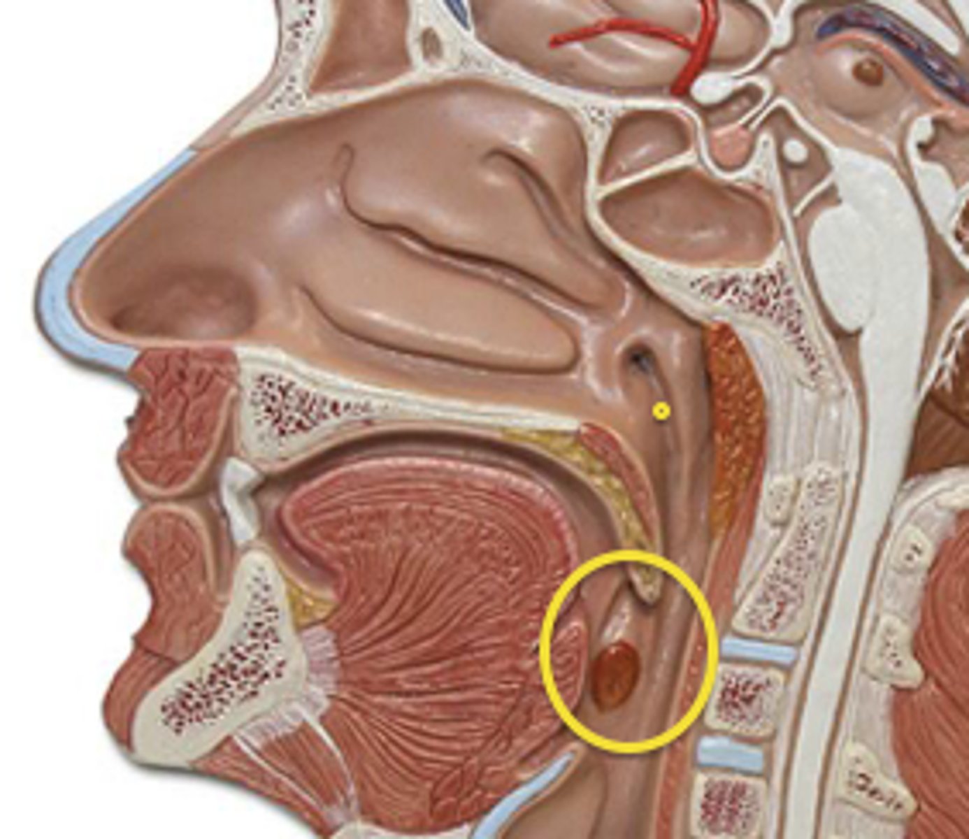
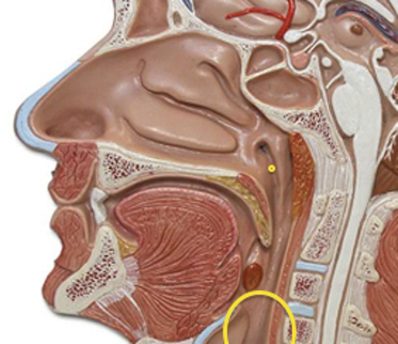
Shared tube to posterior to nose and mouth, for air and food
Nasopharynx
Oropharynx
Laryngopharynx
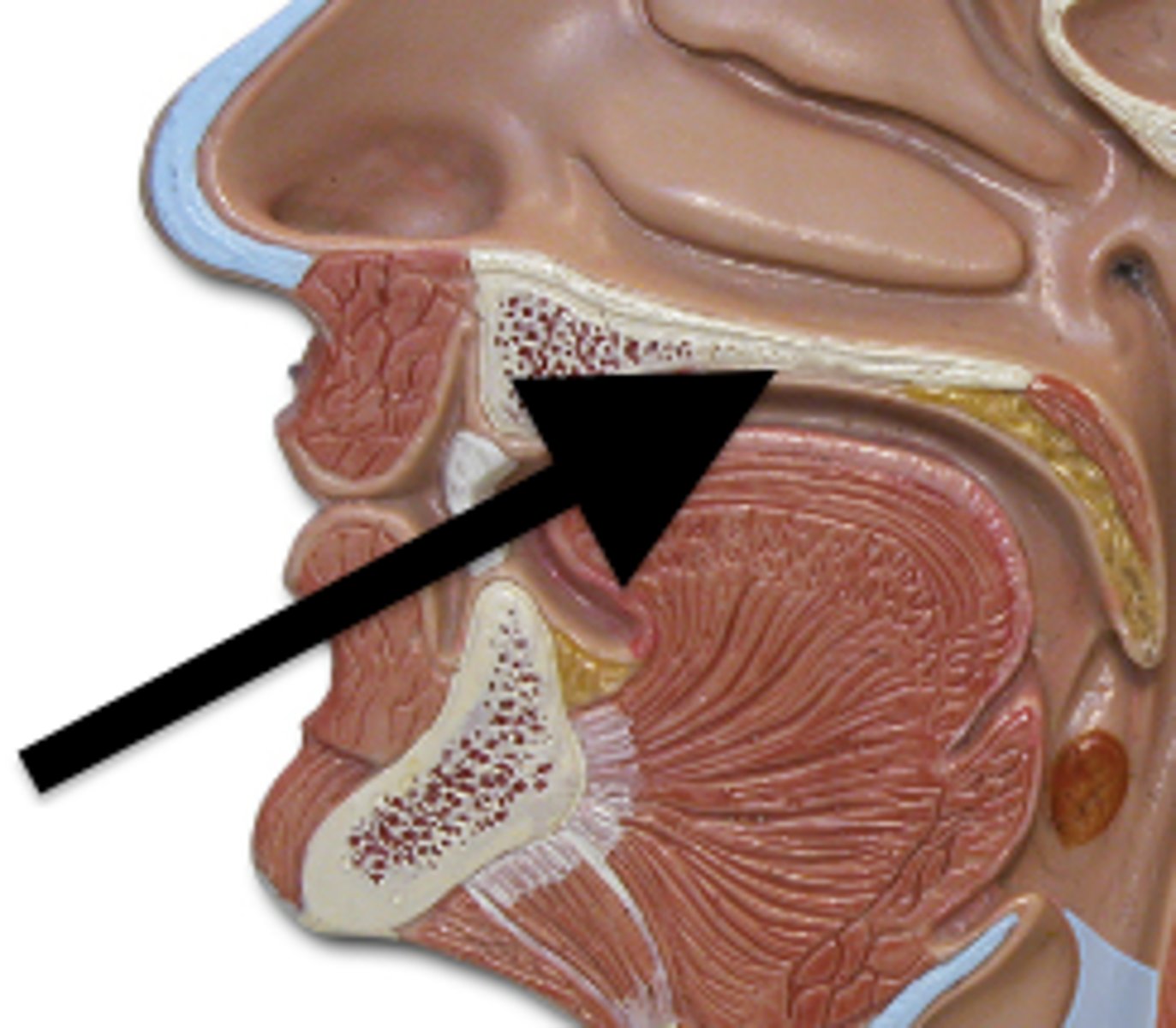
Hard palate
Soft palate
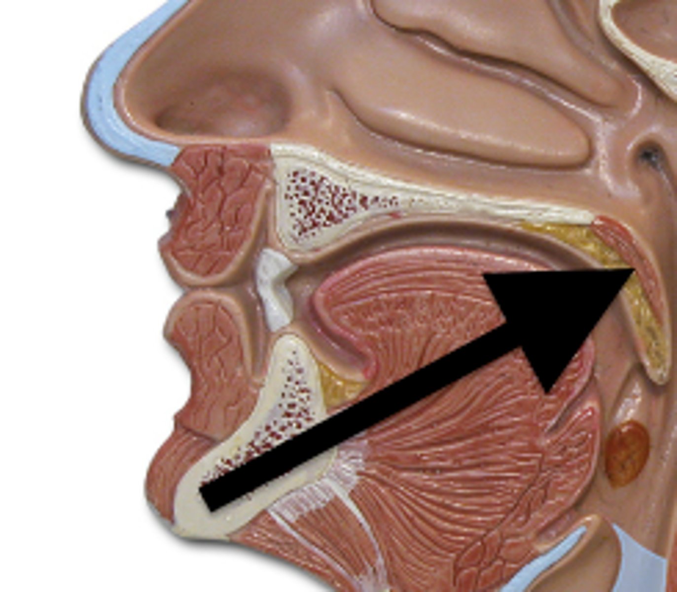
Epiglottis
Covers trachea during swallowing
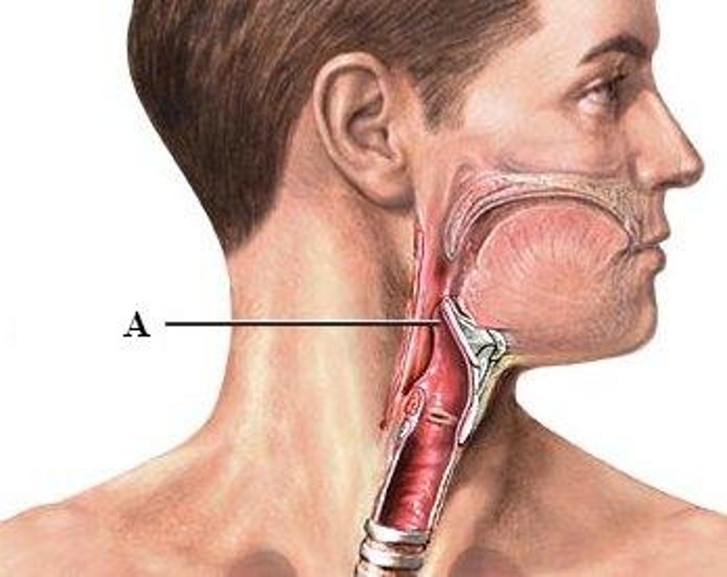
Larynx
Changes shape to make sounds
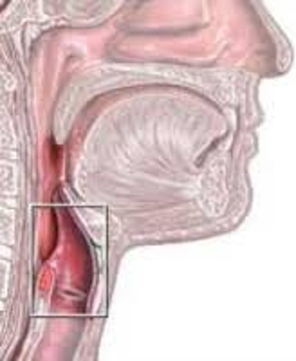
Mediastinum
Contains trachea, bronchi, heart, nerves and esophagus
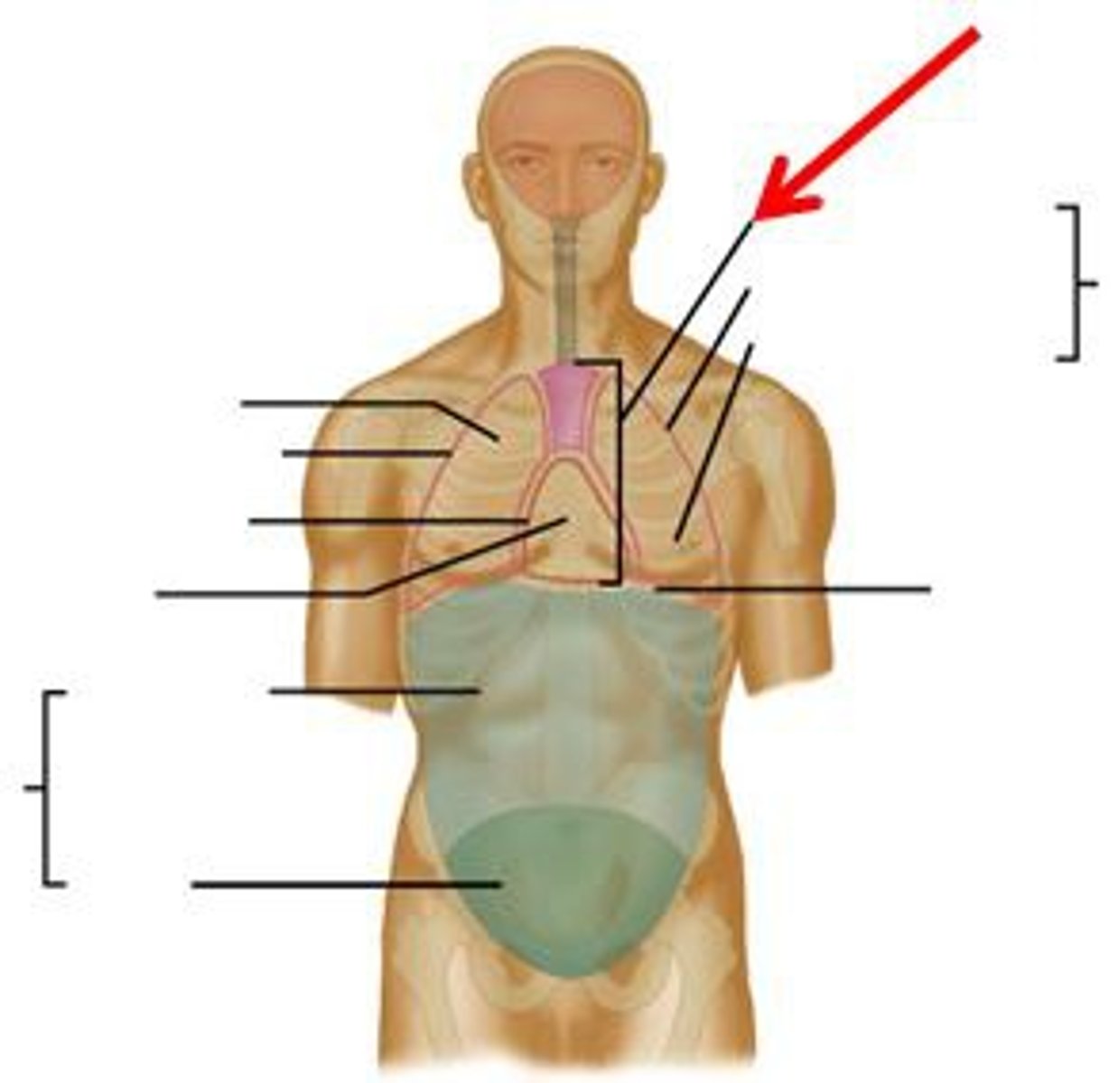
Lungs
Spongy organs, site for gas exchange, left has 2 lobes, right has 3 lobes