BioInst Exam 3
1/64
Earn XP
Description and Tags
Flashcards for BioInst Exam 3
Name | Mastery | Learn | Test | Matching | Spaced | Call with Kai |
|---|
No analytics yet
Send a link to your students to track their progress
65 Terms
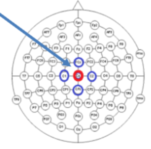
How does a Bipolar Montage work
Each EEG “channel” is the difference between two electrode recordings, such as F7-T3
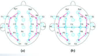
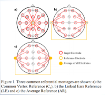
How does a Referential Montage work?
Each channel is the difference between an “active electrode” and a “fixed reference electrode”
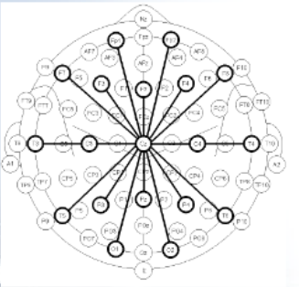
On a referential montage, if Cz is the reference the difference is?
p1-Cz, Fp-Cz, Fz-Cz, F7-Cz, etc.
How does a Common Montage work?
Each channel is the difference between an “active electrode” and the average of all electrodes.
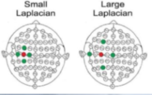
How does a Laplacian Montage work?
Each EEG channel is an active electrode relative to weighted local average. For example, small Laplacian: Cz-(C1+C2+Fcz+Cpz)/4
What is the Temporal Lobe?
The auditory cortex.
What is the Temporal Lobes function?
It is involved in processing sensory input into derived meanings for the appropriate retention of visual memories, language comprehension, and emotion association.
What is the Occipital Lobe?
The visual cortex.
What is the Parietal Lobes function?
It integrates sensory information among various modalities, including proprioception, mechanoreception in the somatosensory cortex, and the dorsal stream of the visual system.
What is the Frontal Lobes function?
It contains most of the dopamine-sensitive neurons in the cerebral cortex. The dopamine system is associated with reward, attention, short-term memory tasks, planning, and motivation.
What is Dopamines function?
Dopamine tends to limit and select sensory information arriving from the thalamus to the forebrain.
What are Synchronous EEG signals?
They are signals driven by external stimulation (sound, light, touch), and it requires no training.
What are Asynchronous EEG signals?
They are signals initiated by the subjects, such as the Mu wave, It requires a lot of training, and it does not depend on external stimulation.
What is the RAS?
The brainstem reticular activating system is a set of connected nuclei in the brains of vertebrates that is responsible for regulating wakefulness, sleep-wake transitions, and providing the pacemaker mechanism.
Where does the EEG come from?
The EEG comes from the total rhythm caused by a large assembly of neurons.
How do neuronal pathways influence EEGs?
Neuronal pathways from columns that resonate with each other and influence EEG frequency.
What domain are Brain waves in?
Brain waves are in the frequency domain.
What domain are Evoked potentials / ERP in?
Evoked potentials / Event-related potentials are in the time domain requiring temporal analysis.
In what applications can EEG Bands serve?
Overall, EEG bands are used in clinical applications that range from monitoring normal wakefulness or arousal states to complex clinical situations involving seizure or coma.
What is the frequency of Delta EEG Bands?
0 - 4 Hz
Where are Delta EEG bands seen?
They are usually seen during sleep, in infancy, or in serious brain diseases.
Where do the Delta EEG bands originate?
Research suggests that these waves originate solely within the cortex, independent of any activity in the deeper brain regions.
What is the frequency of Theta EEG Bands?
4 - 7 Hz
Where are Theta EEG bands seen?
They occur mainly in parietal and temporal regions of children’s brains, but they can additionally be seen shortly after a seizure, as well as in patients with metabolic disorders, white matter encephalopathy, or extensive lesions of the upper brain stem.
What causes Theta EEG bands in healthy and alert adults?
They appear during emotional stress or certain stages of sleep but are otherwise generally absent.
What is the frequency of Alpha EEG Bands?
8 - 13 Hz
Where are Alpha EEG bands seen?
Alpha EEG bands are often recorded from the occipital region during consciousness, and are reduced by visual and other sensory stimulus. This means Alpha waves are typically seen in awake but relaxed patients when the eyes are closed.
When do Alpha EEG waves disappear in patients?
Alpha EEG waves disappear in sleeping or attentive patients.
What is the frequency of Beta I EEG waves?
13 - 20 Hz
What is the frequency of Beta II EEG waves?
20 - 30 or 50 Hz
Where are Beta EEG bands seen?
Beta EEG bands are recorded from the frontal and parietal lobes.
Where do Beta I EEG bands appear?
Beta I EEG bands appear together with the alpha wave.
Where do Beta II EEG bands appear?
Beta I EEG bands appear only during intense mental activity and tension.
What affects Beta I EEG bands?
Beta I EEG are affected by mental activity in much the same way as an alpha wave, but is independently generated.
How does mental activity affect Beta EEG bands?
Beta II EEG bands are a response by mental activity, whereas Beta I EEG bands are inhibited by mental activity.
Where is the Mu wave found?
The Mu wave is found over the motor cortex.
What is the frequency of the Mu wave?
7.5 - 12.5 and primarily 9 -11 Hz
How does a person suppress Mu waves?
A person suppresses Mu waves when they perform a motor action or, with practice, when they visualize performing a motor action. It can even be suppressed when one observes another performing a motor action.
What is the suppression of Mu waves called?
The suppression is called desynchronization because EEG waveforms are caused by a large number of neurons firing in synchrony.
What kind of neurons produce the electrical activity that creates the EEG?
Pyramidal neurons produce the electrical activity of the brain that creates the EEG.
What is the function of pyramidal neurons?
Pyramidal neurons carry the information over long distances.
How many pyramidal neurons does it take to create a signal large enough to be picked up by scalp electrodes?
For pyramidal neurons to create a signal large enough to be picked up by scalp electrodes there need to be thousands of pyramidal neurons sitting in parallel contributing their small voltages.
What does the differential amplifier do in Electrode Montages?
The differential amplifiers cancels out the far-field common activity in electrode montages.
What regulates the resistance of blood flow?
The resistance of blood flow is regulated by arterioles.
What are the Semilunar valves?
The Semilunar valves are the aortic valve (left) and the pulmonary valve (right).
What are the Atrioventricular valves?
The Atrioventricular valves are the mitral valve (left) and tricuspid valve (right).
When do heart sounds reflect turbulence?
The heart sounds reflect turbulence when the heart valves snap shut.
A heart murmur is?
The “murmur is the sound of blood flowing. They can be innocent or can be linked to a damaged or overworked heart valve.
Where does the first heart sound originate?
The first heart sound originates from the movement of blood during a ventricular systole. Further, it originates from the oscillations of blood between descending root of the aorta and the ventricle, and from vibrations of blood turbulence at the aortic and pulmonary valves.
What is the second heart sound?
The second heart sound is a low-frequency vibration associated with the deceleration and reversal of flow in the aorta and pulmonary artery, as well as with the closure of the semilunar valves.
What is the early systolic murmur and when does it occur?
The early systemic murmur is a high-frequency decrescendo murmur, and it begins with the first heart sound and ends before the second heart sound.
When are the third and fourth heart sounds heard?
The rarer extra heart sounds form gallop rhythms and are heard in both normal and abnormal situations.
What is the third heart sound?
The third heart sound, also known as the ventricular gallop,” occurs when the mitral valve opens, allowing passive filling of the left ventricle. The sound can be an important sign of systolic heart failure as the myocardium is overly compliant.
What is the fourth heart sound?
The forth heart sound, also known as the “atrial gallop,” occurs just before the atria contracts to force blood into the left ventricle.
What frequencies can a stethoscope hear?
Stethoscopes can hear frequencies form 0.1 Hz to 2000 Hz.
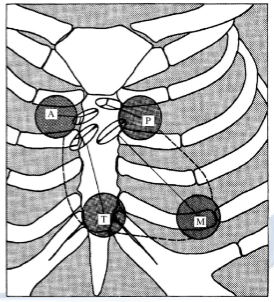
What are the four sites that stethoscopes are used at?
The four sites are the aortic (A), pulmonary (P), tricuspid (T), and mitral area (M).
What is a phonocardiogram?
A PCG is a plot of high fidelity recordings of the sounds and murmurs of the heart.
How does the phonocardiograph pick up sounds?
The phonocardiograph uses a chest microphone that is introduced via the blood vessels into one of the heart chambers.
What does a extravascular sensor do?
A extravascular sensor couples the vascular pressure to an external sensor element via a liquid-filled catheter.
What does a intravascular sensor do?
A intravascular sensor couples the vascular pressure to a sensor placed at the tip of a catheter in the vascular system.
What is underdamped waveform distortion?
increased peaks and a time delay, High frequency response is amplified.
What is overdamped waveform distortion?
big time delay, small amplitude attenuation. Caused by air bubbles or blood clots.
What is catheter whip waveform distortion?
low-frequency oscillations; when an aortic ventricular catheter, in a region of high pulsatile flow, is bent and whipped about by the accelerating blood.
What is aortic stenosis?
the aortic valve does not open fully. This decreases blood flow from the heart.
Why does aortic stenosis occur?
It mainly occurs due to the buildup of calcium deposits that narrow the valve.