Kidney Anatomy and Enlargement
1/26
Earn XP
Description and Tags
Flashcards on the anatomy and enlargement of the kidney.
Name | Mastery | Learn | Test | Matching | Spaced | Call with Kai |
|---|
No analytics yet
Send a link to your students to track their progress
27 Terms
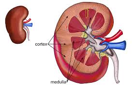
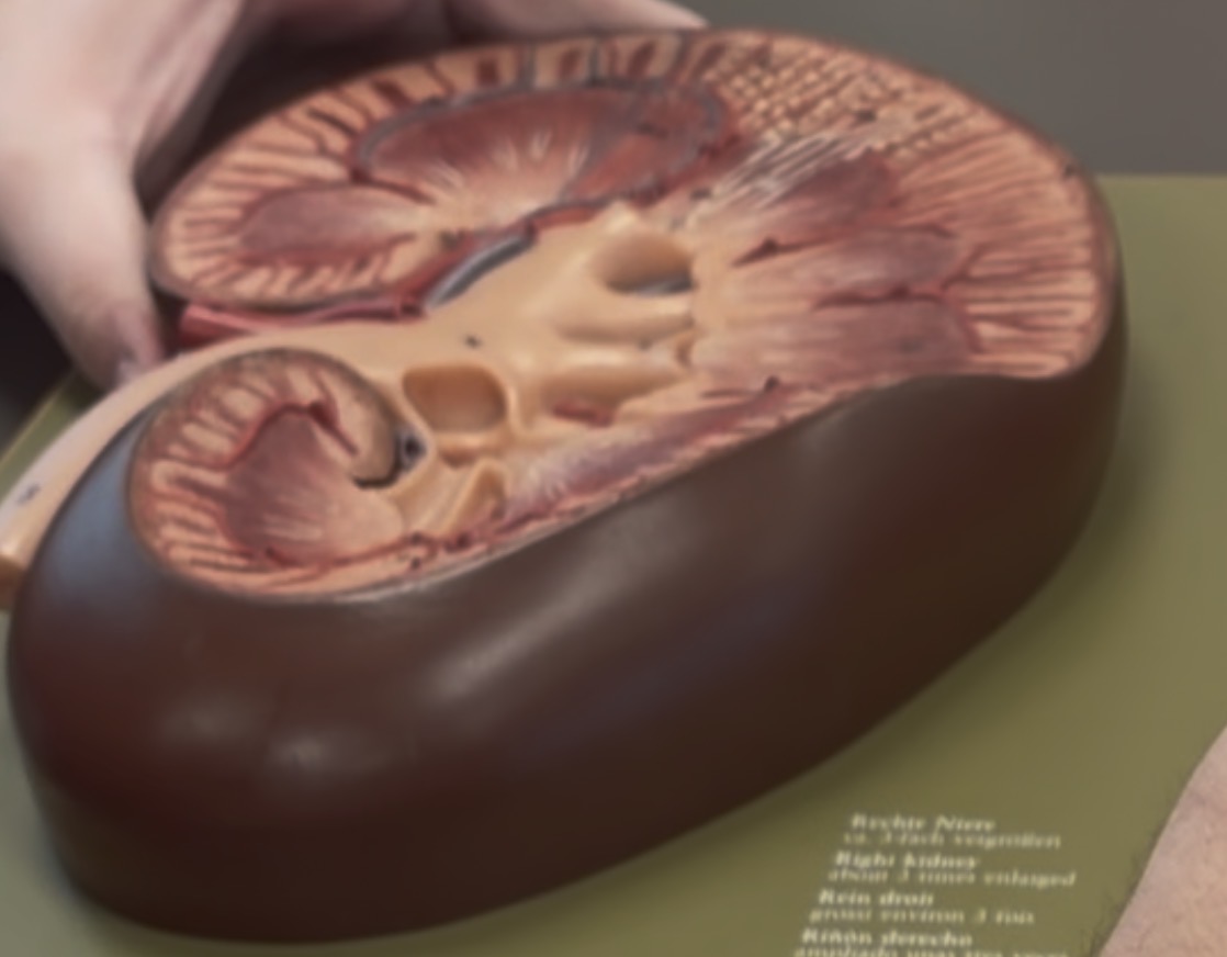
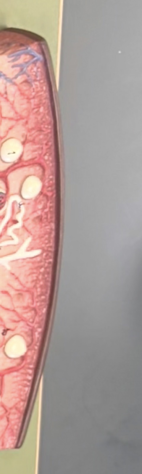
Renal Cortex
the surface around the kidney

Renal Medulla
Center of kidney; closer to renal pyramids

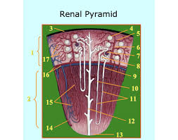
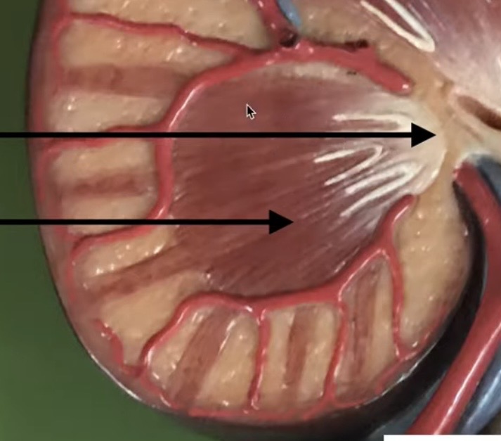
Renal pyramids
Inside the kidney it looks like a pyramid
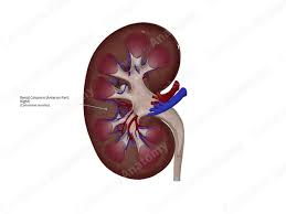
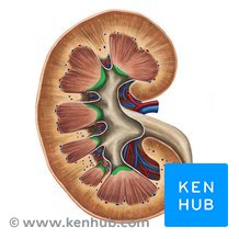
Renal columns
Spaces between the renal pyramids
Renal papilla
where urine exits into calyx;underneath the renal pyramid

Minor calyx
sits underneath the Renal papilla; has 6 branches
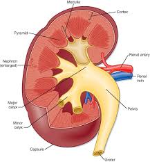
major calyx
formed by merging minor calyx into a big branch; has 3 big branches
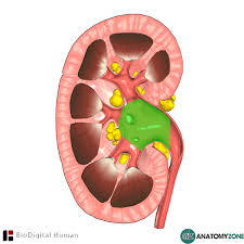
Renal pelvis
blank space in the middle of kidney
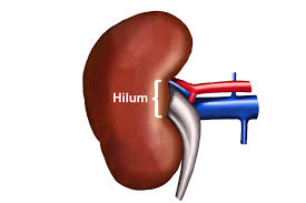
Hilium
Start of renal artery and vein
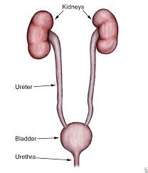
ureter
2 thin lines on both sides
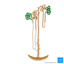
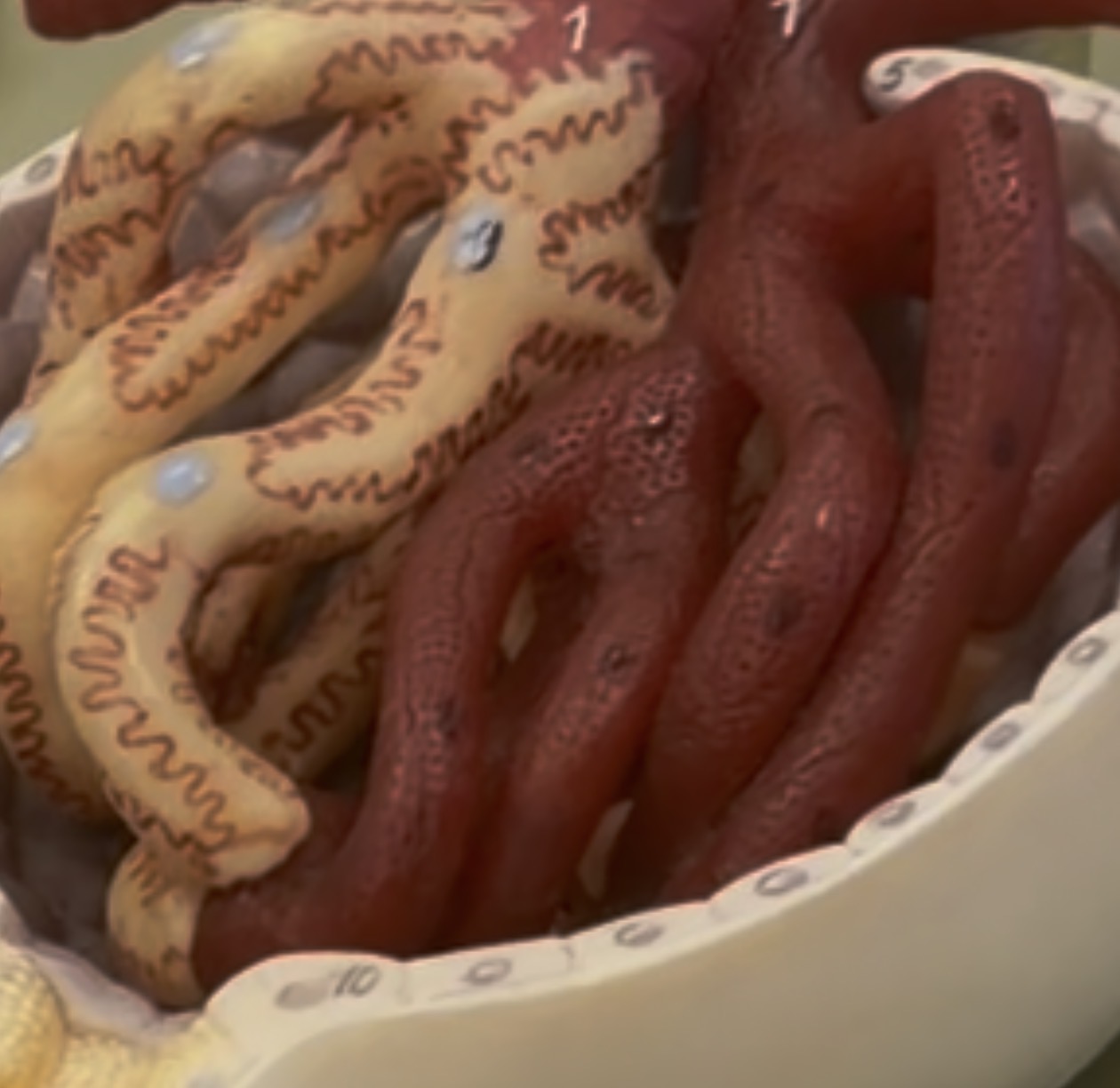
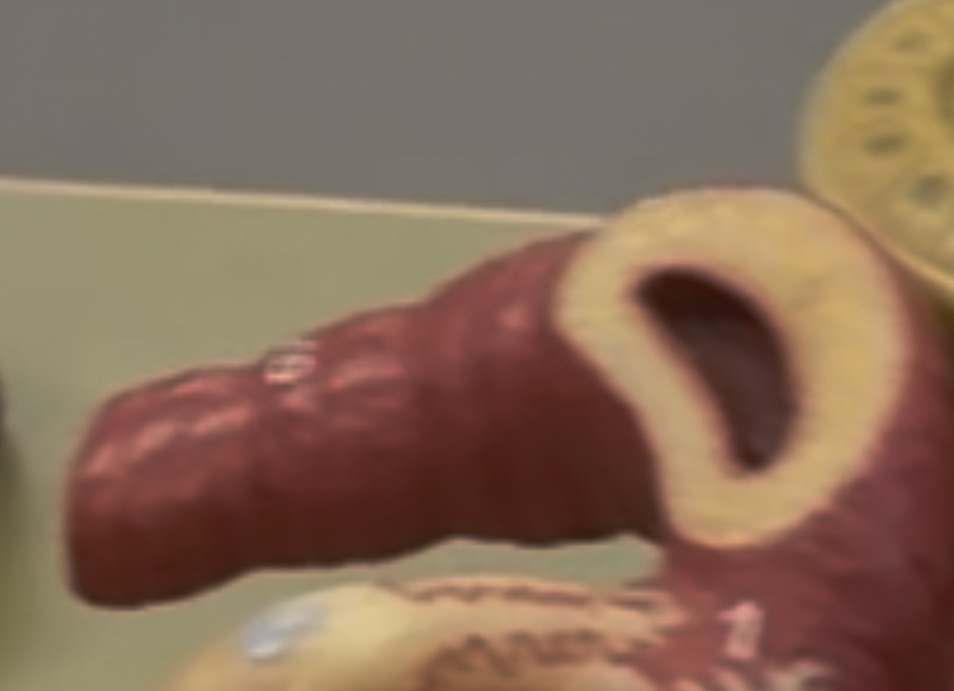
proximal convoluted tubule
Aka PCT on nephron model
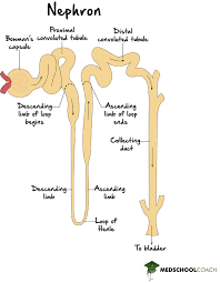
Nephron loop
has a descending and ascending limb
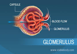
Glomerulus
a small squiggly ball on model

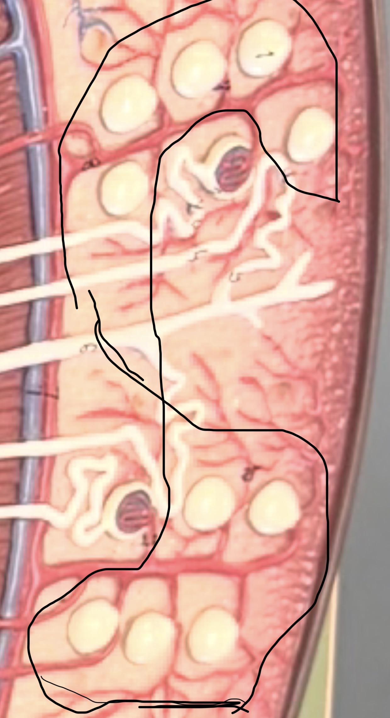
corpuscle is a simple squamous tissue
also this is a corpuscle blown up and is shown on the kidney model

Distal convoluted tubule shown blown in corpuscle
Shown on nephron model
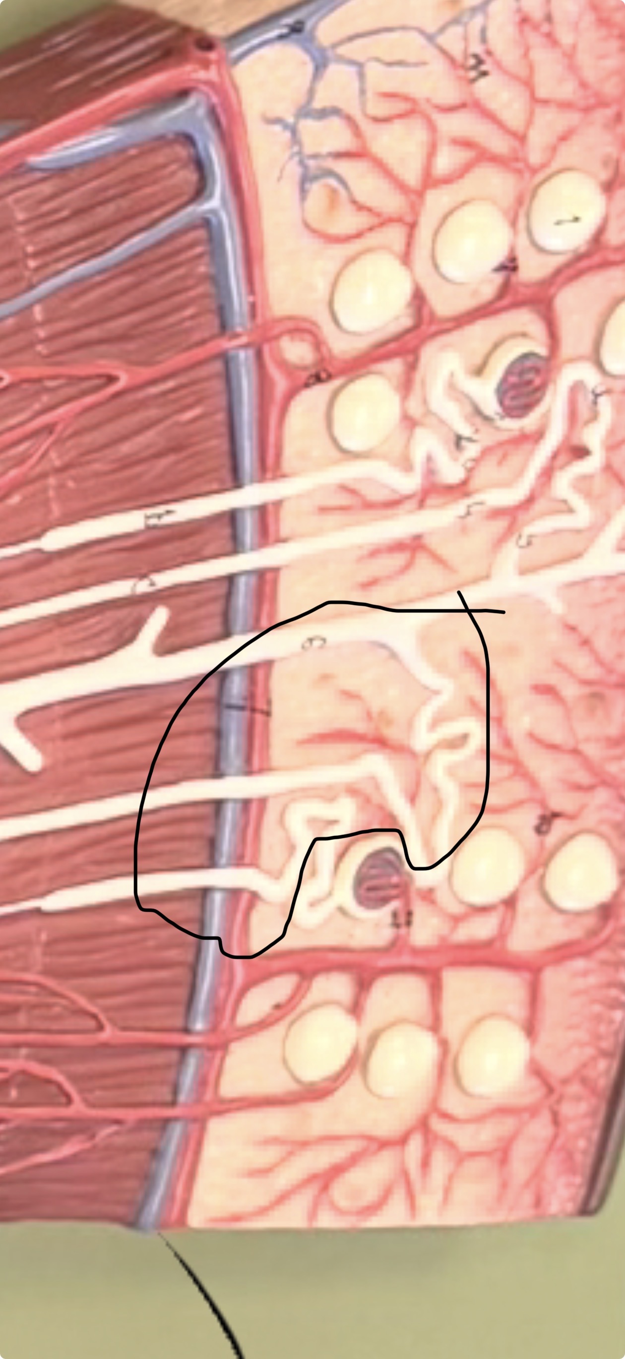
Podocytes
squiggly lines inside the corpuscle
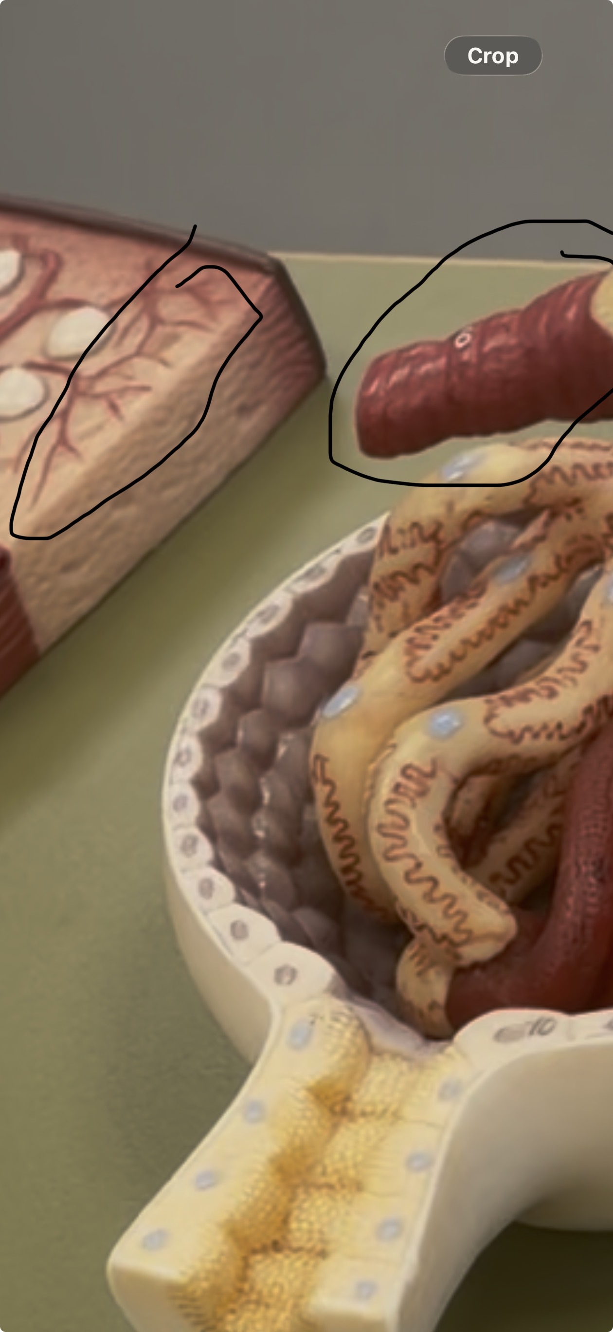
Afferent Arteries
branches off to the corpuscle; shown bigger
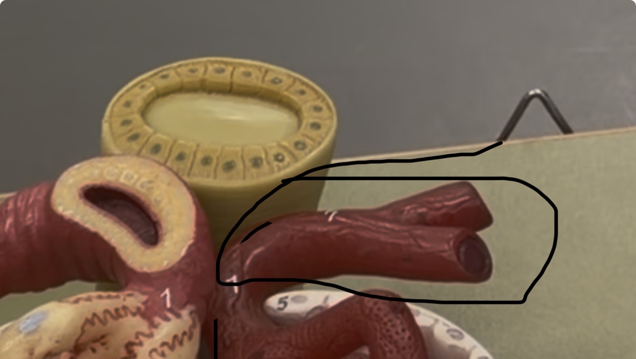
Efferent arteries
shown in the blown up corpuscle
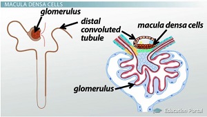
Macula Densa
appears on the afferent artery
Renal Capsule surrounds the kidney
also shown on edge of 2nd model
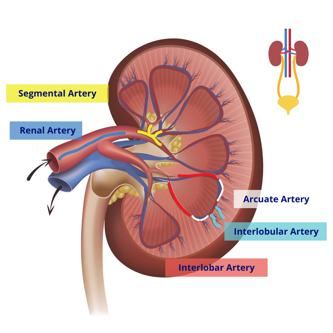
Interlobular artery
it shapes the renal pyramid; the veins on the renal pyramid
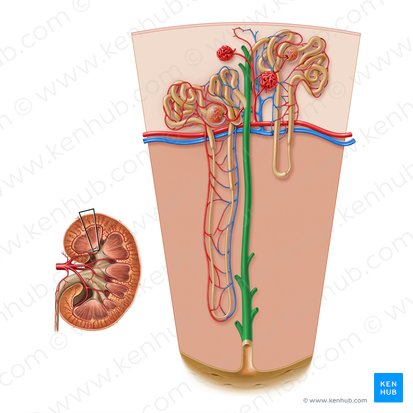
collecting duct
inside the renal pyramid

juxtamedullary vs cortex
names comes from the word
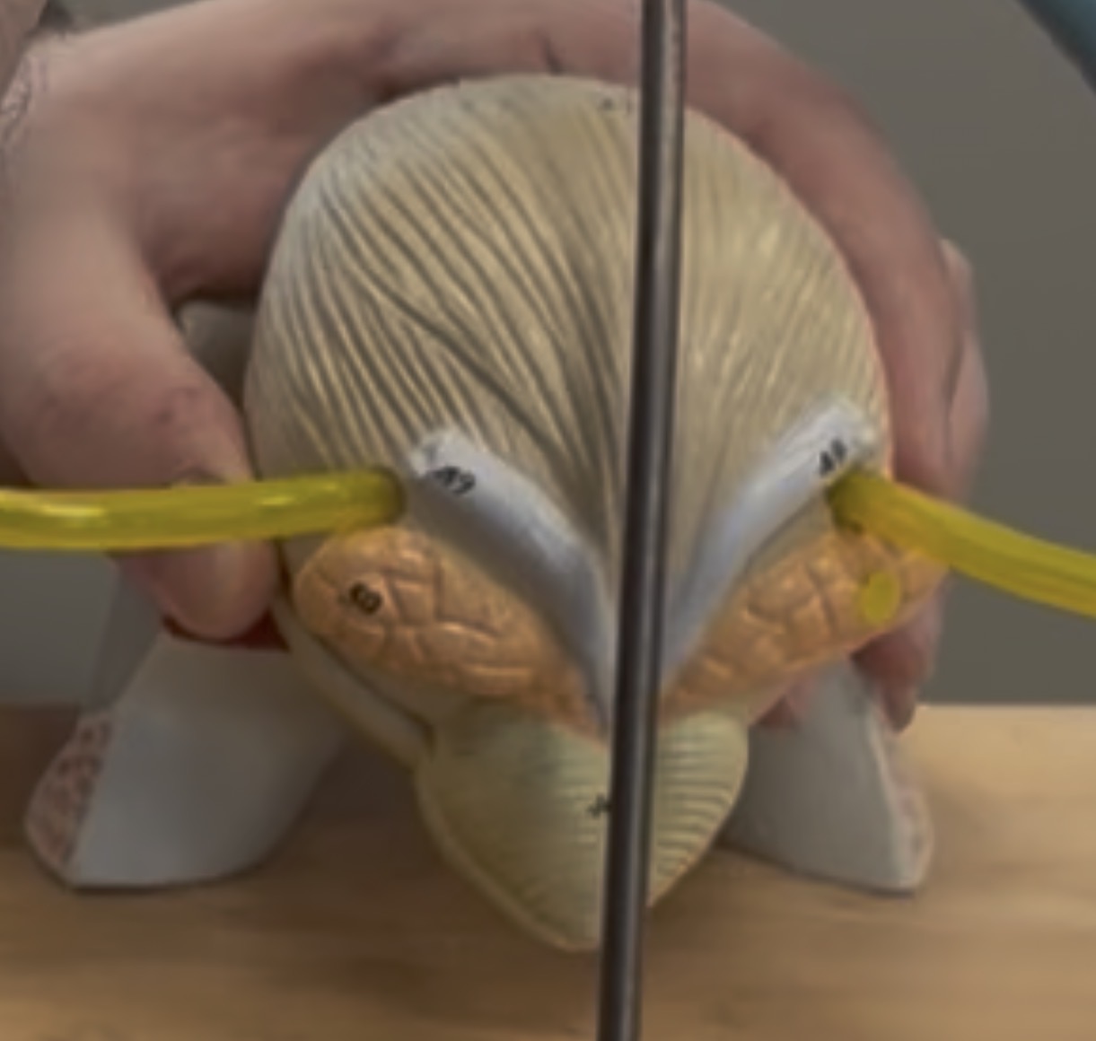
bladder
shown on this model
trigone
its shown blue inside bladder
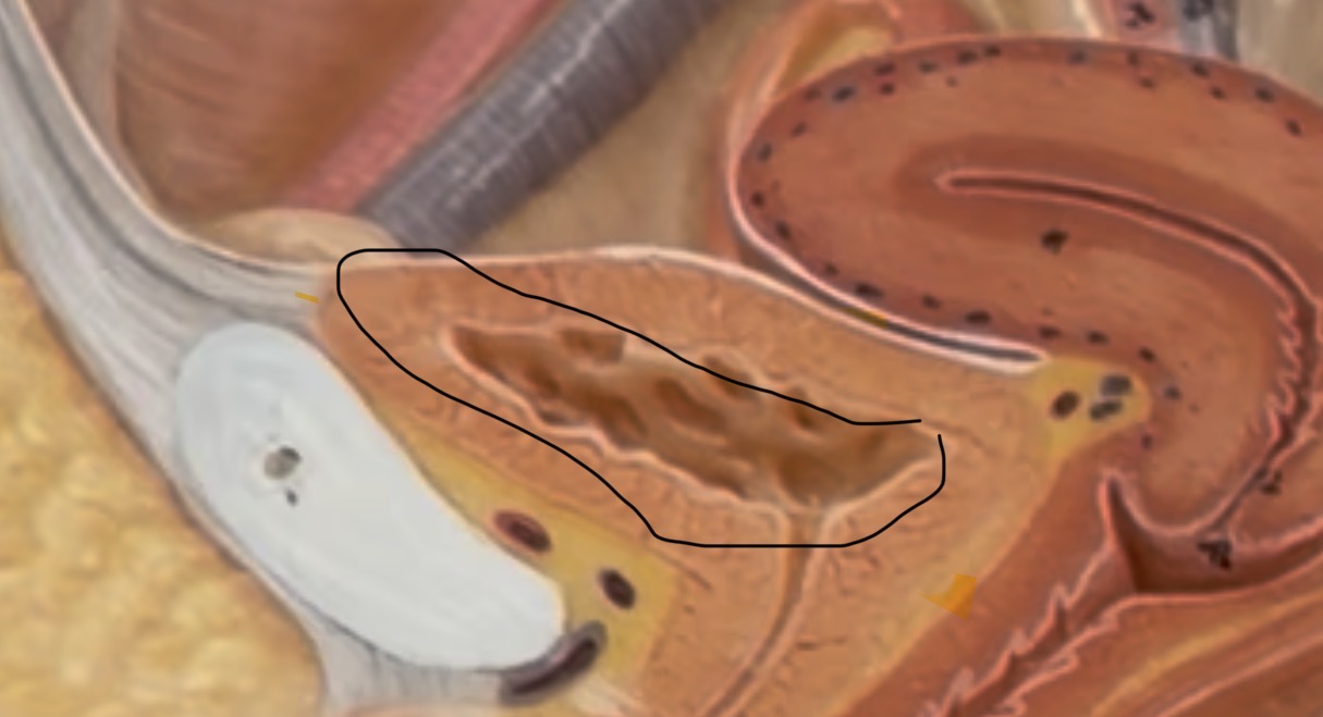
mucosa lining
lining of the rugae
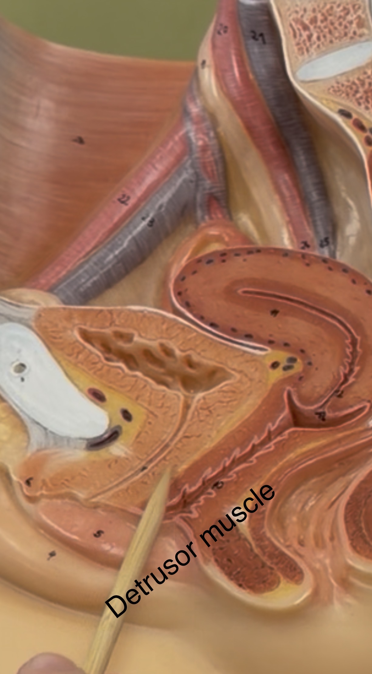
Detrusor muscle
surrounds mucosa lining