3400 Lab Exam 4
1/50
There's no tags or description
Looks like no tags are added yet.
Name | Mastery | Learn | Test | Matching | Spaced | Call with Kai |
|---|
No analytics yet
Send a link to your students to track their progress
51 Terms
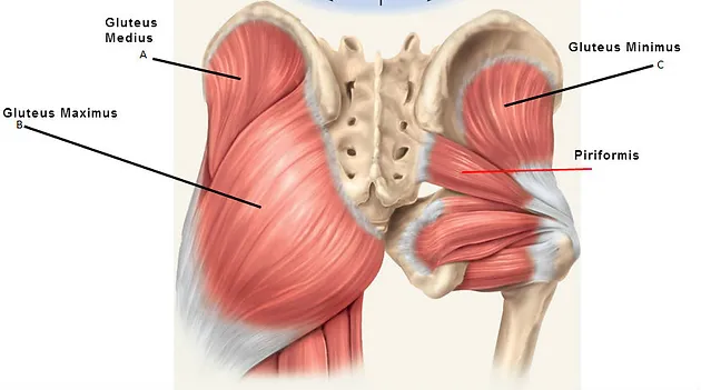
Gluteus maximus
Proximal Attachment (O): Ilium posterior to posterior gluteal line; dorsal surface of sacrum and coccyx; sacrotuberous ligament
Distal Attachment (I): Most fibers end in iliotibial tract, which inserts into lateral condyle of tibia; some fibers insert on gluteal tuberosity.
Nerve: Inferior gluteal nerve (L5, S1, S2)
Action: Extends hip joint (especially from flexed position) and assists in lateral rotation; fixes hip joint and assists in rising from sitting position

Gluteus medius
Proximal Attachment (O): External surface of ilium between anterior and posterior gluteal lines
Distal Attachment (I): Lateral surface of greater trochanter of femur
Nerve: Superior gluteal nerve (L5, S1)
Action: Abduct and medially rotate hip joint; keep pelvis level when ipsilateral limb is weight bearing and advance opposite (unsupported) side during its swing phase.

Gluteus minimus
Proximal Attachment (O): External surface of ilium between anterior and inferior gluteal lines
Distal Attachment (I): Anterior surface of greater trochanter of femur
Nerve: Superior gluteal nerve (L5, S1)
Action: Abduct and medially rotate hip joint; keep pelvis level when ipsilateral limb is weight bearing and advance opposite (unsupported) side during its swing phase.
Tensor fascia latae
Proximal Attachment (O): Anterior superior iliac spine; anterior part of iliac crest
Distal Attachment (I): Iliotibial tract, which attaches to lateral condyle of tibia
Nerve: Superior gluteal nerve (L5, S1)
Action: Abduct and medially rotate hip joint; keep pelvis level when ipsilateral limb is weight bearing and advance opposite (unsupported) side during its swing phase.

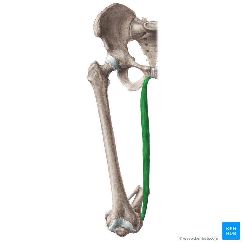
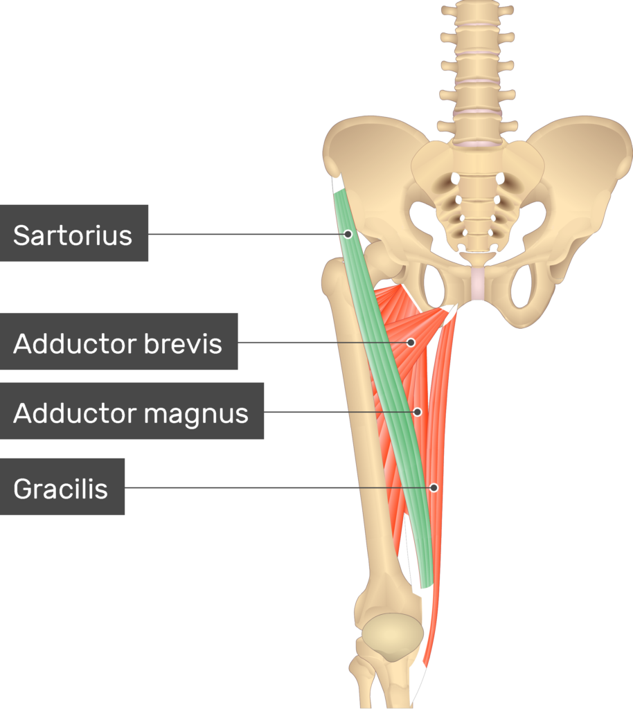
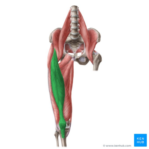
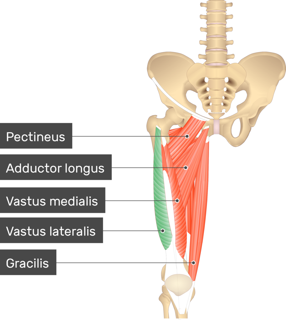
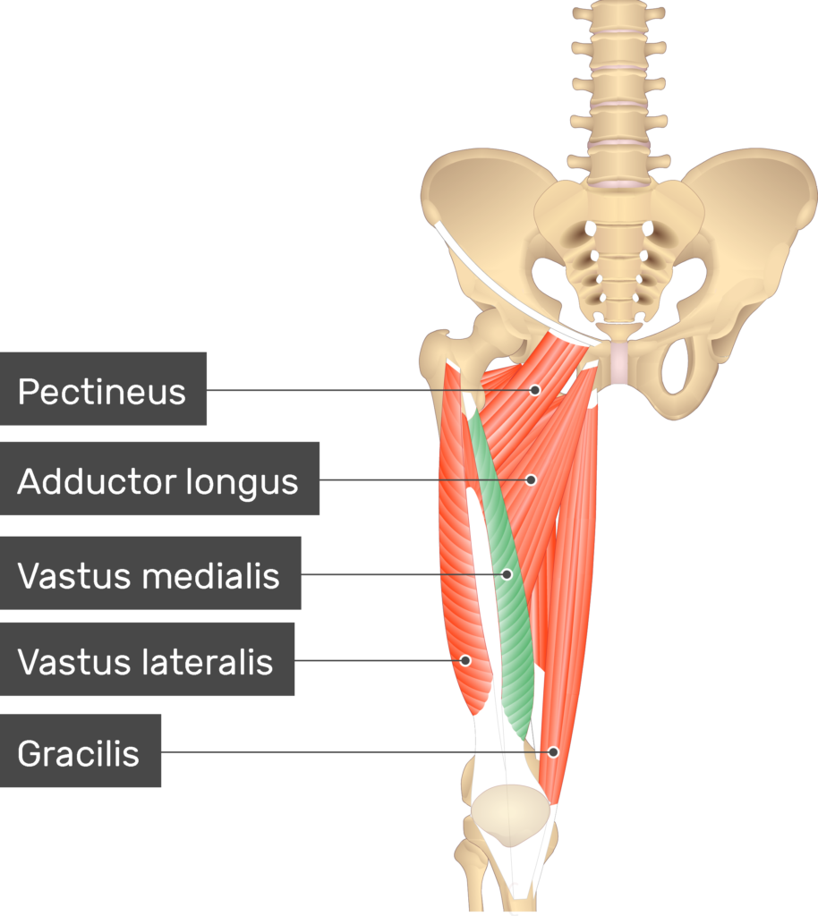
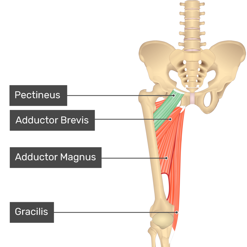
Gracilis
Proximal Attachment (O): Body and inferior ramus of pubis
Distal Attachment (I): Superior part of medial surface of tibia (as part of pes anserinus)
Nerve: Obturator nerve (L2, L3)
Action: Adducts hip joint; flexes knee joint, medially rotating it when flexed
Sartorius
Proximal Attachment (O): Anterior superior iliac spine and superior part of notch inferior to it
Distal Attachment (I): Superior part of medial surface of tibia (as part of pes anserinus)
Nerve: Femoral nerve (L2, L3)
Action: Flexes, abducts, and lateral rotates hip joint; flexes knee joint (medially rotates leg when knee joint is flexed)
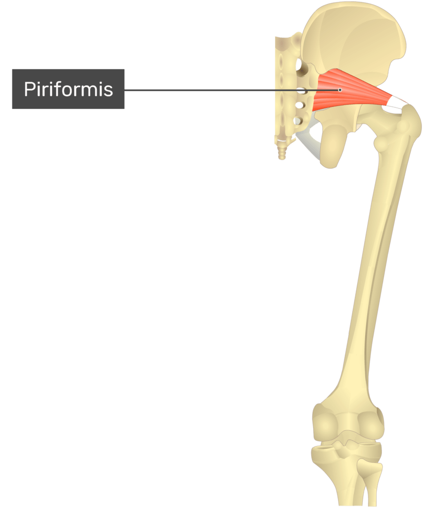
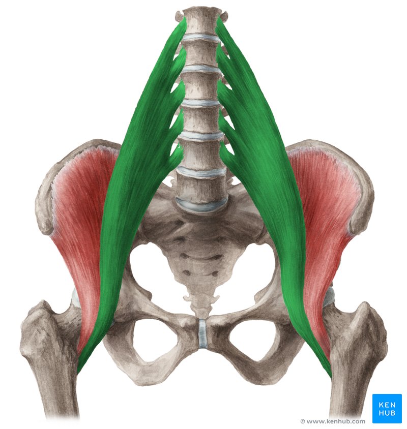
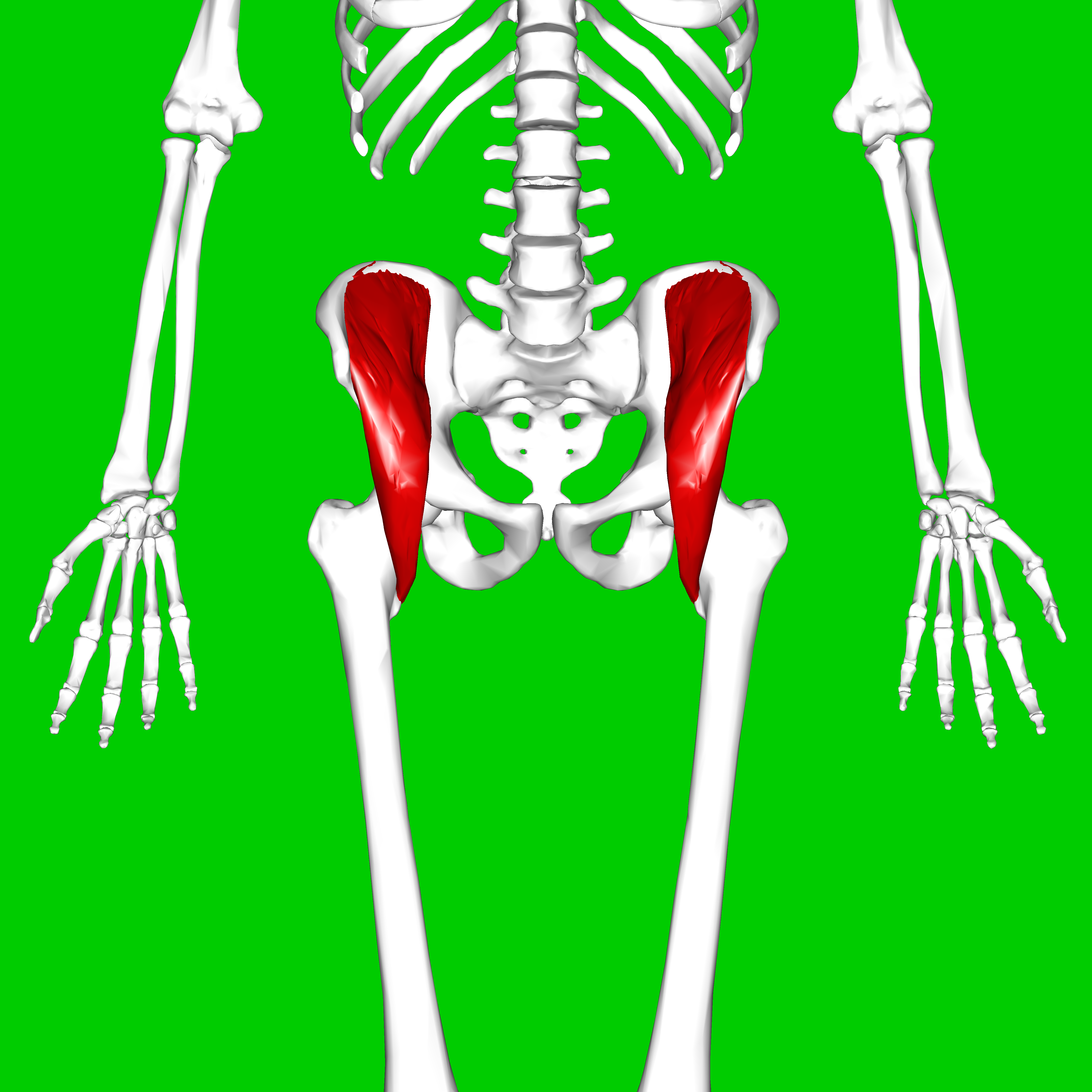
Piriformis
Proximal Attachment (O): Anterior surface of sacrum; sacrotuberous ligament
Distal Attachment (I): Superior border of greater trochanter of femur
Nerve: Branches of anterior rami of S1 and S2
Action: Laterally rotate extended hip joint and abduct hip joint when flexed; stabilize hip joint
Gemellus superior
Proximal Attachment (O): Ischial spine
Distal Attachment (I): Medial surface of greater trochanter (trochanteric fossa) of femurb
Nerve: same nerve supply as obturator internus (Nerve to obturator internus (L5, S1))
Action: Laterally rotate extended hip joint and abduct hip joint when flexed; stabilize hip joint

Gemellus inferior
Proximal Attachment (O): Ischial tuberosity
Distal Attachment (I): Medial surface of greater trochanter (trochanteric fossa) of femur
Nerve: same nerve supply as quadratus femoris (Nerve to quadratus femoris (L5, S1))
Action: Laterally rotate extended hip joint and abduct hip joint when flexed; stabilize hip joint
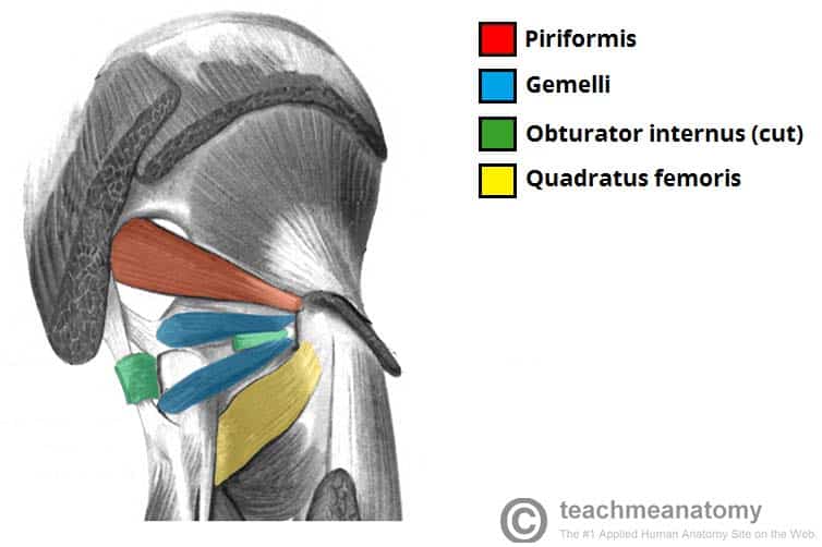
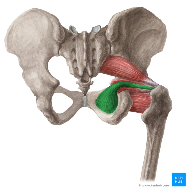
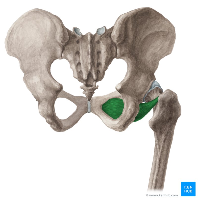
Obturator internus
Proximal Attachment (O): Pelvic surface of obturator membrane and surrounding bones
Distal Attachment (I): Medial surface of greater trochanter (trochanteric fossa) of femur
Nerve: Nerve to obturator internus (L5, S1)
Action: Laterally rotate extended hip joint and abduct hip joint when flexed; stabilize hip joint
Obturator externus
Proximal Attachment (O): Margins of obturator foramen and obturator membrane
Distal Attachment (I): Trochanteric fossa of femur
Nerve: Obturator nerve (L3, L4)
Action: Laterally rotates hip joint; stabilizes hip joint
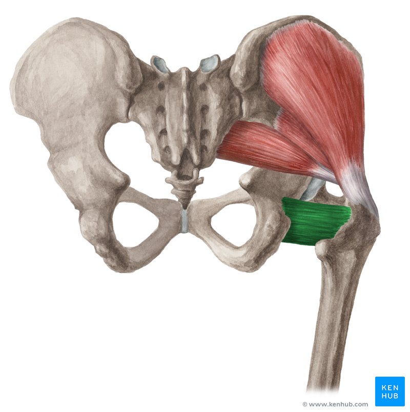
Quadratus femoris
Proximal Attachment (O): Lateral border of ischial tuberosity
Distal Attachment (I): Quadrate tubercle on intertrochanteric crest of femur and area inferior to it
Nerve: Nerve to quadratus femoris (L5, S1)
Action: Laterally rotates hip jointc; stabilizes hip joint
Biceps femoris
Proximal Attachment (O):
Long head: ischial tuberosity
Short head: linea aspera and lateral supracondylar line of femur
Distal Attachment (I): Lateral side of head of fibula. Tendon is split at this site by fibular collateral ligament of knee.
Nerve:
Long head: tibial division of sciatic nerve (L5, S1, S2)
Short head: common fibular division of sciatic nerve (L5, S1, S2)
Action: Flexes knee joint and laterally rotates it when flexed; long head extends hip joint (e.g., accelerating mass during first step of gait and when rising from sitting position).

Semimembranosus
Proximal Attachment (O): Ischial tuberosity
Distal Attachment (I): Posterior part of medial condyle of tibia. Reflected attachment forms oblique popliteal ligament (to lateral femoral condyle).
Nerve: Tibial division of sciatic nerve part of tibia (L5, S1, S2)
Action: Extend hip joint; flex knee joint and medially rotate it when flexed. When hip and knee joints are flexed (as when sitting), these muscles can extend trunk at hip joint (to rise).

Semitendinosus
Proximal Attachment (O): Ischial tuberosity
Distal Attachment (I): Superior part of media surface of tibia (as part of pes anserinus)
Nerve: Tibial division of sciatic nerve part of tibia (L5, S1, S2)
Action: Extend hip joint; flex knee joint and medially rotate it when flexed. When hip and knee joints are flexed (as when sitting), these muscles can extend trunk at hip joint (to rise).

Rectus femoris
Proximal Attachment (O): Anterior inferior iliac spine and ilium superior to acetabulum
Distal Attachment (I): Via common tendinous (quadriceps tendon) and independent attachments to base of patella; indirectly via patellar ligament to tibial tuberosity; medial and lateral vasti also attach to tibia and patella via aponeuroses (medial and lateral patellar retinacula).
Nerve: Femoral nerve (L2, L3, L4)
Action: Extends knee joint; rectus femoris also steadies hip joint and helps iliopsoas flex hip joint.
Vastus lateralis
Proximal Attachment (O): Greater trochanter and lateral lip of linea aspera of femur
Distal Attachment (I): Via common tendinous (quadriceps tendon) and independent attachments to base of patella; indirectly via patellar ligament to tibial tuberosity; medial and lateral vasti also attach to tibia and patella via aponeuroses (medial and lateral patellar retinacula).
Nerve: Femoral nerve (L2, L3, L4)
Action: Extends knee joint; rectus femoris also steadies hip joint and helps iliopsoas flex hip joint.
Vastus intermedius
Proximal Attachment (O): Anterior and lateral surfaces of shaft of femur
Distal Attachment (I): Via common tendinous (quadriceps tendon) and independent attachments to base of patella; indirectly via patellar ligament to tibial tuberosity; medial and lateral vasti also attach to tibia and patella via aponeuroses (medial and lateral patellar retinacula).
Nerve: Femoral nerve (L2, L3, L4)
Action: Extends knee joint; rectus femoris also steadies hip joint and helps iliopsoas flex hip joint.
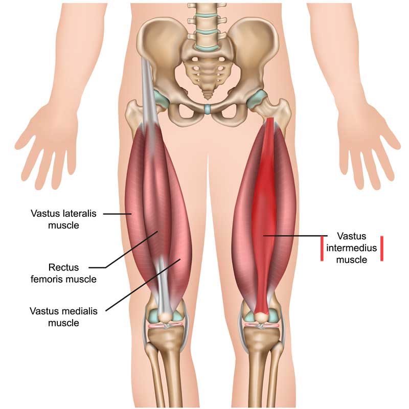
Vastus medialis
Proximal Attachment (O): Intertrochanteric line and medial lip of linea aspera of femur
Distal Attachment (I): Via common tendinous (quadriceps tendon) and independent attachments to base of patella; indirectly via patellar ligament to tibial tuberosity; medial and lateral vasti also attach to tibia and patella via aponeuroses (medial and lateral patellar retinacula).
Nerve: Femoral nerve (L2, L3, L4)
Action: Extends knee joint; rectus femoris also steadies hip joint and helps iliopsoas flex hip joint.
Adductor brevis
Proximal Attachment (O): Body and inferior ramus of pubis
Distal Attachment (I): Pectineal line and proximal part of linea aspera of femur
Nerve: Obturator nerve and branch of anterior division (L2, L3, L4)
Action: Adducts hip joint and, to some extent, flexes it
Adductor longus
Proximal Attachment (O): Body of pubis inferior to pubic crest
Distal Attachment (I): Middle third of linea aspera of femur
Nerve: Obturator nerve and branch of anterior division (L2, L3, L4)
Action: Adducts hip joint
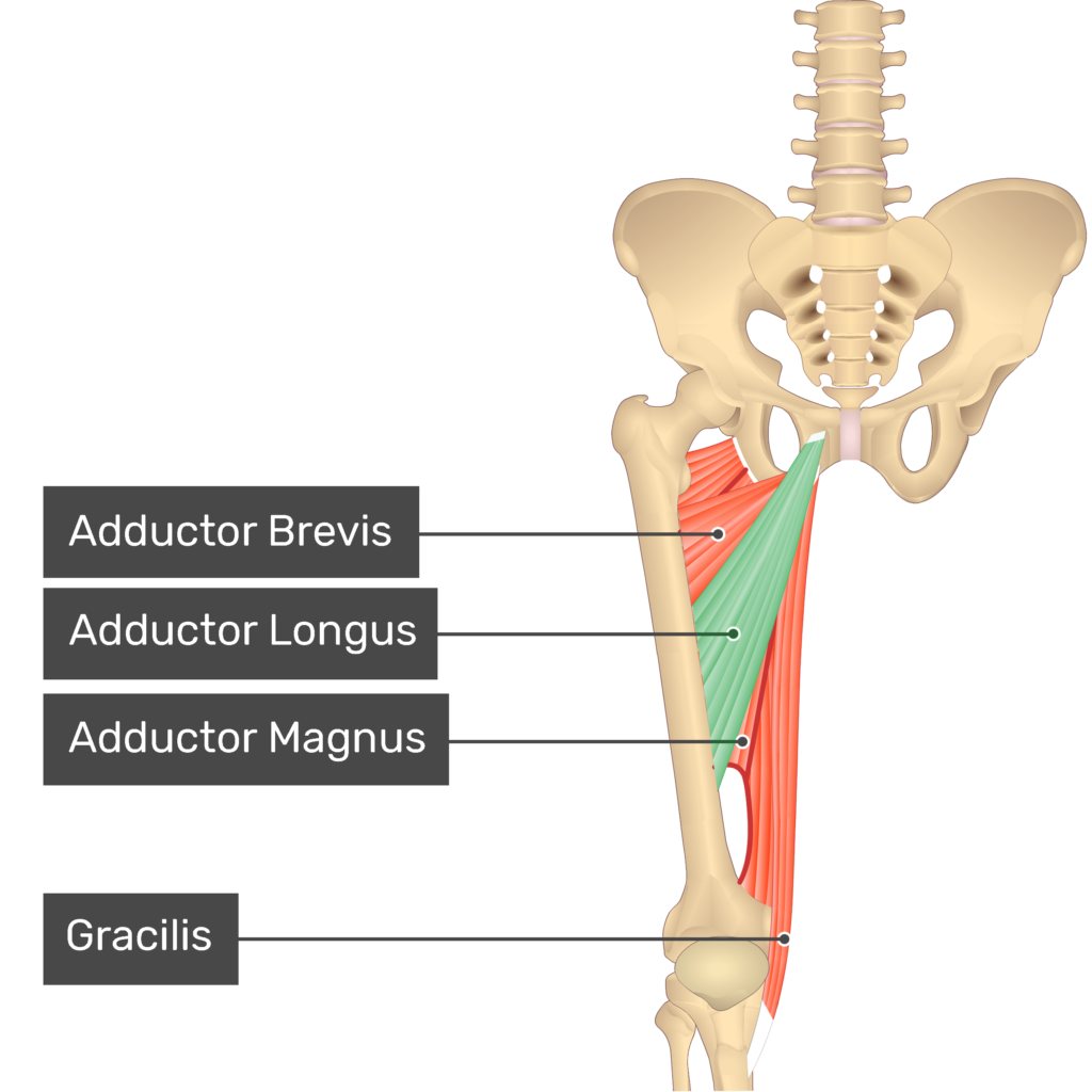
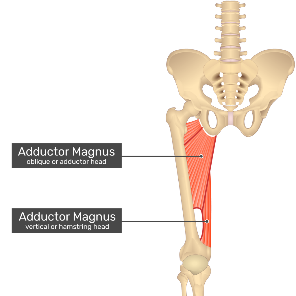
Adductor magnus
Proximal Attachment (O):
Adductor part: inferior ramus of pubis and ramus of ischium
Hamstring part: ischial tuberosity
Distal Attachment (I):
Adductor part: gluteal tuberosity, linea aspera, and medial supracondylar line
Hamstring part: adductor tubercle of femur
Nerve:
Adductor part: obturator nerve (L2, L3, L4) and branches of posterior division
Hamstring part: tibial part of sciatic nerve (L4)
Action:
Adducts hip joint
Adductor part: flexes hip joint
Hamstring part: extends hip joint
Pectineus
Proximal Attachment (O): Superior ramus of pubis
Distal Attachment (I): Pectineal line of femur, just inferior to lesser trochanter
Nerve: Femoral nerve (L2, L3); may receive a branch from obturator nerve
Action: Adducts and slightly flexes hip joint; assists with lateral rotation
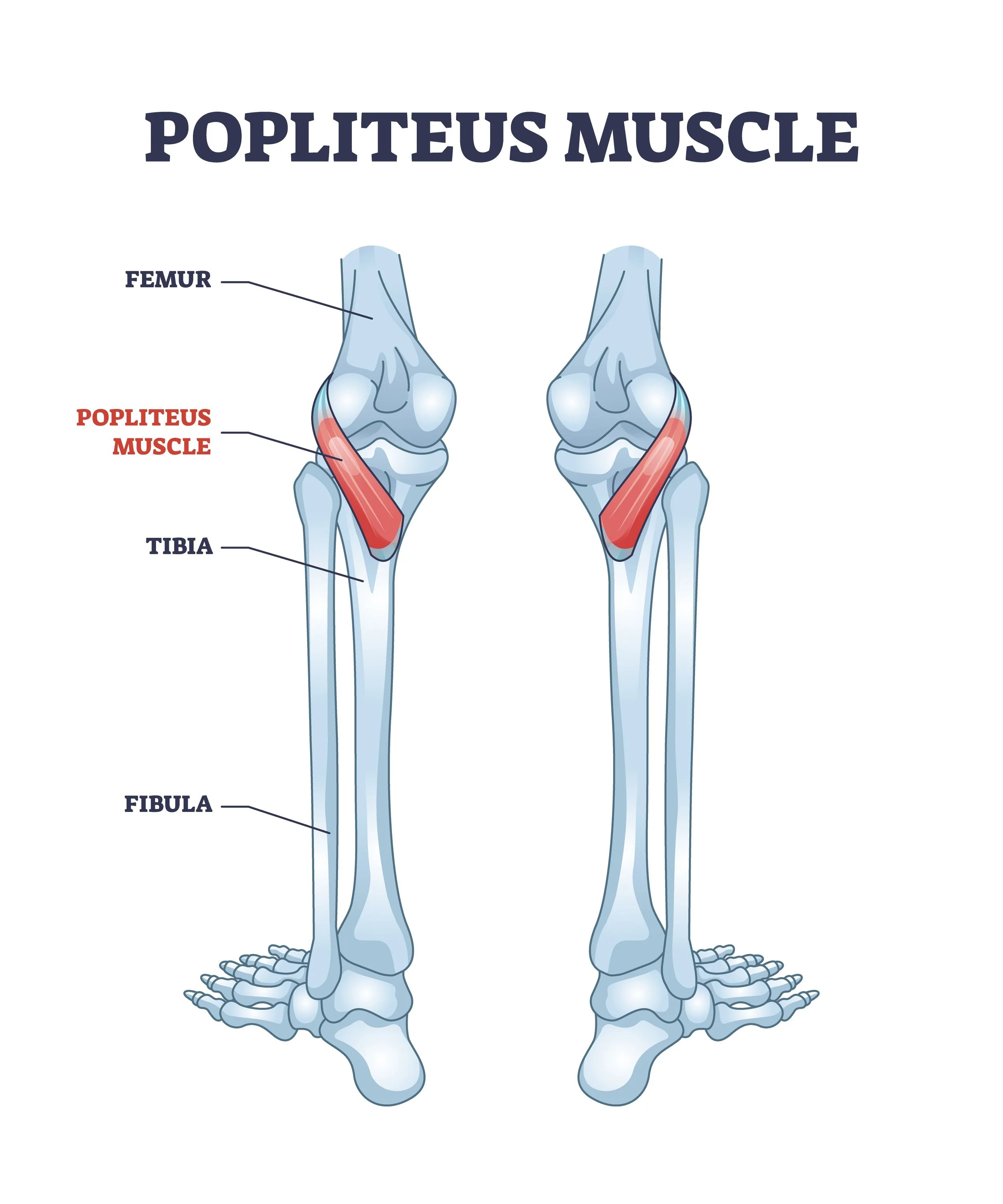
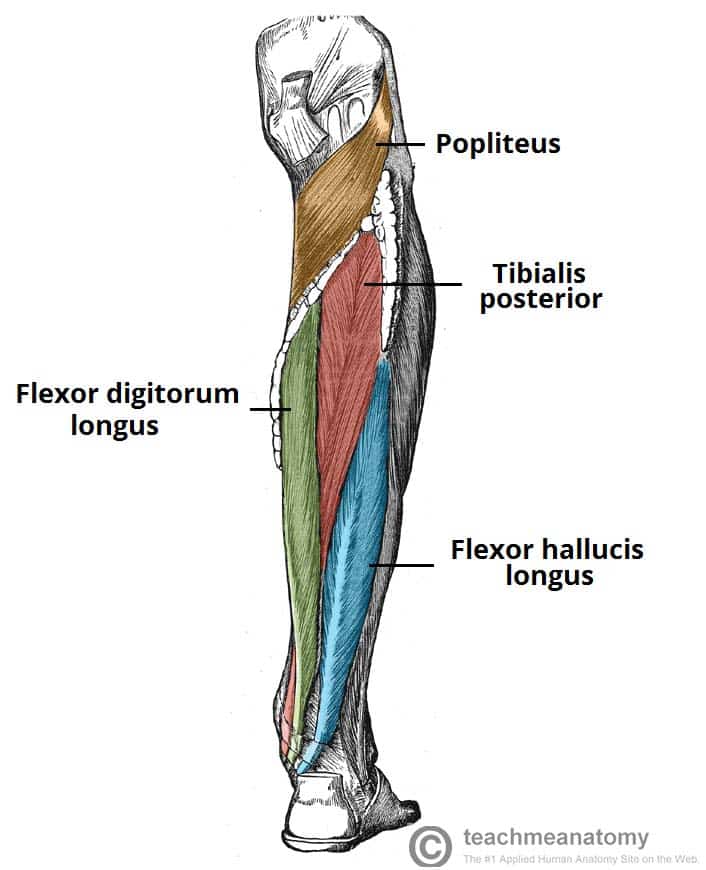
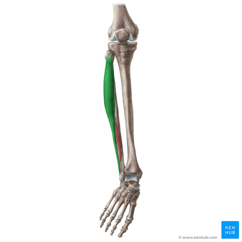
Popliteus
Proximal Attachment (O): Lateral surface of lateral condyle of femur and lateral meniscus
Distal Attachment (I): Posterior surface of tibia and superior to soleal line
Nerve: Tibial nerve (L4, L5, S1)
Action: Weakly flexes knee joint and unlocks it by rotating femur 5° on fixed tibia; medially rotates tibia of unplanted limb
Plantaris
Proximal Attachment (O): Inferior end of lateral supracondylar line of femur; oblique popliteal ligament
Distal Attachment (I): Posterior surface of calcaneus via calcaneal tendon
Nerve: Tibial nerve (S1, S2)
Action: Weakly assists gastrocnemius in plantarflexing ankle joint
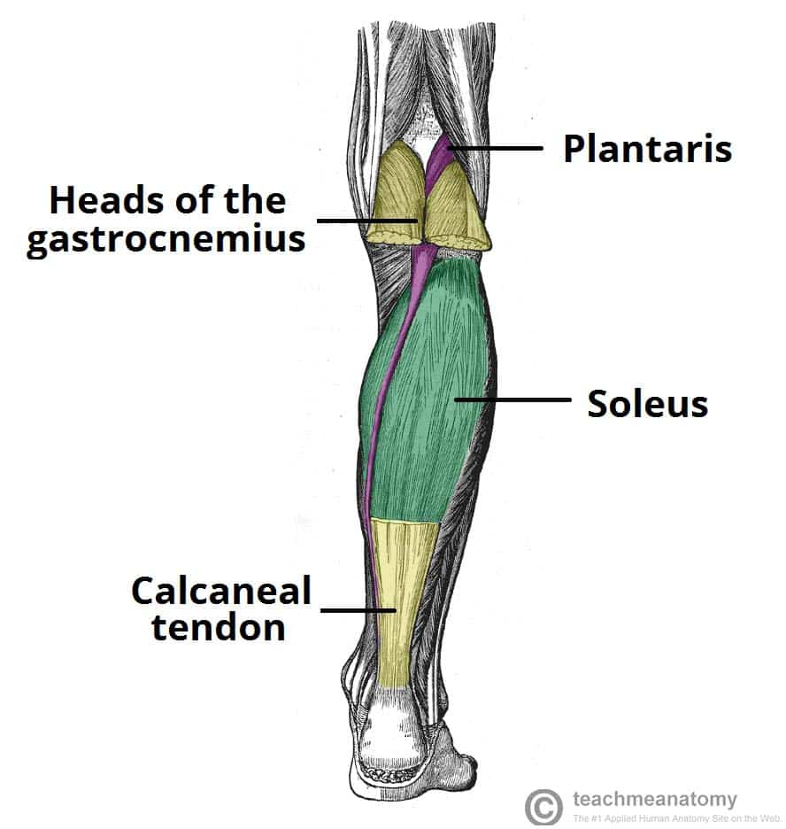
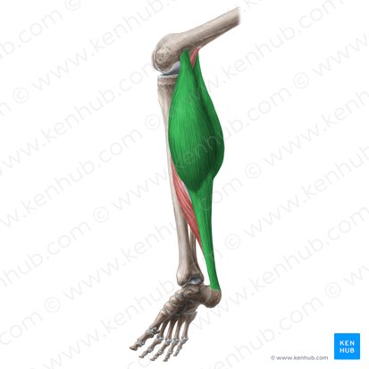
Gastrocnemius
Proximal Attachment (O):
Lateral head: lateral aspect of lateral condyle of femur
Medial head: popliteal surface of femur; superior to medial condyle
Distal Attachment (I): Posterior surface of calcaneus via calcaneal tendon
Nerve: Tibial nerve (S1, S2)
Action: Plantarflexes ankle joint when knee joint is extended; raises heel during walking; flexes knee joint
Soleus
Proximal Attachment (O): Posterior aspect of head and superior quarter of posterior surface of fibula; soleal line and middle third of medial border of tibia; tendinous arch extending between the bony attachments
Distal Attachment (I): Posterior surface of calcaneus via calcaneal tendon
Nerve: Tibial nerve (S1, S2)
Action: Plantarflexes ankle joint independent of position of knee; stabilizes ankle joint
Tibialis posterior
Proximal Attachment (O): Interosseous membrane; posterior surface of tibia inferior to soleal line; posterior surface of fibula
Distal Attachment (I): Tuberosity of navicular, cuneiform, cuboid, and sustentaculum tali of calcaneus; bases of 2nd, 3rd, and 4th metatarsals
Nerve: Tibial nerve (L4, L5)
Action: Plantarflexes ankle joint; inverts foot; maintains medial longitudinal arch

Flexor digitorum longus
Proximal Attachment (O): Medial part of posterior surface of tibia inferior to soleal line; by a broad tendon to fibula
Distal Attachment (I): Bases of distal phalanges of lateral four digits
Nerve: Tibial nerve (S2, S3)
Action: Flexes lateral four digits; plantarflexes ankle joint; supports longitudinal arches of foot

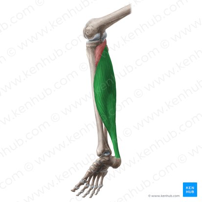
Flexor hallucis longus
Proximal Attachment (O): Inferior two thirds of posterior surface of fibula; inferior part of interosseous membrane
Distal Attachment (I): Base of distal phalanx of great toe (hallux)
Nerve: Tibial nerve (S2, S3)
Action: Flexes great toe at all joints; weakly plantarflexes ankle joint; supports medial longitudinal arch of foot
Tibialis anterior
Proximal Attachment (O): Lateral condyle and superior half of lateral surface of tibia and interosseous membrane
Distal Attachment (I): Medial and inferior surfaces of medial cuneiform and base of 1st metatarsal
Nerve: Deep fibular nerve (L4, L5)
Action: Dorsiflexes ankle joint and inverts subtalar joint
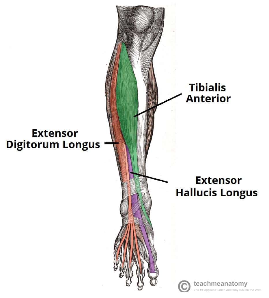
Extensor digitorum longus
Proximal Attachment (O): Lateral condyle of tibia and superior three quarters of medial surface of fibula and interosseous membrane
Distal Attachment (I): Middle and distal phalanges of lateral four digits
Nerve: Deep fibular nerve (L5, S1)
Action: Extends lateral four digits and dorsiflexes ankle joint

Extensor hallucis longus
Proximal Attachment (O): Middle part of anterior surface of fibula and interosseous membrane
Distal Attachment (I): Dorsal aspect of base of distal phalanx of great toe (hallux)
Nerve: Deep fibular nerve (L5, S1)
Action: Extends great toe and dorsiflexes ankle joint

Peroneus longus (fibularis longus)
Proximal Attachment (O): Head and superior two thirds of lateral surface of fibula
Distal Attachment (I): Base of 1st metatarsal and medial cuneiform
Nerve: Superficial fibular nerve (L5, S1, S2)
Action: Everts subtalar joint and weakly plantarflexes ankle joint
Peroneus brevis (fibularis brevis)
Proximal Attachment (O): Inferior two thirds of lateral surface of fibula
Distal Attachment (I): Dorsal surface of tuberosity on lateral side of base of 5th metatarsal
Nerve: Superficial fibular nerve (L5, S1, S2)
Action: Everts subtalar joint and weakly plantarflexes ankle joint
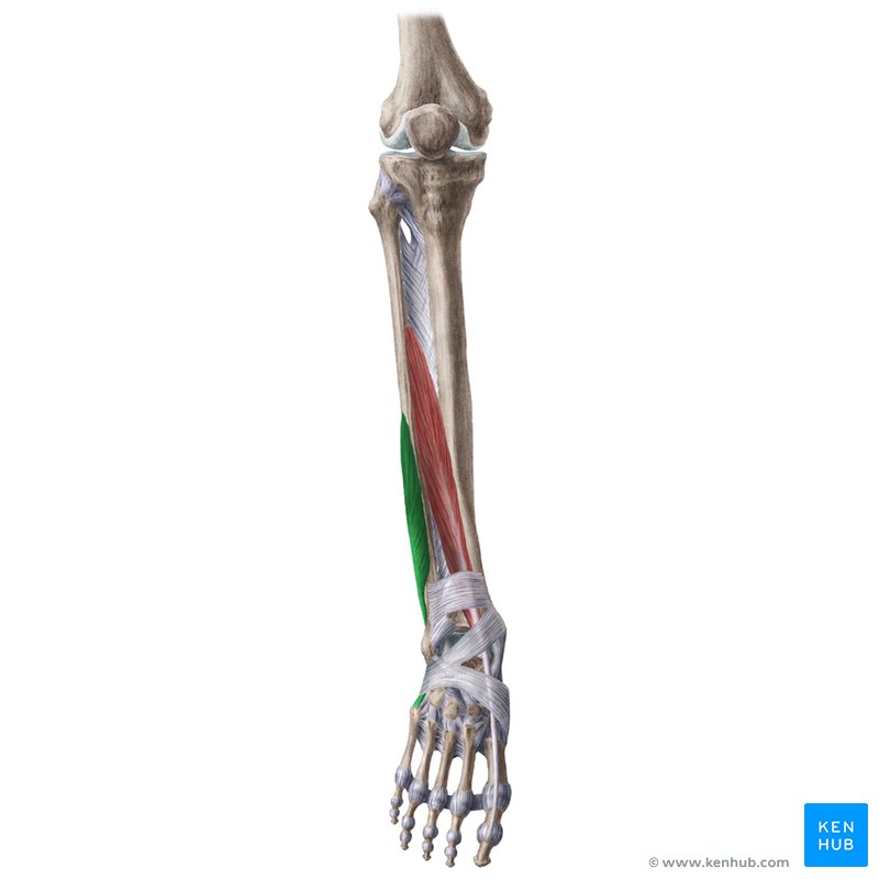
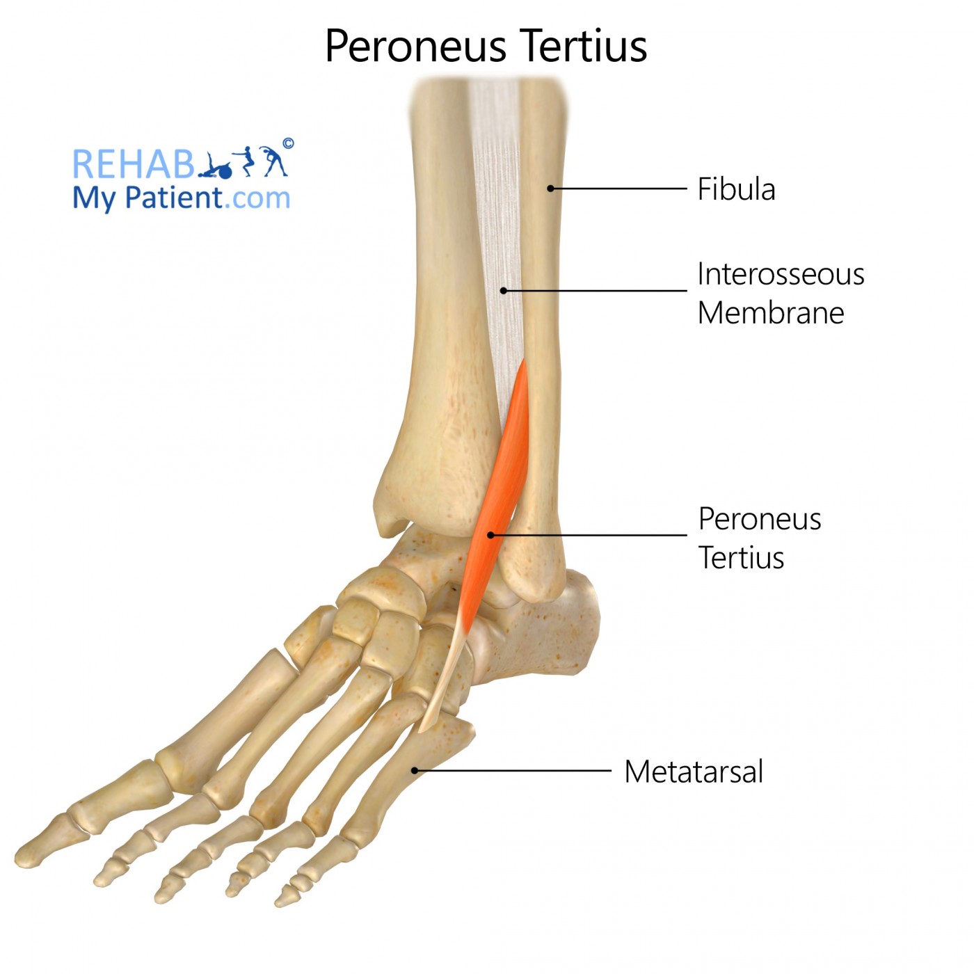
Peroneus Tertius (fibularis tertius)
Proximal Attachment (O): Inferior third of anterior surface of fibula and interosseous membrane
Distal Attachment (I): Dorsum of base of 5th metatarsal
Nerve: Deep fibular nerve (L5, S1)
Action: Dorsiflexes ankle joint and aids in eversion of subtalar joint
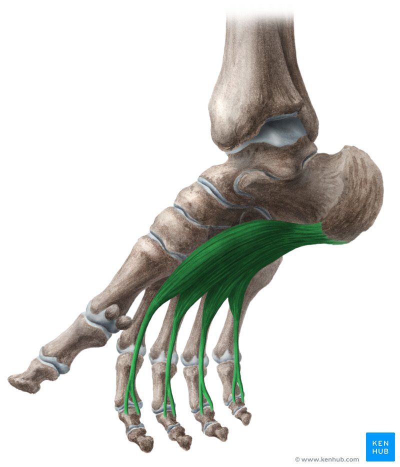
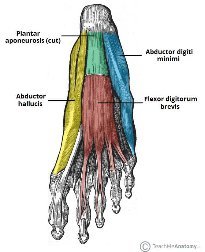
Flexor digitorum brevis
Proximal Attachment (O): Medial tubercle of tuberosity of calcaneus; plantar aponeurosis; intermuscular septa
Distal Attachment (I): Both sides of middle phalanges of lateral four digits
Nerve: Medial plantar nerve (L5, S1)
Action: Flexes lateral four digits at MTP and IP joints
Abductor digiti minimi
Proximal Attachment (O): Medial and lateral tubercles of tuberosity of calcaneus; plantar aponeurosis; intermuscular septa
Distal Attachment (I): Lateral side of base of proximal phalanx of 5th digit
Nerve: Lateral plantar nerve (S1–S3)
Action: Abducts and flexes little toe (5th digit) at MTP and IP joints
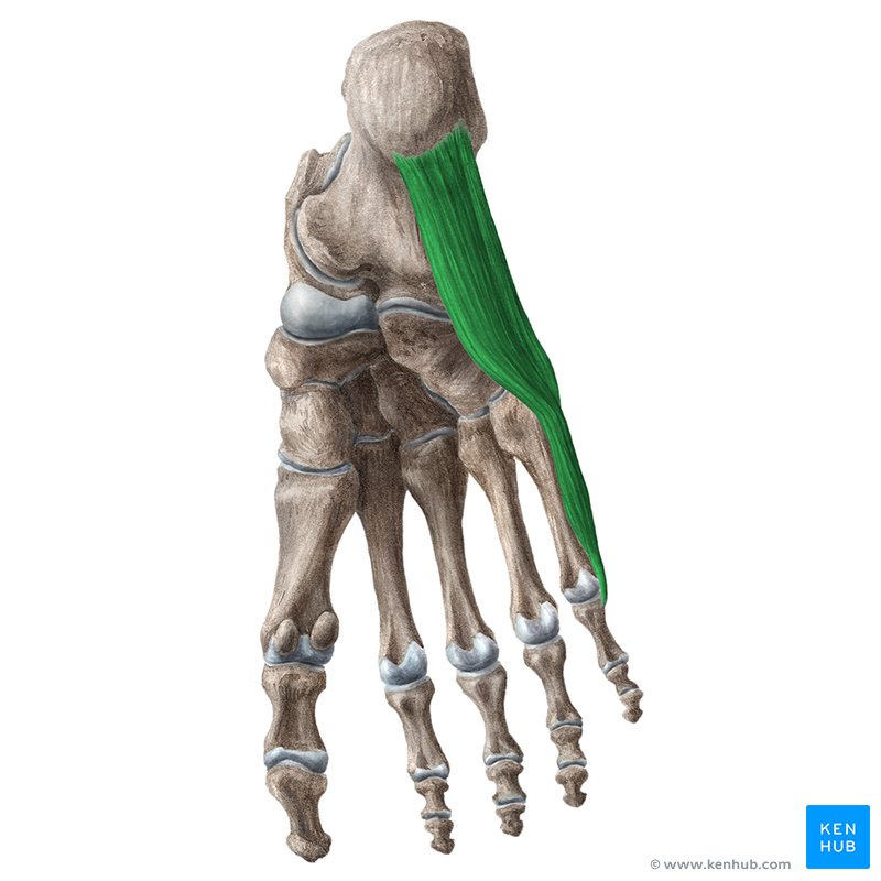
Abductor hallucis
Proximal Attachment (O): Medial tubercle of tuberosity of calcaneus; flexor retinaculum; plantar aponeurosis
Distal Attachment (I): Medial side of base of proximal phalanx of 1st digit
Nerve: Medial plantar nerve (L5, S1)
Action: Abducts and flexes 1st digit (great toe, hallux) at metatarsophalangeal (MTP) and interphalangeal (IP) joints
Adductor hallucis
Proximal Attachment (O):
Oblique head: bases of metatarsals 2–4
Transverse head: plantar ligaments of metatarsophalangeal (MTP) joints
Distal Attachment (I): Tendons of both heads attach to lateral side of base of proximal phalanx of 1st digit.
Nerve: Deep branch of lateral plantar nerve (S1–S3)
Action: Traditionally said to adduct 1st digit; assists in maintaining transverse arch of foot by pulling metatarsals medially
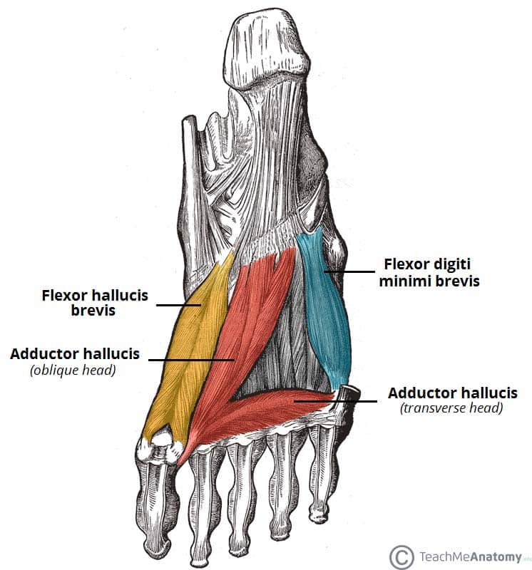
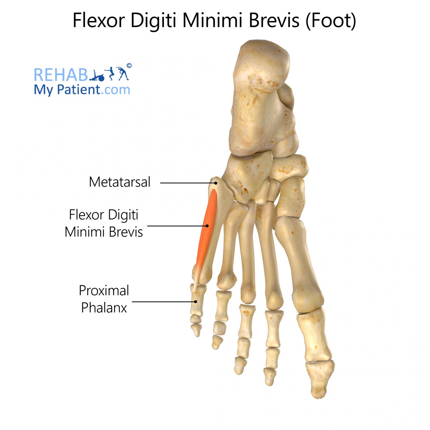
Flexor digiti minimi brevis
Proximal Attachment (O): Base of 5th metatarsal
Distal Attachment (I): Base of proximal phalanx of 5th digit
Nerve: Superficial branch of lateral plantar nerve (S1–S3)
Action: Flexes proximal IP joint of 5th digit, thereby assisting with flexion of digit
Flexor hallucis brevis
Proximal Attachment (O): Plantar surfaces of cuboid and lateral cuneiforms
Distal Attachment (I): Both sides of base of proximal phalanx of 1st digit
Nerve: Medial plantar nerve (L5, S1)
Action: Flexes proximal interphalangeal (IP) joint of 1st digit

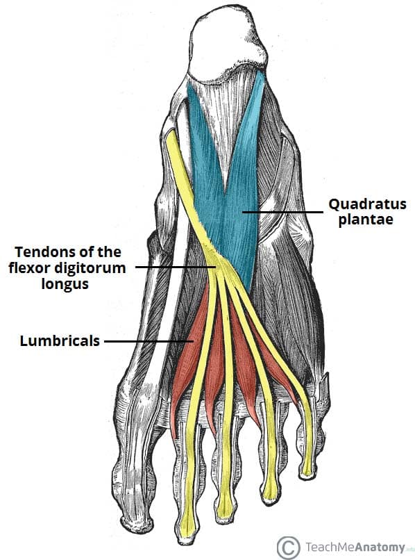
Quadratus plantae
Proximal Attachment (O): Medial surface and lateral margin of plantar surface of calcaneus
Distal Attachment (I): Posterolateral margin of tendon of flexor digitorum longus
Nerve: Lateral plantar nerve (S1–S3)
Action: Assists flexor digitorum longus in flexing lateral four digits at MTP and IP joints
Psoas minor
Proximal Attachment (O): Sides of T12–L1 vertebrae and intervertebral discs
Distal Attachment (I): Pectineal line and iliopubic eminence via iliopectineal arch
Nerve: Anterior rami of lumbar nerves (L1, L2)
Action: Act conjointly in flexion and lateral rotation of hip joint and in stabilizing this joint when standing
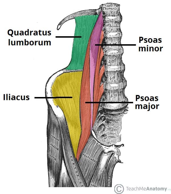
Psoas major
(Posterior Abdominal Wall)
Superior Attachment (O): Transverse processes of lumbar vertebrae; sides of bodies of T12–L5 vertebrae and intervening intervertebral discs
Inferior Attachment (I): By a strong tendon to lesser trochanter of femur
Nerve: Anterior rami of lumbar nerves L1, L2, and L3
Action: Acting inferiorly with iliacus, it flexes the thigh; acting superiorly, it flexes the vertebral column laterally; it is used to balance the trunk; when sitting, it acts inferiorly with the iliacus to flex the trunk
Iliacus
(Posteiror abdominal wall)
Superior Attachment (O): Superior two thirds of iliac fossa, ala of sacrum, and anterior sacro-iliac ligaments
Inferior Attachment (I): Lesser trochanter of femur and shaft inferior to it and to psoas major tendon
Nerve: Femoral nerve (L2-L4)
Action: Flexes thigh and stabilizes hip joint; acts with psoas major
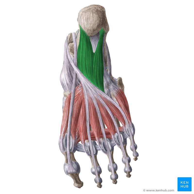
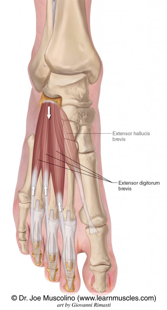
Extensor digitorum Brevis
Proximal Attachment (O): Calcaneus (floor of tarsal sinus); interosseous talocalcaneal ligament; stem of inferior extensor retinaculum
Distal Attachment (I): Long extensor tendons of three intermediate digits (toes 2–4)
Innervation: Deep fibular nerve (L5 or S1, or both)
Action: Aids the extensor digitorum longus in extending the three intermediate toes at the metatarsophalangeal (MTP) and interphalangeal joints

Extensor hallucis brevis
Proximal Attachment (O): Calcaneus (floor of tarsal sinus); interosseous talocalcaneal ligament; stem of inferior extensor retinaculum
Distal Attachment (I): Dorsal aspect of base of proximal phalanx of great toe (digit 1)
Innervation: Deep fibular nerve (L5 or S1, or both)
Action: Aids the extensor hallucis longus in extending the great toe at the MTP joint
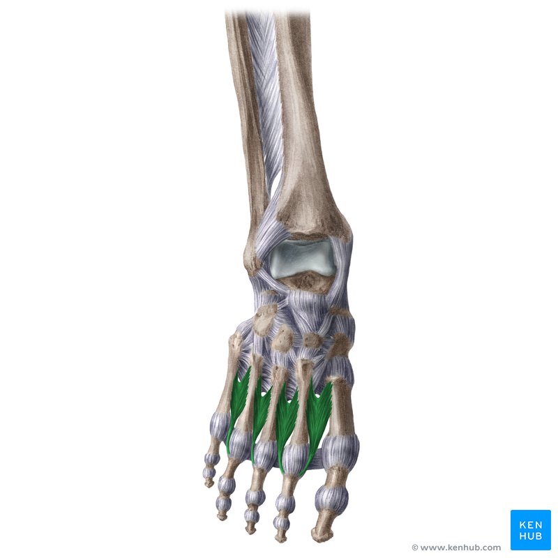
Lumbricals
Proximal Attachment (O): Tendons of flexor digitorum longus
Distal Attachment (I): Medial aspect of expansion over lateral four digits
Innervation:
Medial one: medial plantar nerve (L5–S1)
Lateral three: lateral plantar nerve (S1–S3)
Action: Flex proximal IP joint; extend middle and distal IP joints of lateral four digits
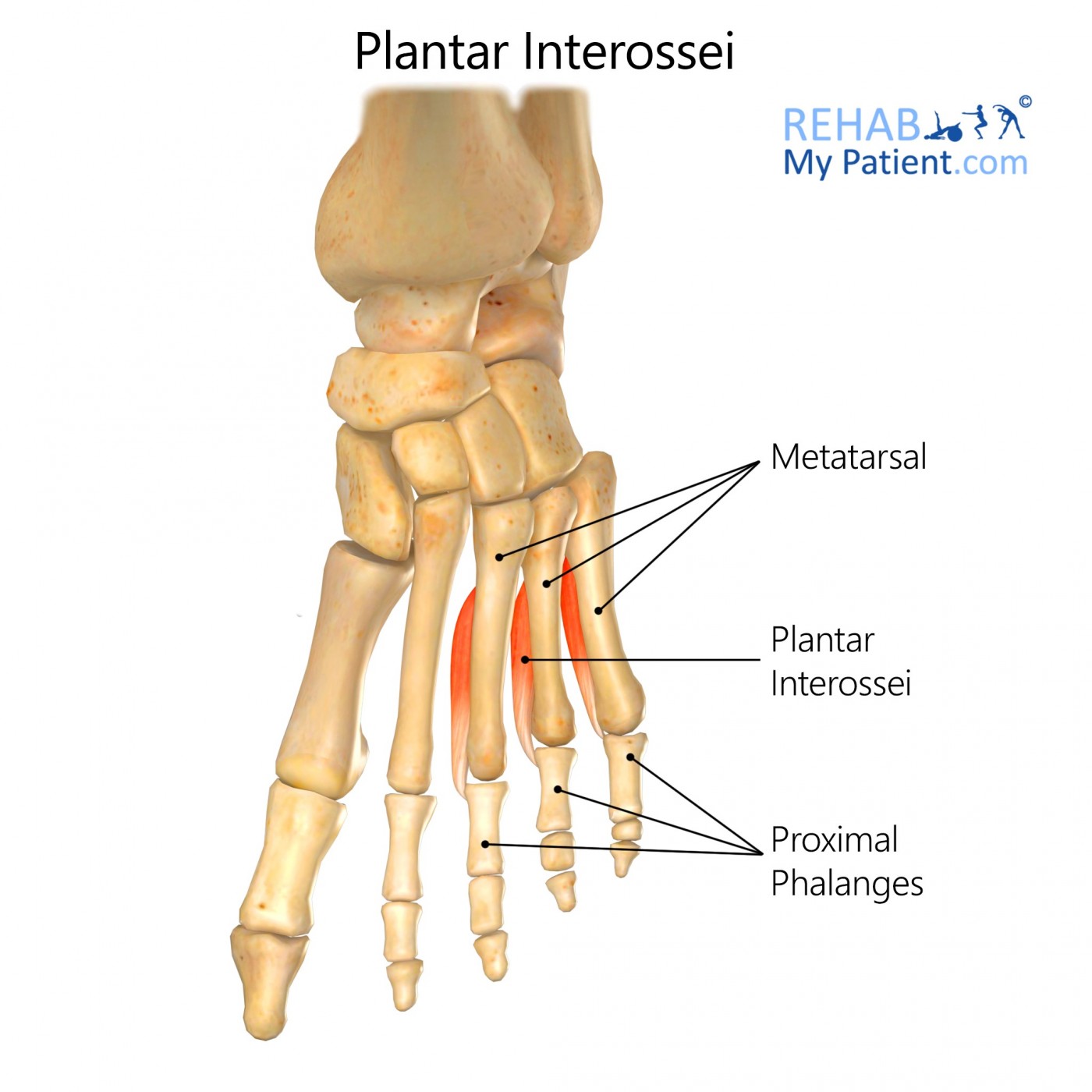
Plantar Interossei (3)
Proximal Attachment (O): Plantar aspect of medial sides of shafts of metatarsals 3–5
Distal Attachment (I): Medial sides of bases of phalanges of 3rd–5th digits
Innervation: Lateral plantar nerve (S1–S3)
Action: Adduct and flex digits 3–5 at MTP joints
Dorsal Interossei (4)
Proximal Attachment (O): Adjacent sides of shafts of metatarsals 1–5
Distal Attachment (I):
1st: medial side of proximal phalanx of 2nd digit
2nd–4th: lateral sides of 2nd–4th digits
Innervation: Lateral plantar nerve (S1–S3)
Action: Abduct and flex digits 2–4 at MTP joints