Neuroanatomy Exam 1
1/84
There's no tags or description
Looks like no tags are added yet.
Name | Mastery | Learn | Test | Matching | Spaced | Call with Kai |
|---|
No analytics yet
Send a link to your students to track their progress
85 Terms
What level of section?
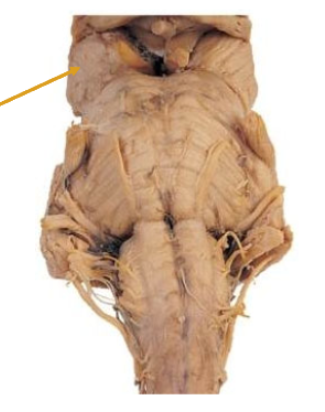
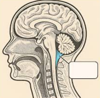
Rostral medulla
White matter is comprised of…
Fibers
Grey matter is comprised of…
Cells
Outer layer: cerebral cortex
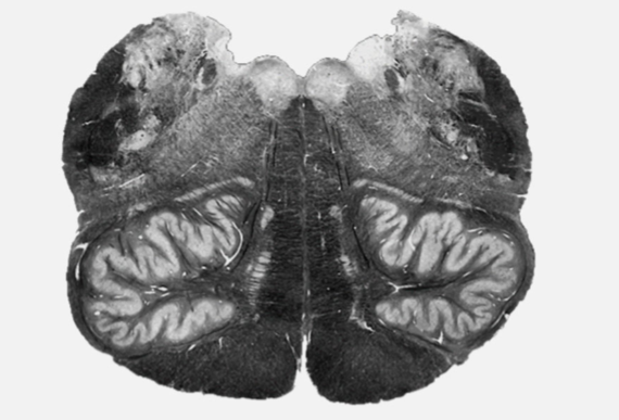
What kind of stain is this?
Myelin stain
White matter is dark, grey matter is light
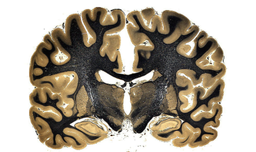
What kind of stain is this?
Cell stain
White matter is light, grey matter is dark - useful for highlighting cell bodies
Collection of white matter joining different areas of the central nervous system
Peduncle
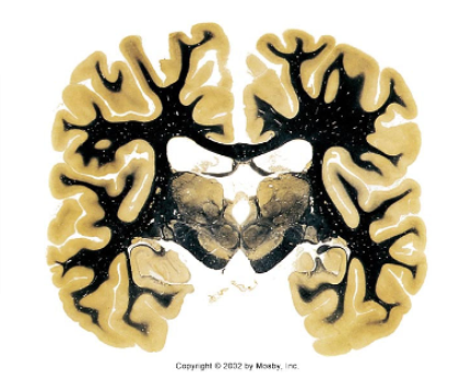
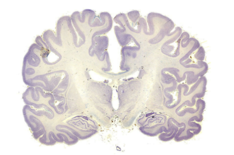
What plane of section?
Coronal
Looking head-on at an upright patient
Divides front and back
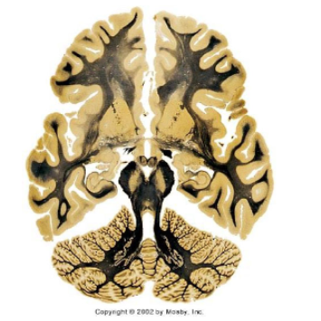
What plane of section?
Horizontal, axial
Patient is lying down, nose up
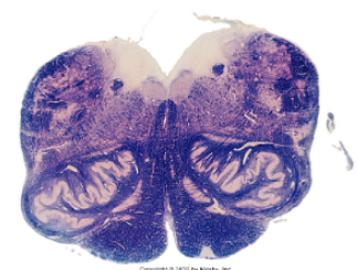
What plane of section?
Horizontal/transverse- brainstem
Anatomical orientation (clinical orientation is flipped)
What plane of section?
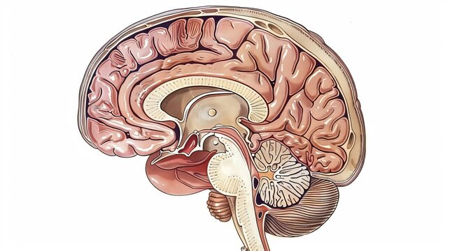
Sagittal
Looking at an upright patient from the side
Front to back
Anterior to posterior
Top, closest to top of the head
Dorsal or superior
Bottom, closest to neck
Ventral or inferior
The frontal section of the brain closest to the face, nose, and mouth
Rostral (anterior)
The rear section of the brain closest to the back of the head
Caudal (posterior)
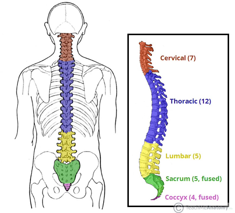
Regions of the spinal cord
Cervical, thorasic, lumbar, sacral
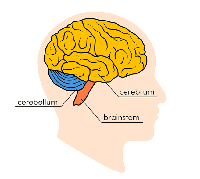
Main anatomical/functional divisions of the brain
Cerebrum, cerebellum, brainstem
Forebrain structures (cerebrum + what?)
Cerebrum, basal ganglia, limbic system, thalamus, hypothalamus, pineal gland
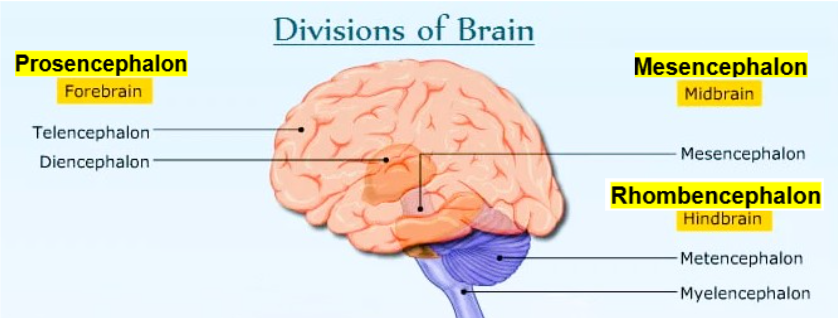
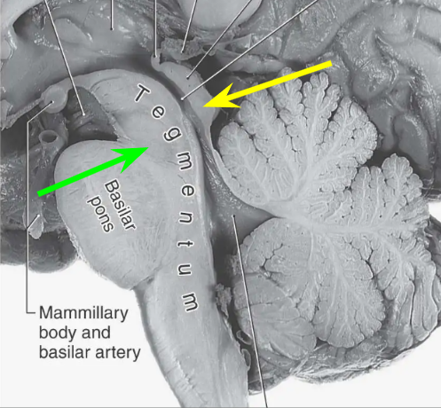
Main anatomical/functional divisions of the midbrain
Tectum, tegmentum
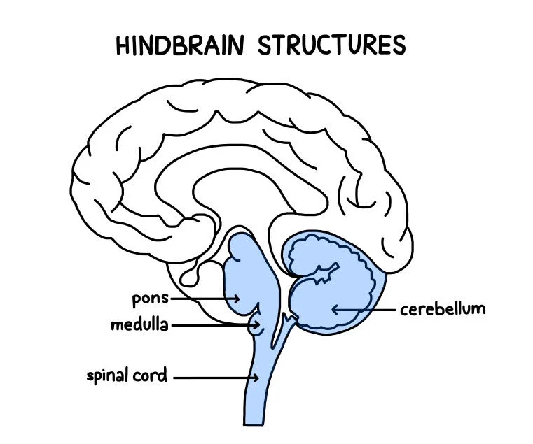
Structures of the hindbrain
Pons, cerebellum, medulla, spinal cord
Fissure dividing left and right hemispheres
Median longitudinal fissure
Lobe and major areas
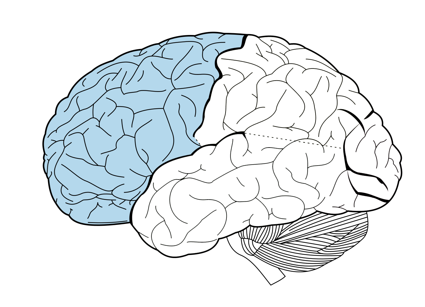
Frontal lobe - in front of central sulcus
Prefrontal cortex, motor cortex, Broca’s area
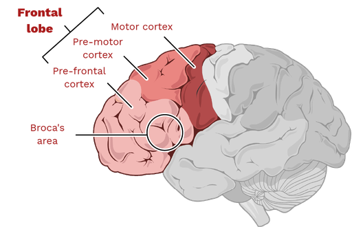
Controls language production
Broca’s area
Controls muscle movement
Motor cortex
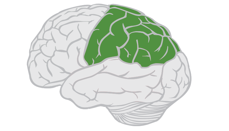
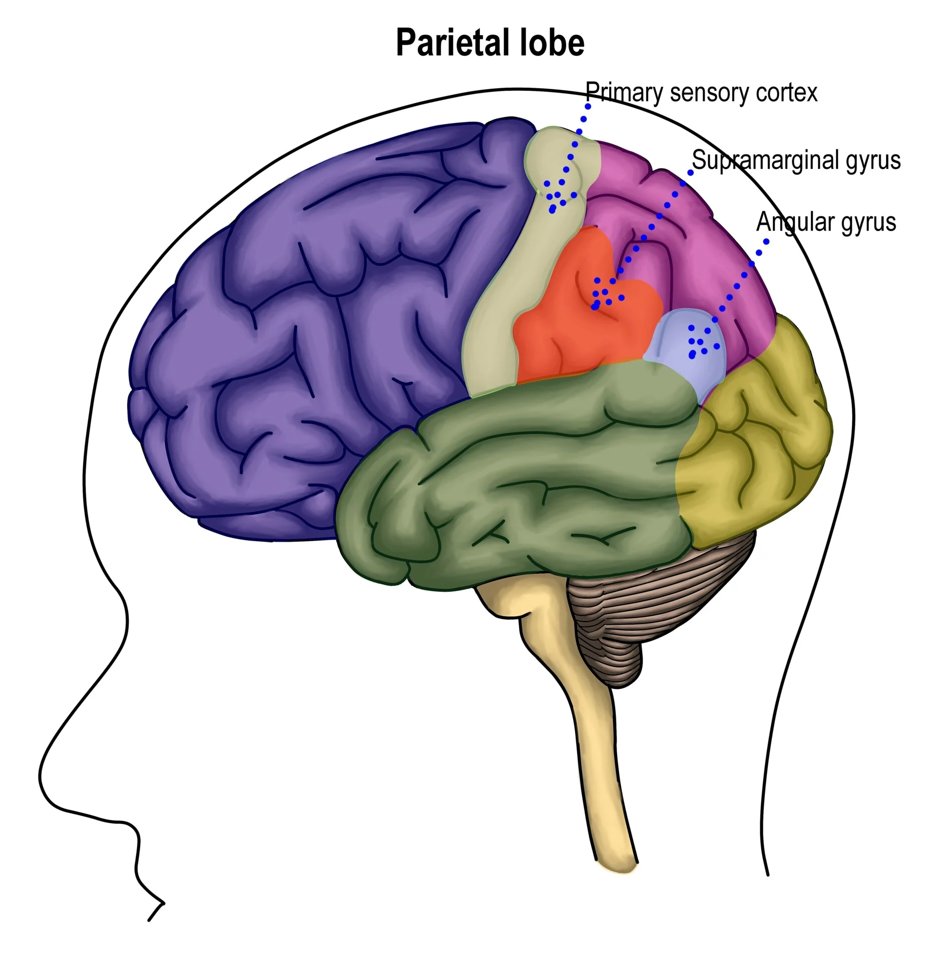
Lobe and major structures
Parietal lobe - behind central sulcus
Somatosensory cortex (sensory processing)
Superior parietal lobule (spatial orientation)
Inferior parietal lobule (language and body image)
Name the structures and functions
Tegmentum: motor coordination, pain management, arousal, autonomic functions
Tectum: visual and auditory reflexes
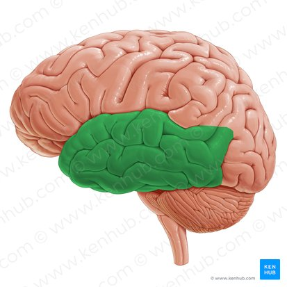
Lobe and functions
Temporal lobe
Processing auditory info, language comp., memory formation
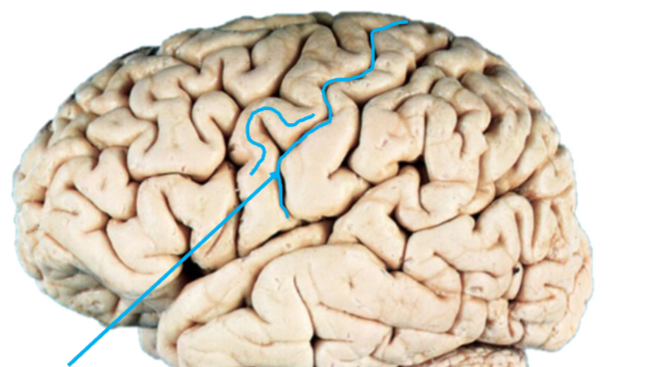
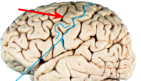
Name the groove
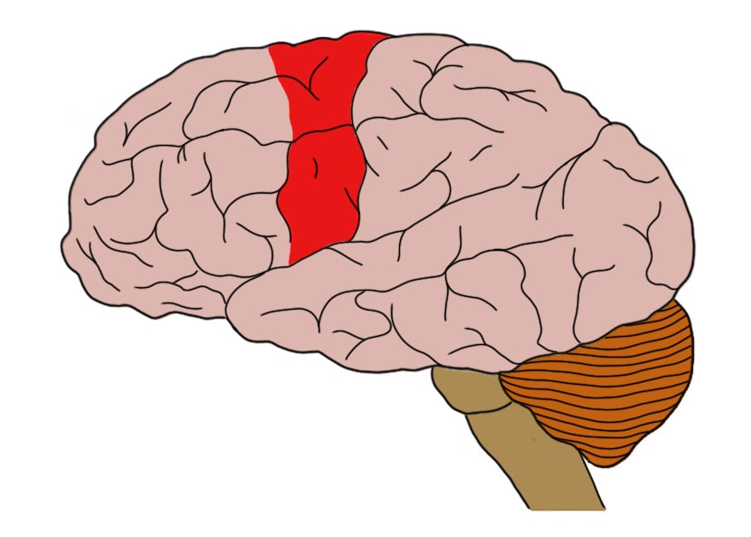
Central sulcus
Divides primary motor cortex from primary somatosensory cortex - separates parietal lobe
Broca’s Aphasia
Stroke in Broca’s area
Functions include executive functions, emotion regulation, personality expression, social behavior
Prefrontal cortex (PFC)
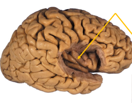
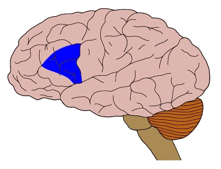
Hidden deep within the lateral sulcus of the brain
Insular cortex - Island of Reil, Insula
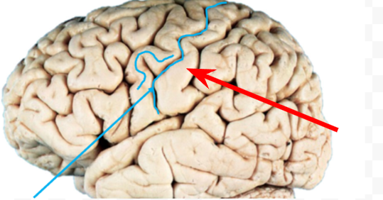
Motor strip of primary motor cortex
Precentral gyrus: executing voluntary movements, anterior to central sulcus
Primary somatosensory cortex
Postcentral gyrus: sensory receptive area for sense of touch, posterior to central sulcus
Two commissures that interconnect the hemispheres of cerebral cortex
Corpus callosum, anterior commissure
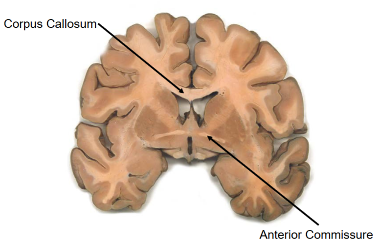
Name the structure
Anterior commissure
Two structures of symptoms are on the same side of the body
Ipsilateral
Two structures of symptoms are on opposite sides of the body
Contralateral
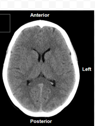
Type of scan, orientation
CT, axial, density of tissue, brain anatomy
* Quick scan
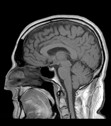
Type of scan, orientation
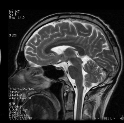
MRI, T1, sagittal
Bright white matter, dark gray matter
Type of scan, orientation
MRI, T2, sagittal
Bright grey matter, dark white matter
Type of scan, orientation
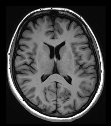
MRI, T1, axial
Type of scan, orientation
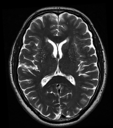
MRI, T2, axial
MRI T2 is best at…
Defining fluids, pathology sequence
MRI T1 is best at…
Defining anatomy, anatomical sequence
Type of scan, orientation
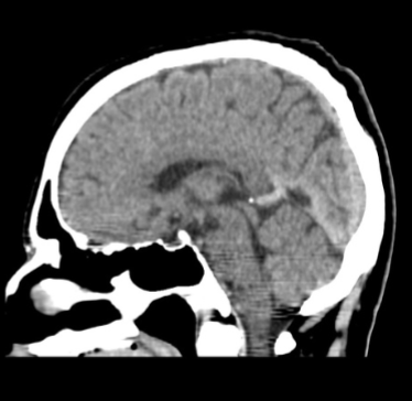
CT, sagittal
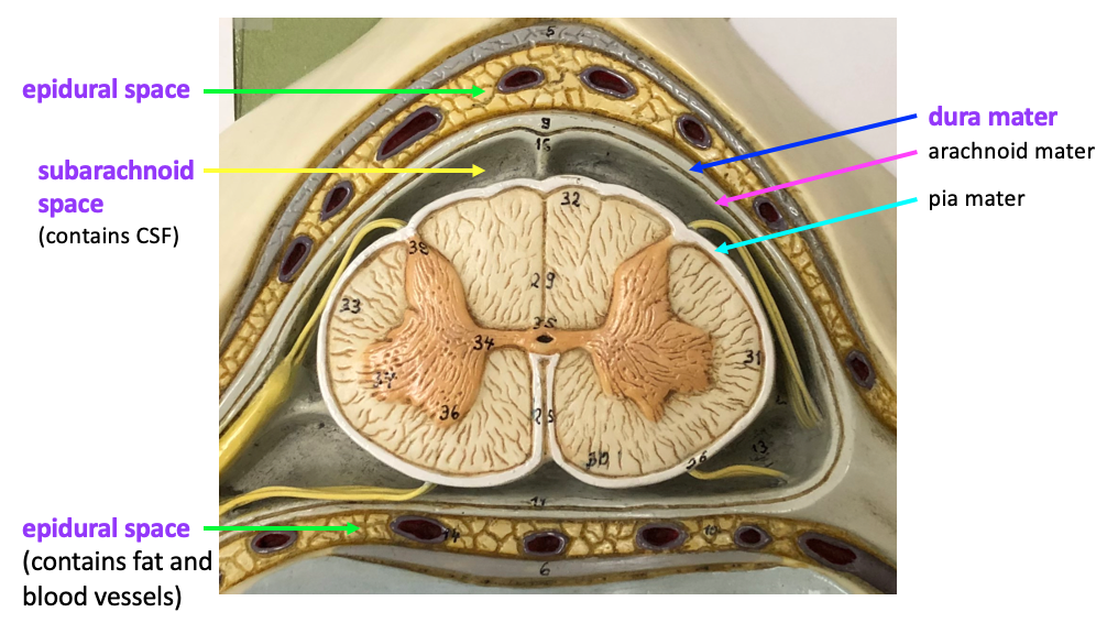
Meninges layers (top of skull, down)
Dura mater (2 layers)
Arachnoid mater (3 layers)
Pia mater (2 layers)
Middle membrane
Contains blood vessels and CSF
Arachnoid mater
Allow CSF to pass/exit the brain from subarachnoid space → venous system
Outermost membrane
Contains dural sinuses, forms dural reflections
Dura mater
Epidural space in the spinal meninges
Normal space between vertebra/discs, houses spinal nerve roots and the dura
Dura reflections
Created by dura mater folding inwards upon itself
Forms four dural reflections → reflect into cranial cavity, contains venous sinuses
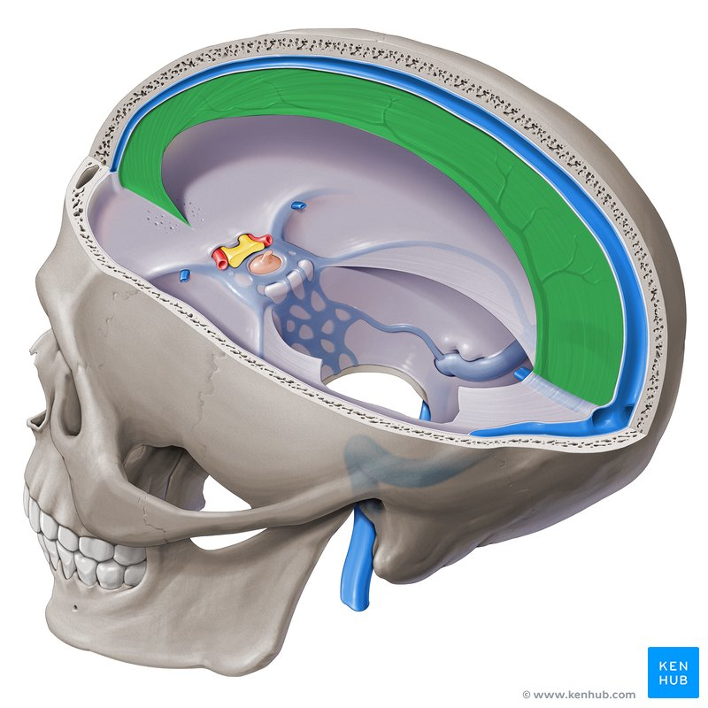
Falx cerebri
(vertical)
Separates the two hemispheres of the brain, largest dural fold
Dural venous sinuses
Located between dura and the brain
Twelve that drain the brain predominately into the internal jugular vein
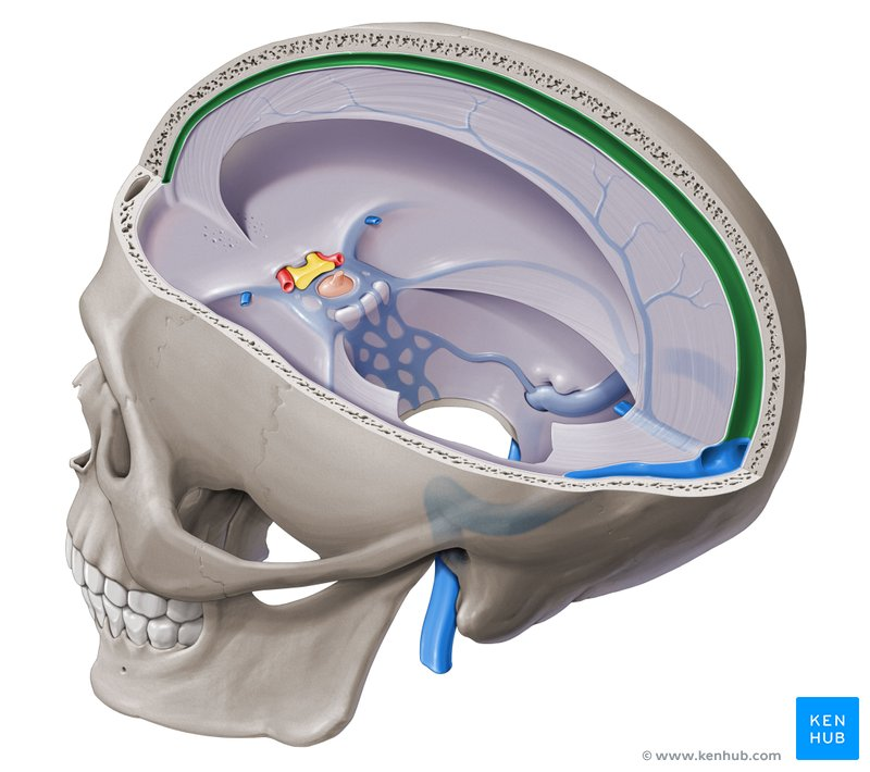
Superior sagittal sinus (SSS)
Largest dural venous sinus
Single venous channel spanning midline and terminating at confluences of the sinuses (junction of four sinuses)
Where is epidural anesthesia injected into?
Epidural space of the spine
Meningitis
Inflammation of the membranes that surround the brain and spinal cord
Ventricular system and major functions
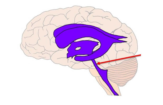
Lateral ventricles (2), third ventricle, cerebral aqueduct, fourth ventricle, central canal
Production, transport and removal of CSF
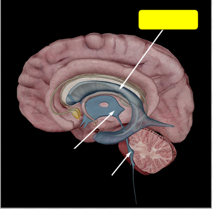
Name the structure
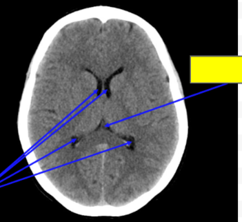
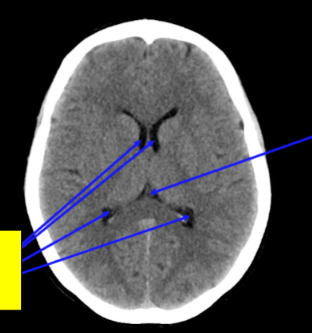
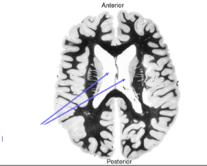
Lateral ventricle
Name the structure
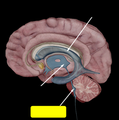
Third ventricle
Name the structure
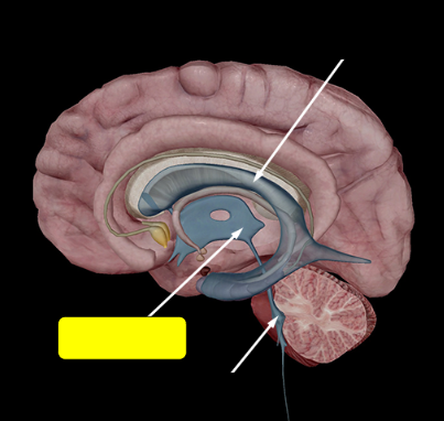
Fourth ventricle
Name the structure
Cerebral aqueduct
Name the structure
3rd ventricle
CSF → 4th ventricle
Name the structure
Lateral ventricles
Produces CSF and transports it → third ventricle
Name the structure
Lateral ventricles
Name the structure
4th ventricle
Name the structure
Cerebral aqueduct
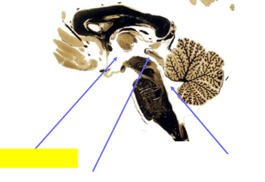
Name the structure
3rd ventricle
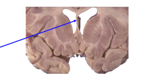
Septum Pellucidum
Partition between a portion of the lateral ventricles
What makes up CSF and where does it reabsorb?
Made by specialized ependymal cells in the choroid plexi
Choroid plexi → ventricular system → subarachnoid space → arachnoid granulations
Name the structure
Choroid plexus
At the roof of lateral, 3rd, 4th ventricles
Name the structure
Central canal
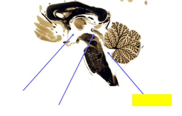
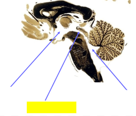
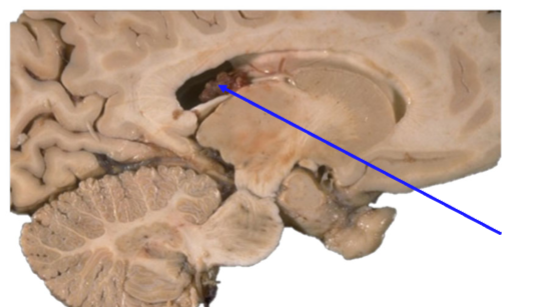
Largest cistern between cerebellum and dorsal surface of medulla oblongata at and above the level of the foramen magnum
Cisterna magna
Enlargement of the ventricles of the brain
Ventriculomegaly
Hydrocephalus
Enlargement of the ventricles caused by an increase in pressure/build up of CSF
* Intercranial pressure

#3
Anterior cerebral artery
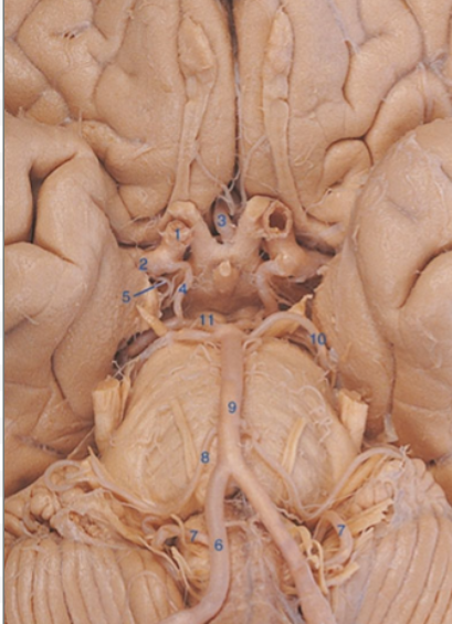
#1
Internal carotid artery

#2
Middle cerebral artery

#4
Posterior communicating artery

#11
Posterior cerebral artery

#6
Vertebral artery

#9
Basilar artery
What comprises the Circle of Willis?
Anterior communicating artery
Anterior cerebral artery
Middle cerebral artery
Internal carotid artery
Posterior communicating artery
Posterior cerebral artery

Vascular territories of the three main cerebral arteries
ACA: medial part of front and parietal lobe, corpus callosum, basal ganglia, internal capsule
MCA: lateral surface of the hemispheres (frontal, temporal & parietal lobe), insula
PCA: Inferior-medial part of the temporal lobe and the occipital lobe, visual cortex, thalamus, corpus callosum, midbrain
* All “C” are cerebral
Name the structure
Anterior cerebral artery (ACA)
Major arterial supply to the spinal cord
Anterior spinal artery (ASA)
Anterior cord syndrom
Interruption of blood supply to anterior portion of spinal cord
Bilateral motor paralysis below the level of the lesion
Bilateral loss of pain and temp sensation
Retained proprioception and vibratory sensation (intact dorsal columns)