Chapter 9-12 AP200
1/199
There's no tags or description
Looks like no tags are added yet.
Name | Mastery | Learn | Test | Matching | Spaced | Call with Kai |
|---|
No analytics yet
Send a link to your students to track their progress
200 Terms
Hem/o hemat/o
blood, pertaining to the blood
Phagocytes
A type of white blood cell that ingests invading microbes. eat or swallow
Red blood cells
Erythrocyte, red
remember, RBCs do have have nucleus and do not have DNA
Composition of blood
55% plasma, 45% formed elements
Component of plasma
Water is most abundant , more than 90%Albumin(most abundant protein).fibrinogen and globulin
Serum
plasma without clotting factors
What Hemopoiesis?
formation of blood cells, as example erythropoiesis
Thrombus vs embolus
Thrombus = stationary clot in arteries
embolus = dislodged traveling clot in arteries
CBC
complete blood count
CBC with differential
look at WBCs monocytes, lymphocytes, granulocytes, neutrophils, eosinophils, basophils
hemoglobin test (H, Hg, Hgb, HGB)
total amount of hemoglobin in a sample of peripheral blood
Hematocrit (Hct)
percentage of erythrocytes in a volume of blood
polycythemia
A disorder characterized by an abnormal increase in the number of red blood cells in the blood
Leukemia
cancer of white blood cells
Thrombocytopenia
low platelet count
Anemia
A condition in which the blood is deficient in red blood cells, in hemoglobin, or in total volume to carry hemoglobin to maintain homeostasis
pernicious anemia
Lack of intrinsic factors
Function of basophil
release histamine and other mediators of inflammation; contain heparin, an anticoagulant
Function of eosinophil
kill parasitic worms; complex role in allergy and asthma
Function of neutrophil
phagocytize bacteria
Function of monocytes
phagocytosis; develop into macrophages in tissues
Erythropoietin (EPO)
hormone secreted by the kidney to stimulate the production of red blood cells by bone marrow
Universal blood donor
Type O
universal reciepient
Type AB
How blood type determined
By the antigen on the RBCs' surface
Blood type A positive may receive blood from what blood type?
A+, A- o+,o-
Blood type A negative may receive blood from what blood type?
A-, o-
What are the two main component of blood?
Plasma and formed elements
What are the three types of blood cells and mention function of each.
RBCs, WBCs, Platelets, see your text book for functions
sickle cell anemia
a genetic disorder that causes abnormal hemoglobin, resulting in some red blood cells assuming an abnormal sickle shape
aplastic anemia
failure of blood cell production in the bone marrow
pernicious anemia
Caused by Vitamin B12 deficiency or intrinsic factor deficiency
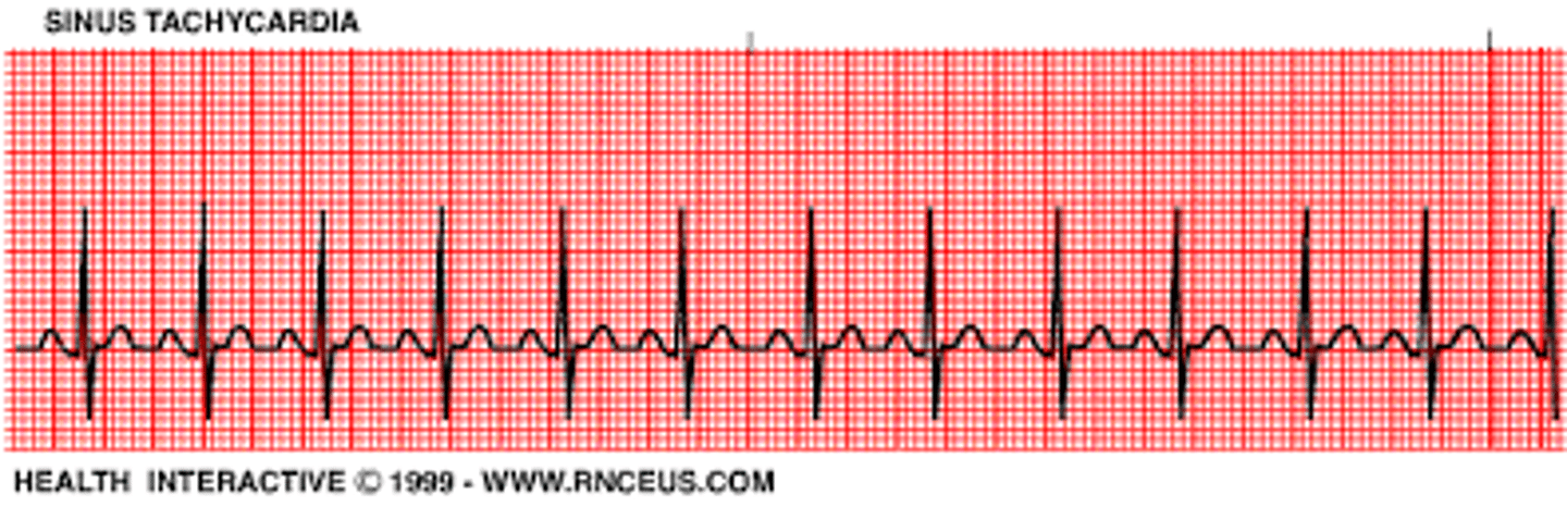
Tachycardia
fast heart rate
Bradycardia
slow heart rate
Endocardium
inner lining of the heart

Myocardium
muscular, middle layer of the heart
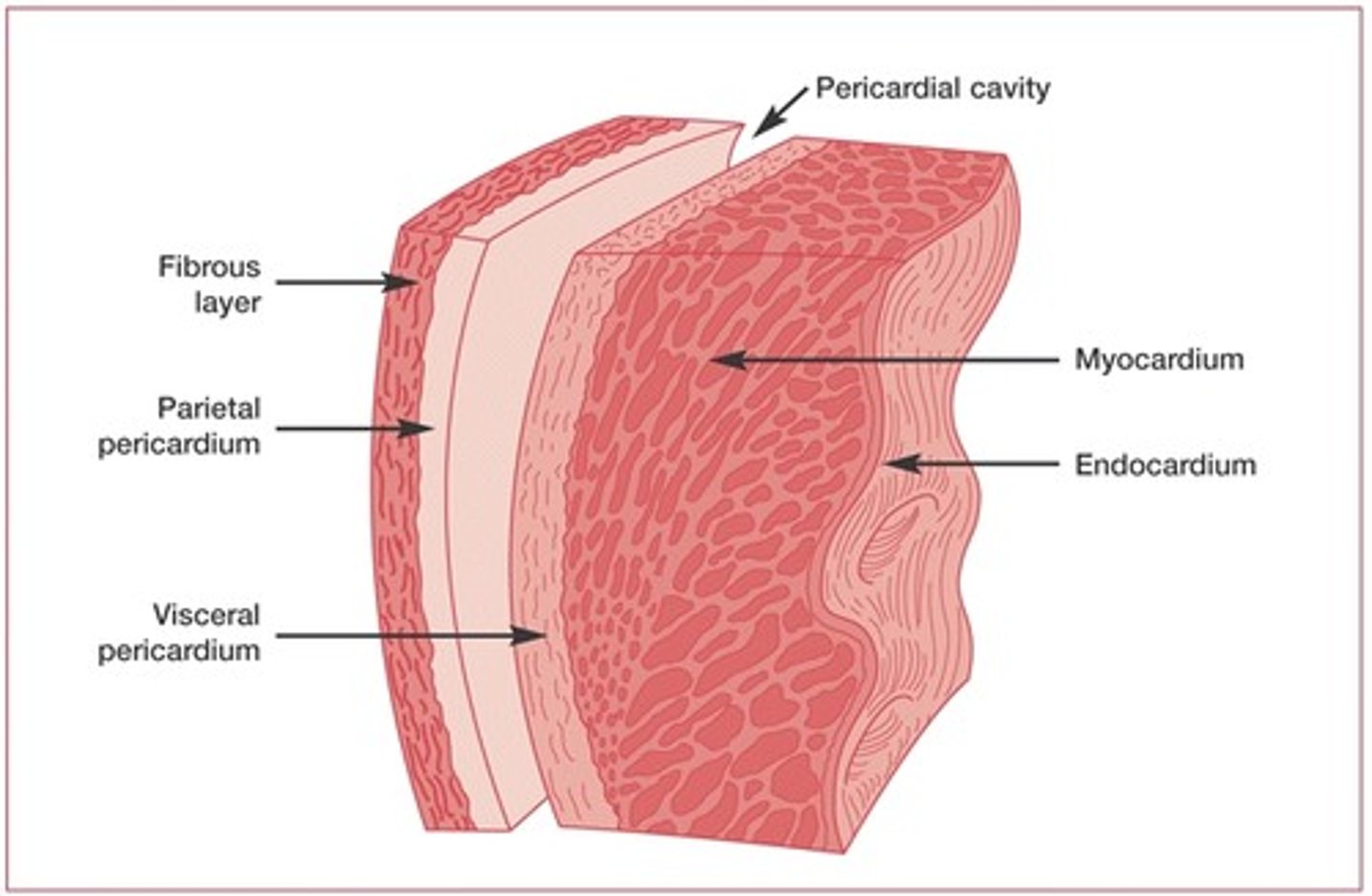
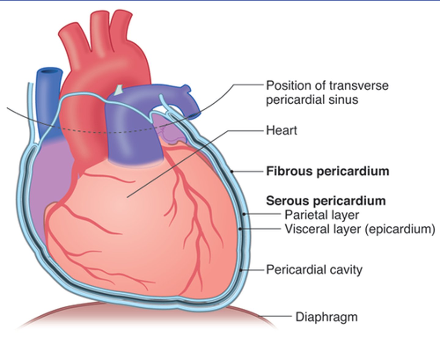
Pericardium
Double-layered membrane surrounding the heart.
serous membrane
inner layer is visceral/ epicardium and it is in direct contact with the heart
outer is parietal pericardium
pericardial cavity is in between
Atria
superior heart chambers
ventricles
the two lower chambers of the heart
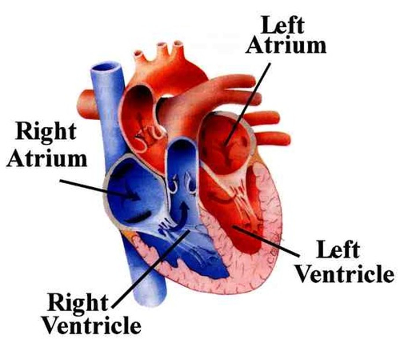
which structure separate the right atrium and the lefty atrium?
interatrial septum
Which structure separate the right and left ventricles?
interventricular septum
What are the AV valves?
Tricuspid and Mitral (bicuspid ) valves
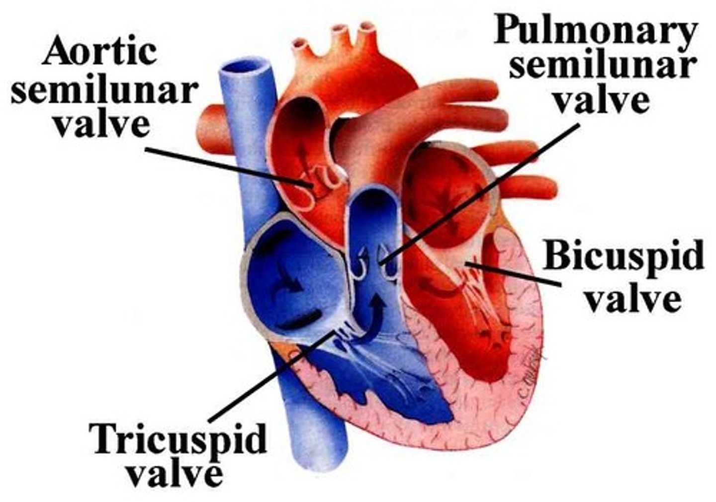
What are the semilunar valves?
pulmonary and aortic valves

What is the function of cardiac valves?
direct flow of blood through the heart chambers and allow one way flow to prevent backup flow
function of papillary muscles
contract when ventricles contract to prevent AV valves from opening
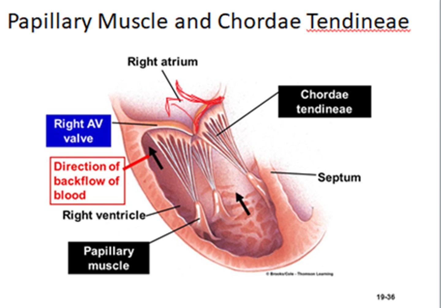
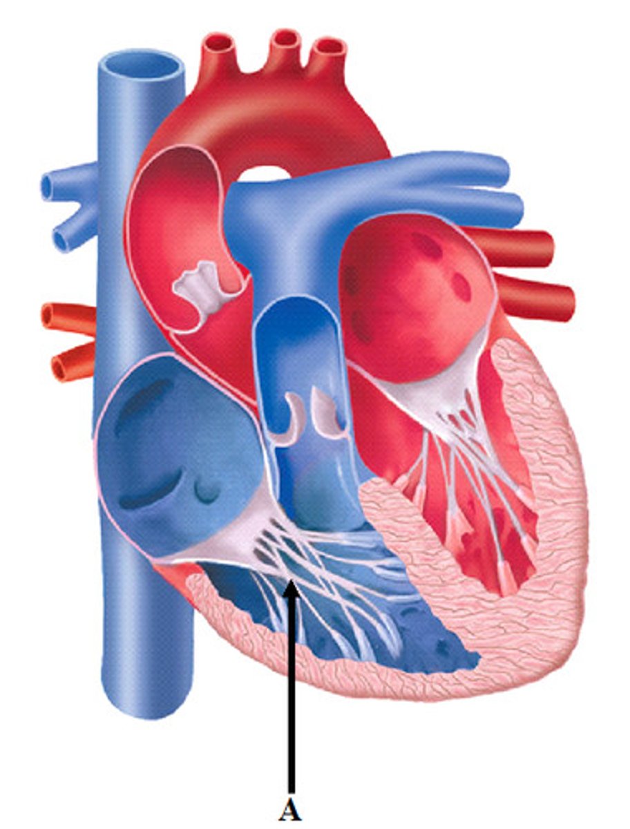
The function of the chordae tendinae is to
anchor the atrioventricular valves to papillary muscle
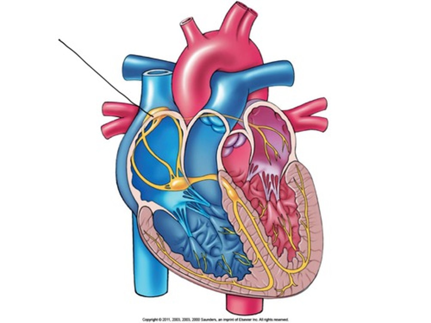
What is the pacemaker of the heart?
sinoatrial node (SA node)
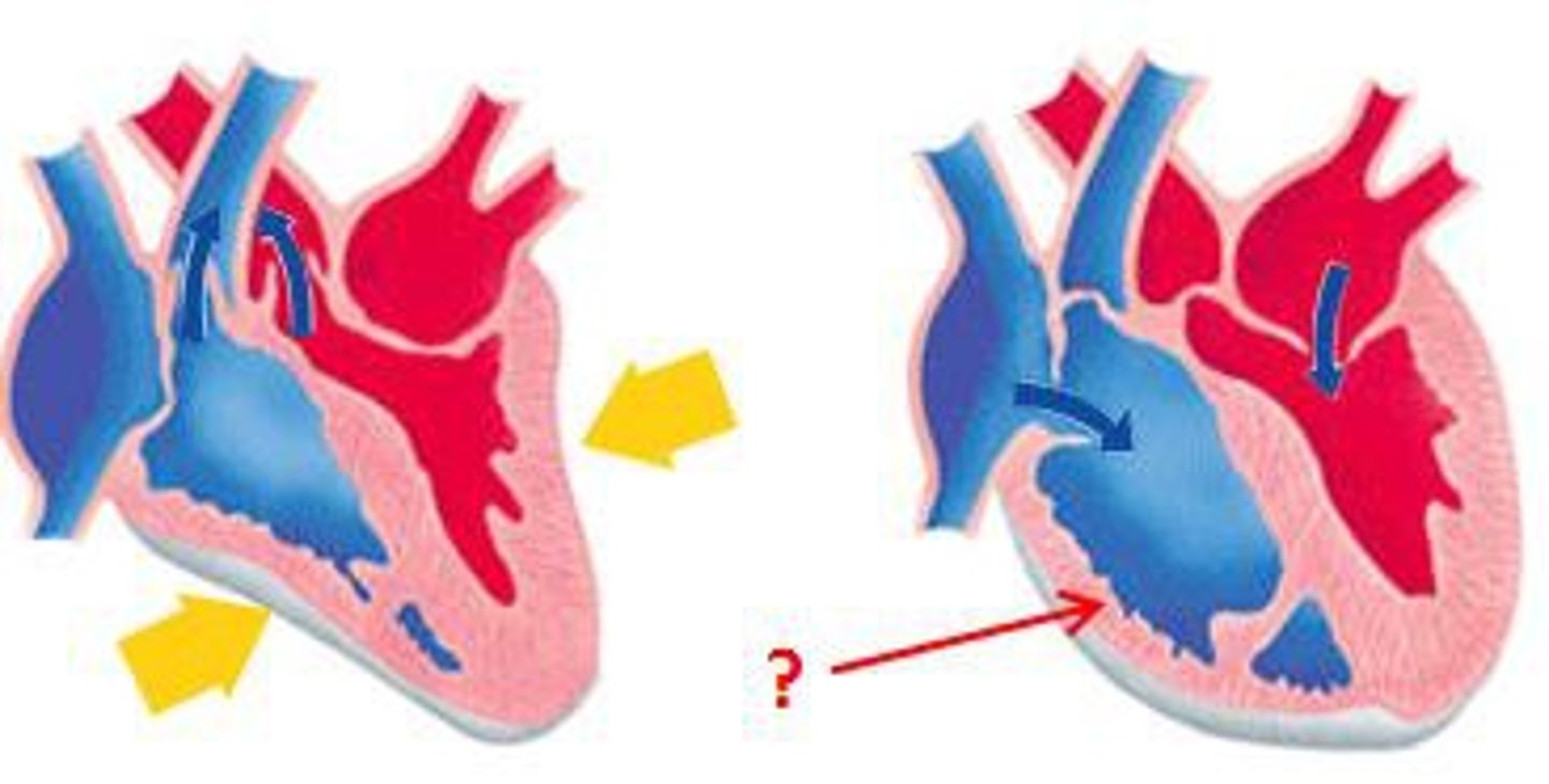
Systole
Contraction of the heart
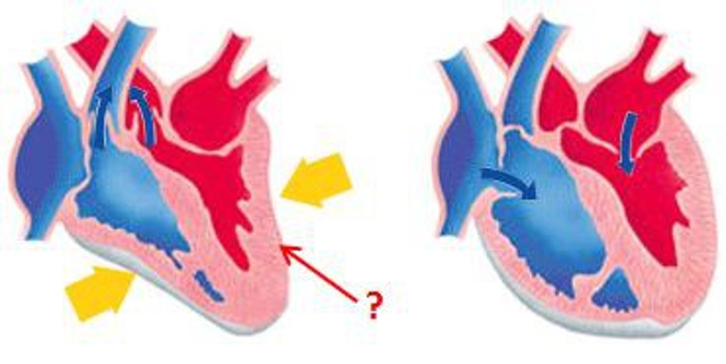
Diastole
Relaxation of the heart
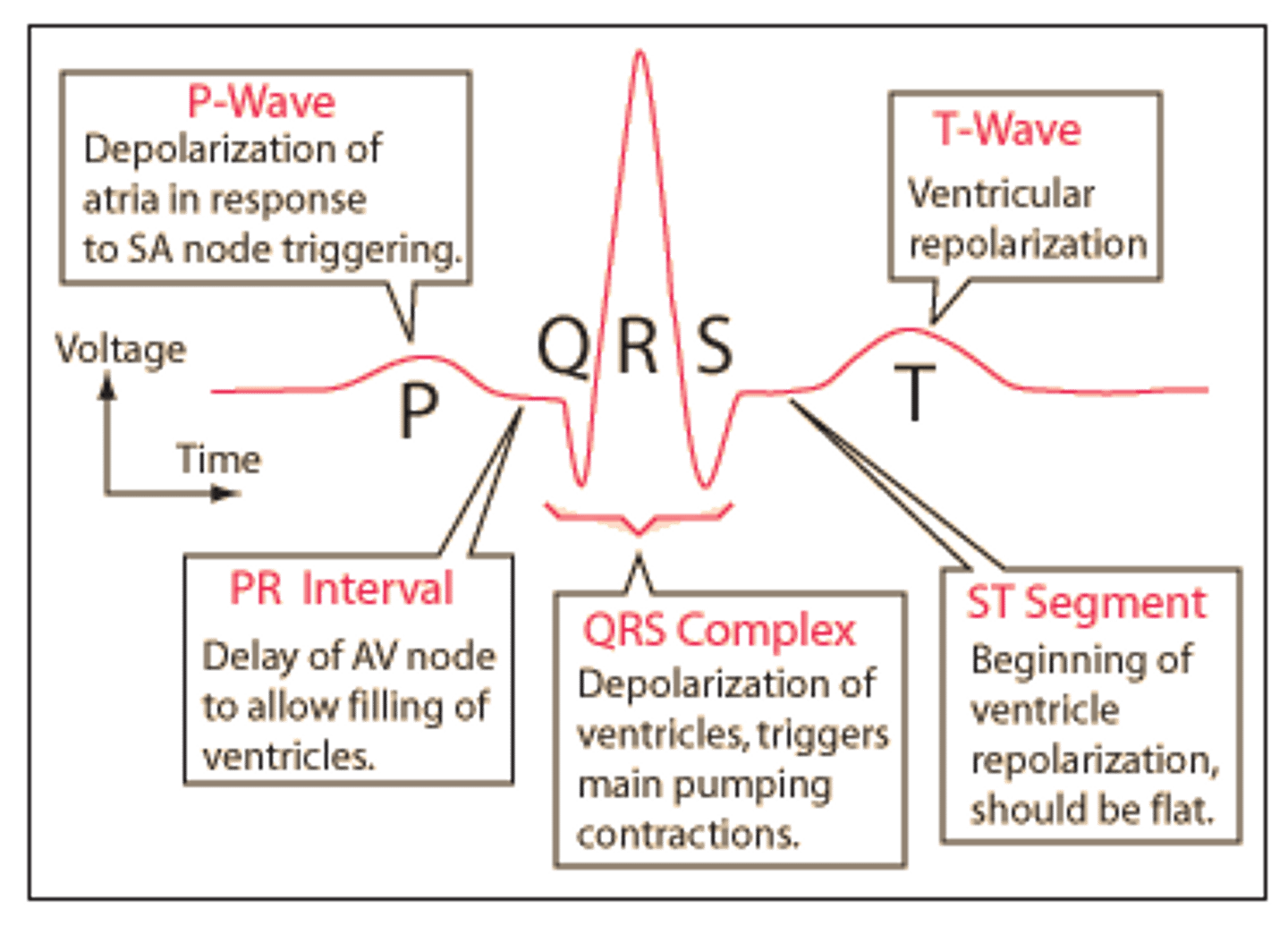
EKG/ECG
electrocardiogram
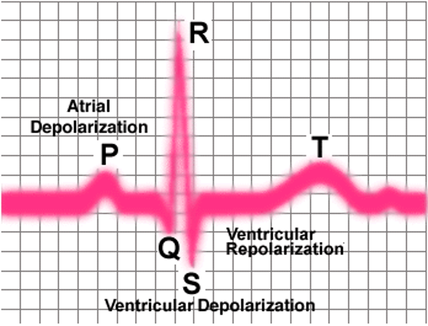
Explain EKG waves
P Wave- SA node fires (Atrial depolarization)
QRS- AV Node fires (Ventricular depolarization)
T- Ventricular repolarization (relaxing) (takes longer than atrial repolarization)
Each small box is 0.04 seconds (know that)
5 small boxes in 1 big box which is 0.2 seconds
Cardiac Output (CO)
Amount of blood pumped in 1 minute (~5 L)
Stroke Volume (SV)
The volume of blood pumped forward with each ventricular contraction/ per beat
Which blood vessels Carry blood to the heart
veins
Which blood vessels carry blood away from the heart?
Arteries
Which type of blood vessels have valves to prevent back flow?
Veins
Compare arteries and veins
Arteries are bigger and carry blood away from the heart. Veins carry blood to the heart. Arteries usually carry oxygenated blood, while veins carry deoxygenated. Arteries usually work with gravity, while veins work against it. Arteries have thicker walls (because they have a higher blood pressure) and are more elastic and muscular. However, both still have the same number of layers (3) in their walls. (Arteries just have a thin elastic layer in their middle layer.) Veins also function as blood reservoirs.
Blood pressure definition
the pressure that is exerted by the blood against the walls of blood vessels
Normal blood pressure in average weight adult
120/80
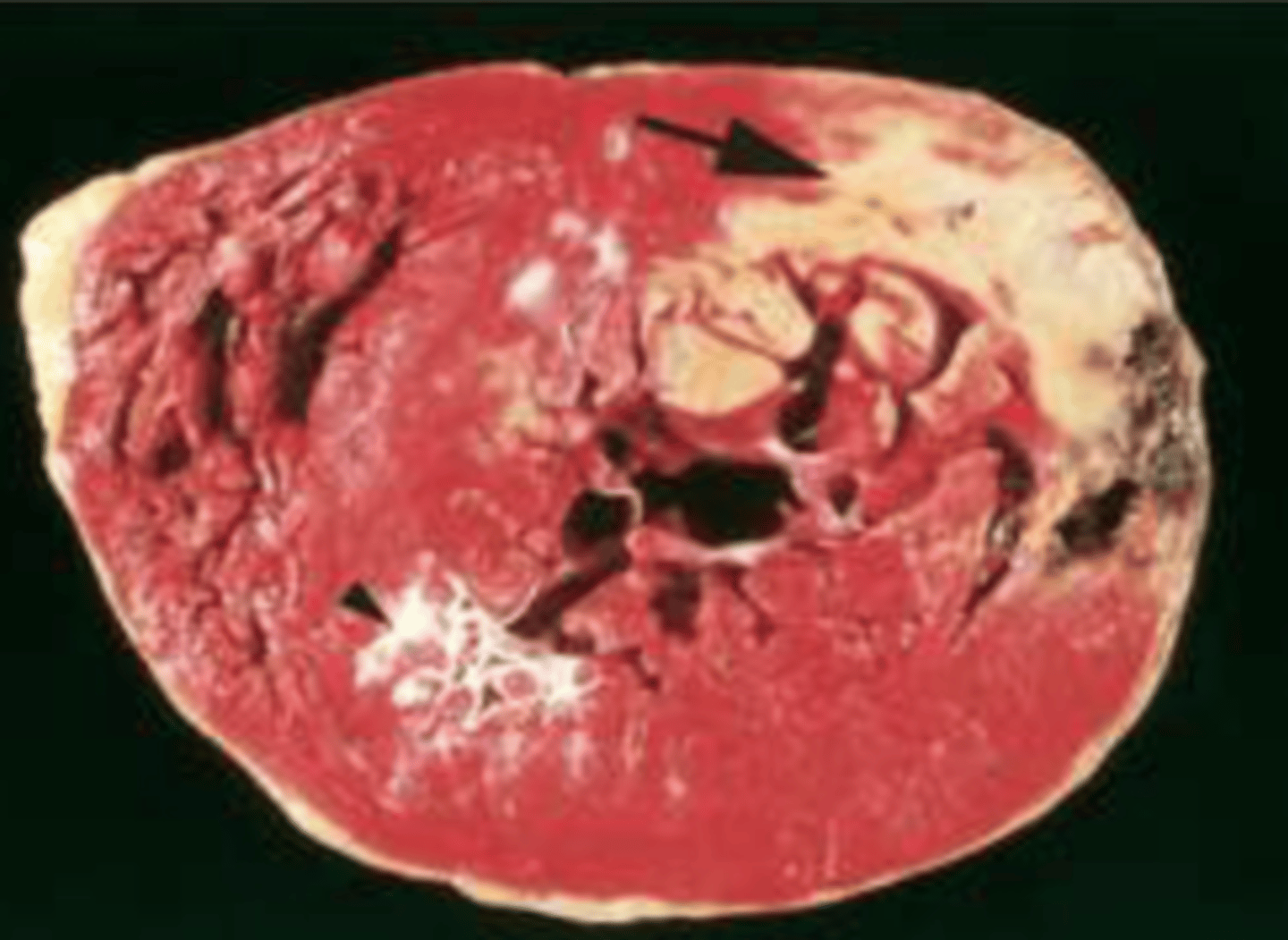
Death of myocardial tissue
myocardial infarction
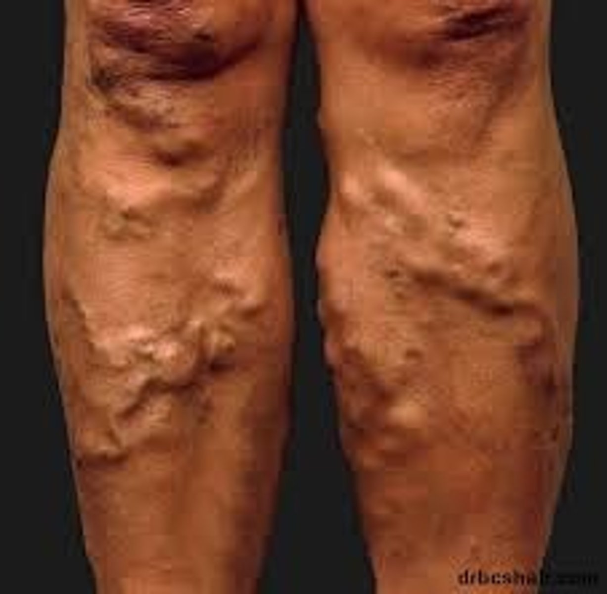
varicose veins
abnormally swollen, twisted veins with defective valves; most often seen in the legs
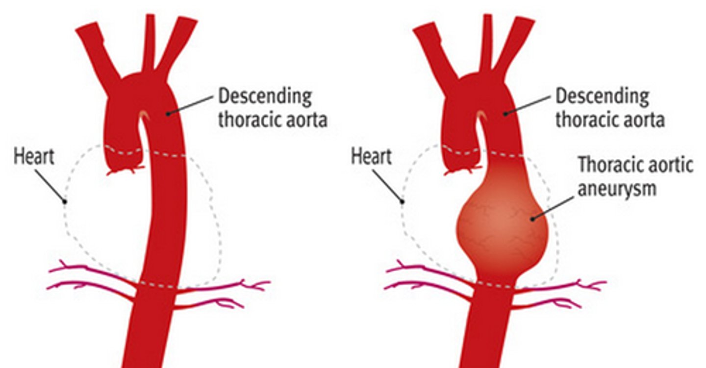
Aneurysm
ballooning of a weakened portion of an arterial wall
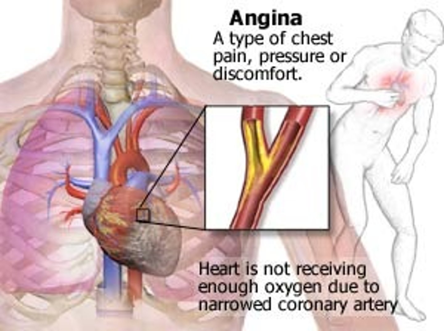
angina pectoris
chest pain that results when the heart does not get enough oxygen
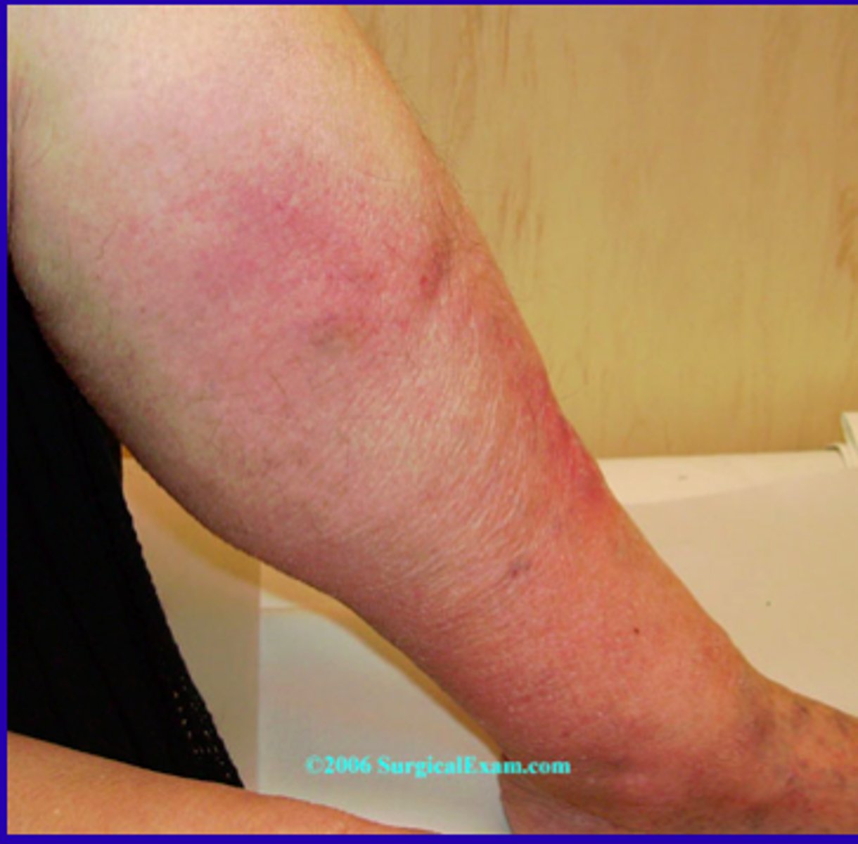
Thrombophlebitis
inflammation of a vein associated with a clot formation
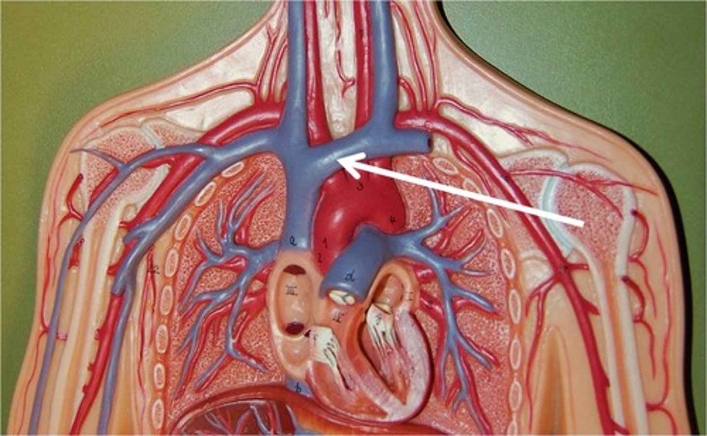
Vein drains the lower part of the body
IVC
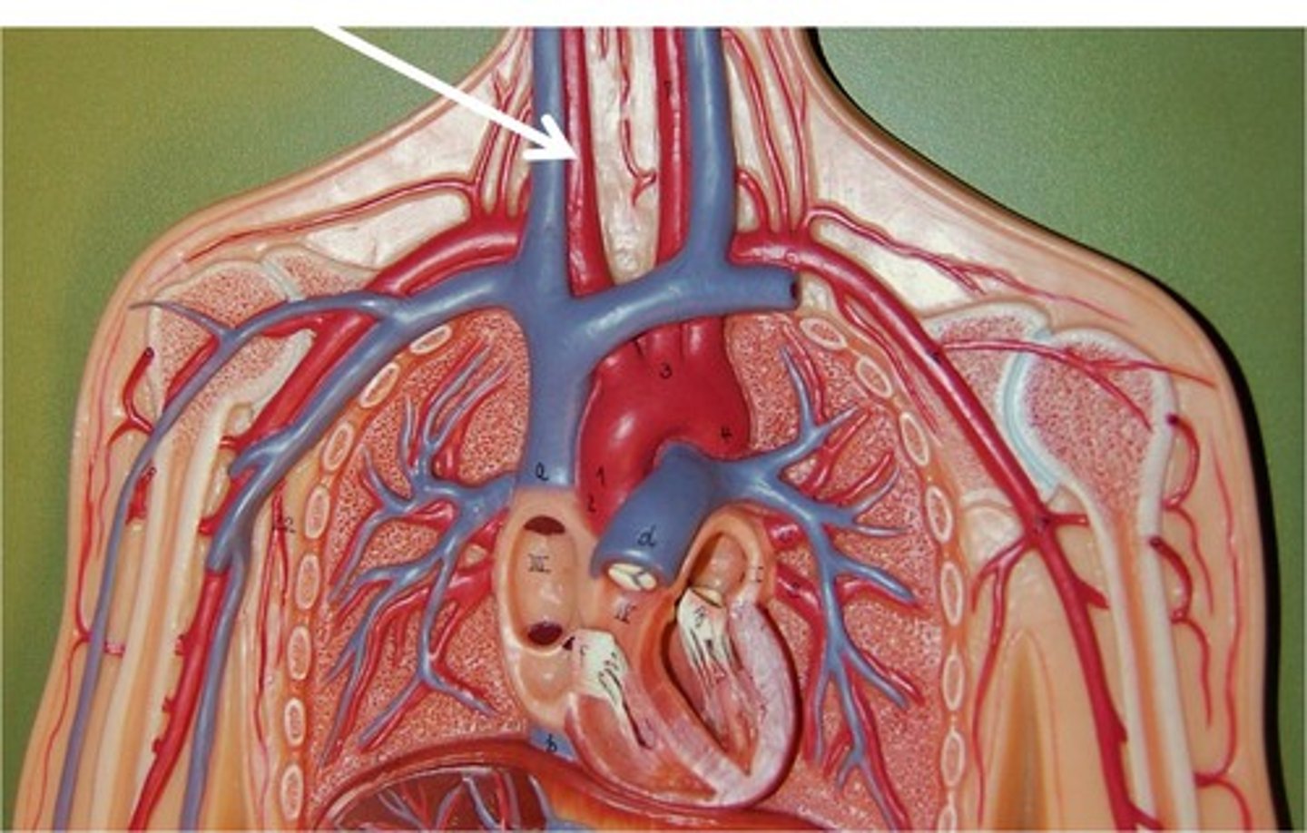
Vein drains the upper part of the body
SVC
Biggest artery in the body
Aorta
Biggest vein in the body
Saphenous vein
carry deoxygenated blood to the heart
SVC and IVC
Receives blood from IVC and SVC
Right Atrium
Receives oxygenated blood from the lungs
left atrium
four pulmonary veins return blood from the lungs
When blood leaves right atrium and path through tricuspid valve will enter in--
Right ventricle
Blood leave the right atrium through
Tricuspid valve
Blood leave the right ventricle through
Pulmonary semilunar valve
Blood leaves the left atrium through
Mitral/ Bicuspid valve
Blood forced to ------- to leave left ventricle
Aorta
Carry oxygenated blood from the heart to the rest of the body
Aorta
What is the correct sequence of vessels that blood travels as it leaves the heart?
Aorta, arteries, arterioles, capillaries, venules, veins, vena cava
Which valve prevents the backflow of blood into the left ventricle?
Aortic semilunar valve
what chamber forms the inferior surface of the heart?
Right ventricle, left ventricle and right atrium
What chamber forms the anterior surface of the heart?
right atrium, right ventricle and part of left ventricle
posterior surface of the heart
left ventricle, left atrium and right atrium
What chamber form the apex of the heart?
Left ventricle
AV valves vs SL valves
AV valves are entry valves, SL valves are exit valves
Angina vs MI
angina-squeezing pain; relieved with NTG, exertion increases pain
MI-sharp pressure pain, more serious than Angina.
CABG
coronary artery bypass graft
angioplasty and stent
balloon inserted in blood vessel to expand it, then a stent is placed inside to hold the vessel up
endartrectomy
removal of plaque from artery
primary pacemaker of the heart
sinoatrial (SA) node
Secondary pacemaker of the heart
atrioventricular (AV) node
carotid arteries
the large neck arteries, one on each side of the neck, that carry blood from the heart to the head
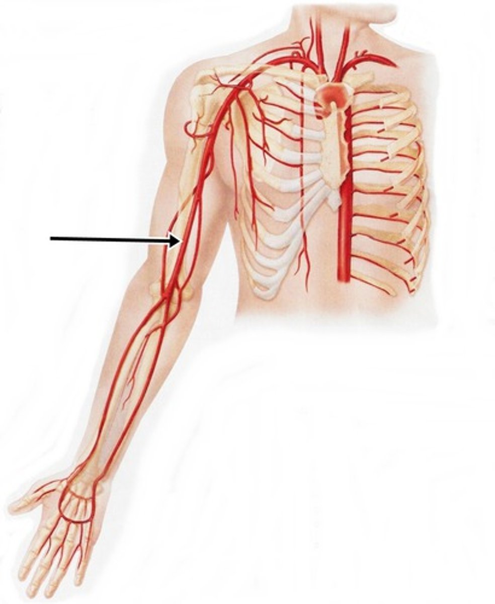
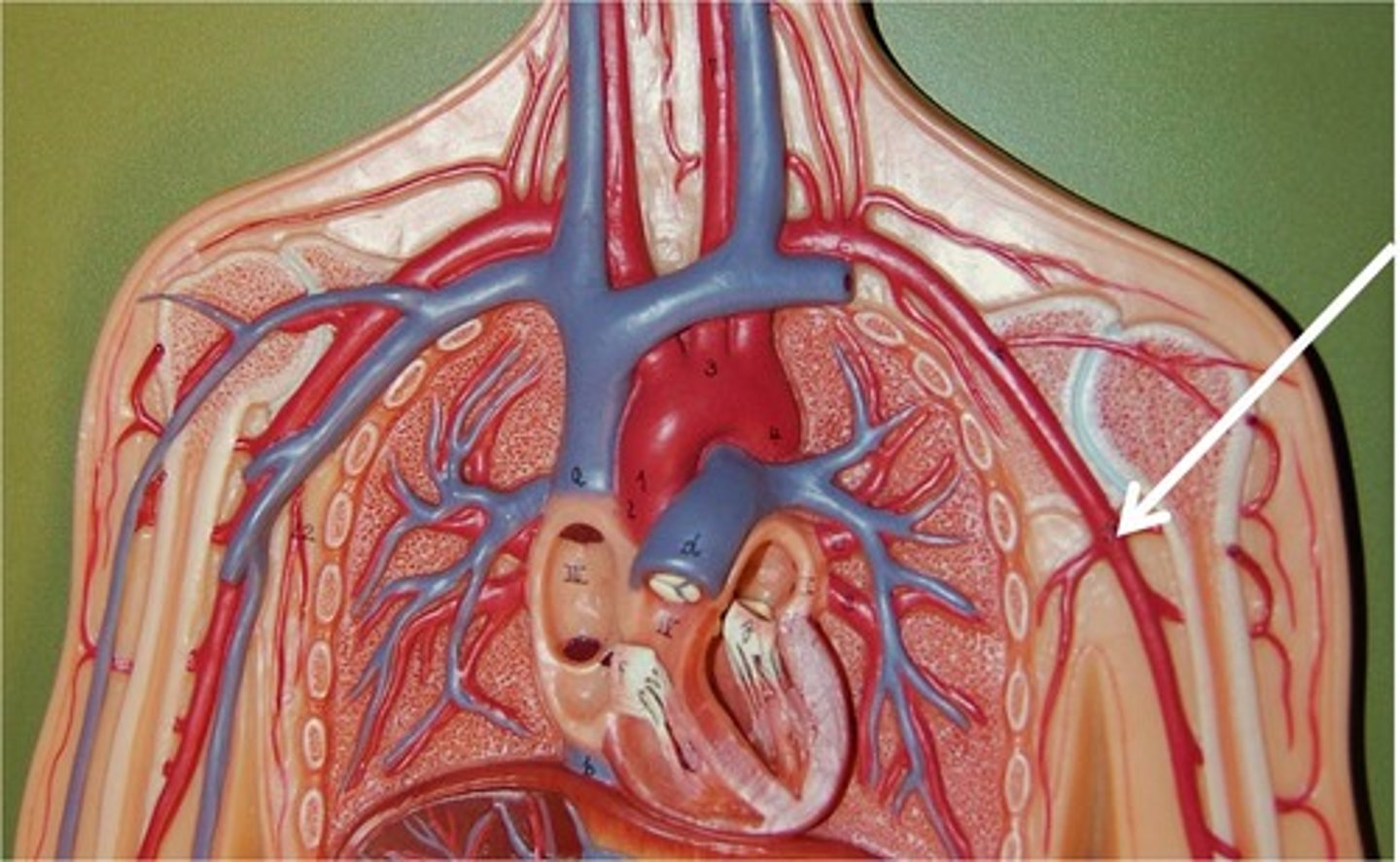
brachial artery
artery of the upper arm; the site of the pulse checked during infant CPR
femoral artery
the major artery supplying the leg
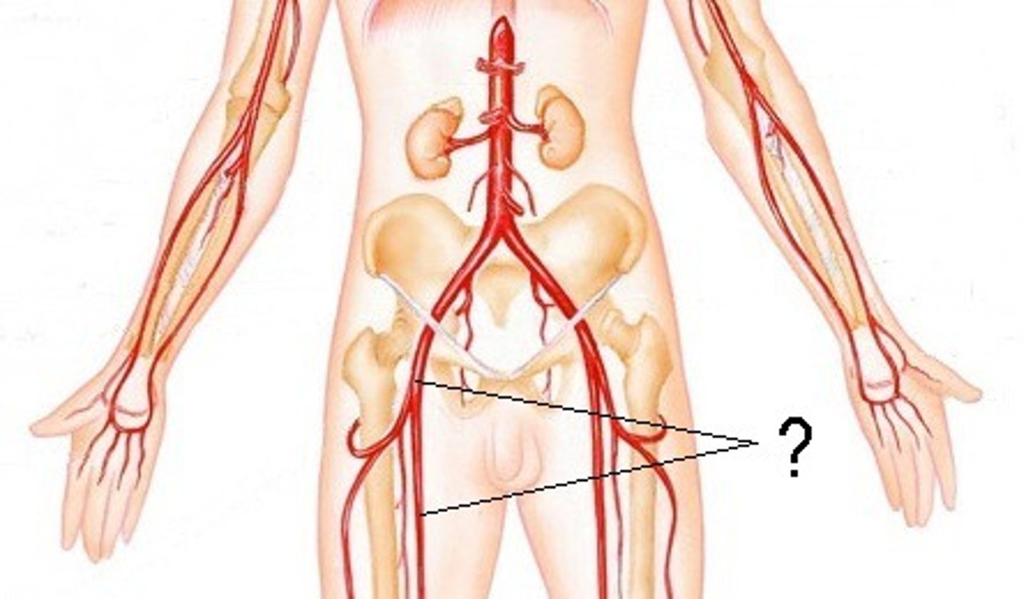
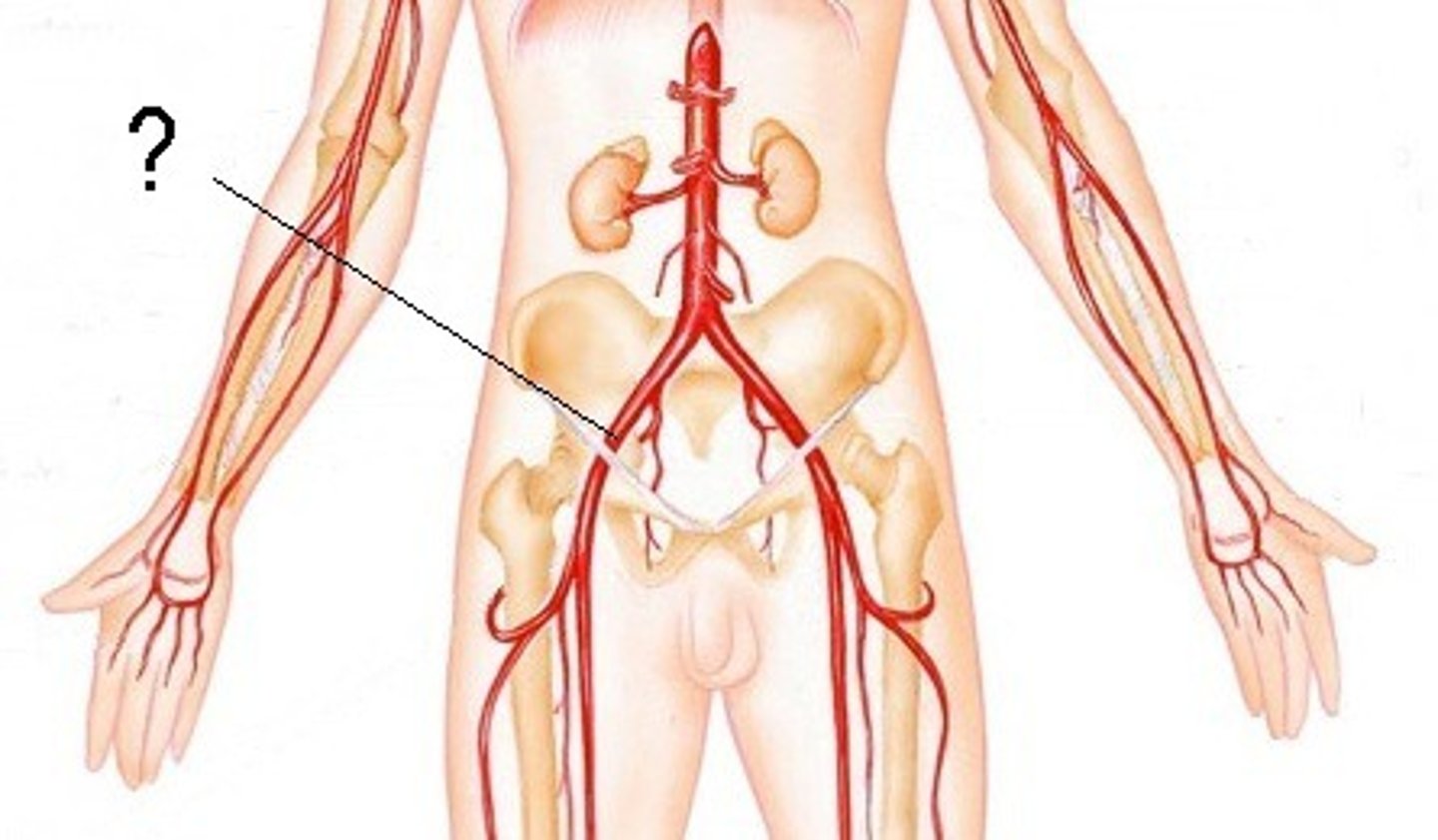
common iliac artery
Terminal branches of the abdominal aorta, supplies pelvic organs
axillary artery
artery that carries oxygenated blood to the axilla (armpit) area
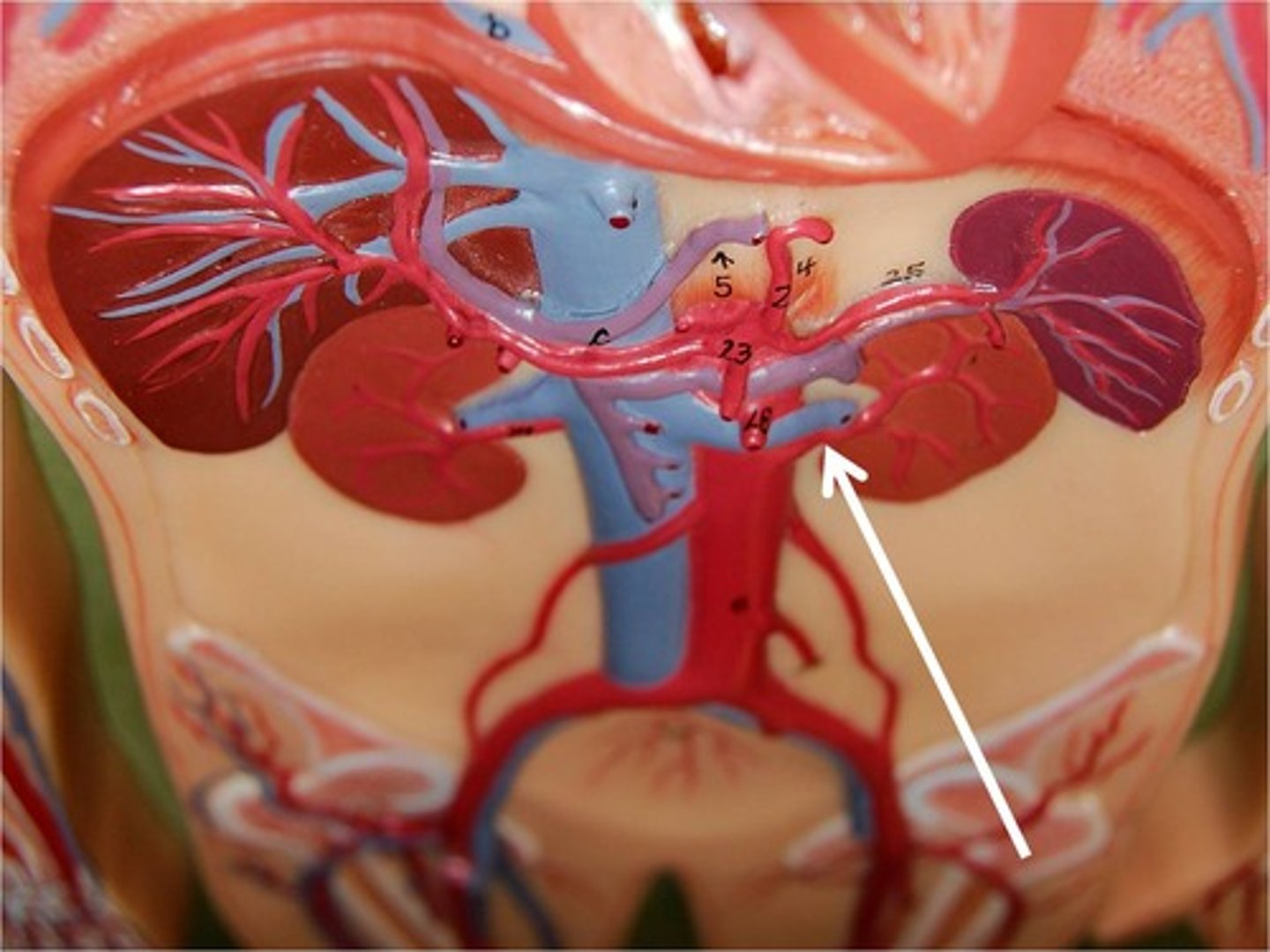
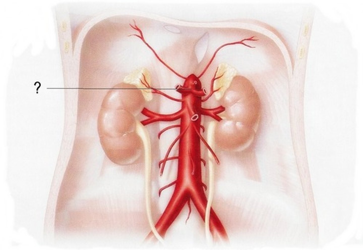
renal artery
blood vessel that carries blood to the kidney
celiac trunk artery
The liver, stomach and spleen receive their blood supply from celiac trunk
Which is not a component of the lymphatic system?
Veins
The fluid circulated by the lymphatic system is called
lymph
The fluid contained in lymphatic vessels, which is derived from plasma, is called
lymph or lymph fluid