14 Response to stimuli
1/11
Earn XP
Description and Tags
A level Biology yr 13 content
Name | Mastery | Learn | Test | Matching | Spaced | Call with Kai |
|---|
No analytics yet
Send a link to your students to track their progress
12 Terms
Define Stimulus, Receptor
Stimulus is a detectable change in the external or internal environment that leads to a response, increasing the chance of survival for organism
Receptor is a specialised group of cells that detects a specific stimulus
Stimulus → receptor → Coordinator → Effector → Response
Taxes (Taxis)
A taxis is a directional movement of an organism in response to external stimulus.
Positive Taxis: Movement towards a stimulus (e.g algea move towards light)
Negative Taxis: Movement away from stimulus (e.g. Earthworms move away from light)
Kineses
Kinesis is a non-directional movement responding to a stimulus where the organism’s speed or rate of turning changes due to the intensity of stimulus
An example, Woodlice loose water in dry conditions, so if they move from damp to dry environment they change their direction often and move quickly, since they are more likely to return to their favourable environemnt since they are closer. However, if they are far away as in they do this regularly, they will slow down and move in straight lines, so they are more likely to come back to origional environment.
Tropisms: Gravitropism and phototropism
Phototropism: is the plant responding to levels of changes in light. SHOOTs IAA is promoting elongation
When the sun is facing one side of the plant shoot, the other side is in darker side, so higher concentration of IAA on darker side, and in shoots this promotes growth (ELONGATION), and the shoot elongated to face the direction of sun.
Gravitropism: is the plant responding to the stimulus gravity. ROOTs IAA is inhibiting elongation
The side of the root facing towards the direction of gravity there is an uneven distribution of IAA, in which the side of gravity will have higher concentration of IAA, so in roots this inhibits elongation. So, the upper side of the roots elongated and causes the turning of the root into the plant.
The differences between Taxis, Kineses and Tropisms:
Taxis:
Mobile organism
Directional movement
Whole organism
Response is directly towards or away from stimulus
Kineses:
Mobile organism
Non-directional, random movement where organism changes speed or rate of turning
Response is random
Response based on the intensity of stimulus
Whole organism
Tropism:
Stationary organisms (plants)
Directional
Direction of growth directly away or towards a stimulus
Direction of a reflex arc, and importance:
Stimulus → Receptor → Sensory Neuron → Intermediate neuron (Coordinator) → Motor neuron → Effector → Response
Protect body from harm
Fast, as direction of impulse through few synapses
Absence of decision making (unconcious) => Action is rapid
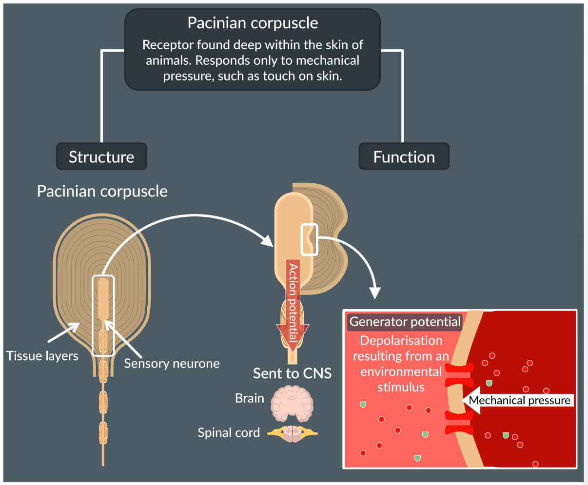
Pacinian Corpuscle
Pacinian Corpuscle has a single sensory neuron surrounded by layers of tissue. It is a transducer in which converts the mechanical energy into generated potential energy
When mechanical pressure is felt on pacinian corpuscle, this causes stretch-mediated sodium ion channels to open, and sodium ions to enter via facilitated diffusion
This causes inside of the sensory neuron to get depolarised, known as a generated potential.
When enough of these generated potentials reach threshold, there is an action potential that passes onto the CNS.
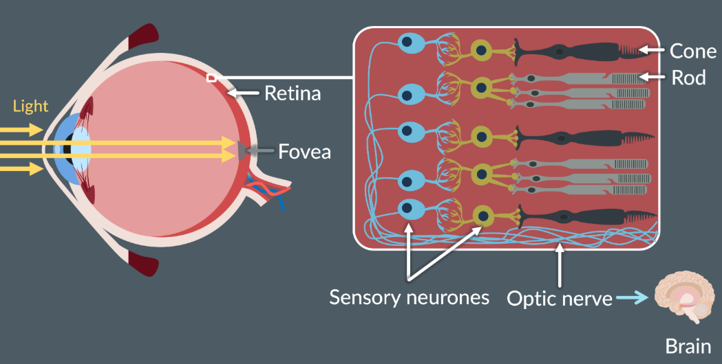
The structure of human Retina
Retina: Layer of tissue at the back of human eye that detects light from the environment.
At the centre there is the fovea, where most of the light falls. At the fovea (Cones are present, rod cells are absent, for sharp, detailed and colour vision)
The cone and rod cells are connected to sensory neurons, which are connected to the optic nerve to send impulses to the brain. They are also transducers (convert light energy to electrical energy)
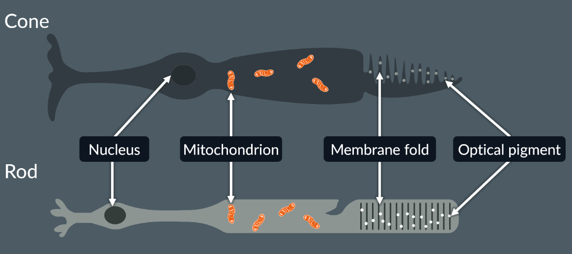
Structures of rod and cone cells
Cone cells are connected to a bipolar cell, then a sensory neuron.
Rode cells are connected to a biopolar cell then many sensory neurons.
The membrane folds are dotted with optical pigments - that are chemicals that enable to respond to different wavelengths of light, so this makes each rod and cones to see different colours.
Differences between Rod and cone cells:
Cone cells:
They have optical pigment to detect red, green and blue (so we can see in colour)
High visual acuity: how clearly you can see (distinguish shapes or clarity)
Low light sensitivity, can see when very bright light.
One cone is connected to one sensory neurone, so separate action potentials are sent to the brain => Temporal summation
Rod cells:
They only have one optical pigment, so interpret colours as shades of black and white.
They have low visual acuity: only pick out rough shapes
High light sensitive, so can detect low levels of light
Many rods are connected to a single sensory neurone, so a single action potential is sent (as detect weak stimulus) =>
Interesting: that to see the pigment needs to be broken down, and with rod cells the pigment needs less energy so we can see in low light intensities, with cone cells the pigment needs more energy so we can see in high light intensities
Controlling resting heart rate
Cardiac muscle is myogenic - can contract by itself without initiating the external nervous impulse
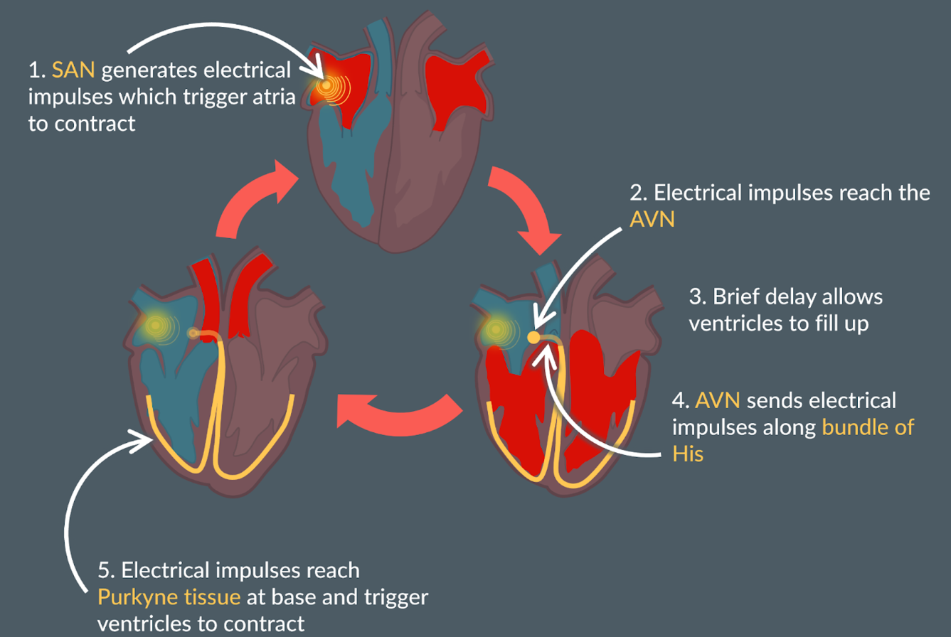
The (SAN) generates electrical impulses, which trigger the atria to contract.
While the artia contract there is a delay of 0.1 second until the electrical impulse is travelled to the atriventricular node (AVN). There is a layer of non-conductive tissue between the atria and ventricles, preventing the electrical impulse from crossing.
There is a delay so that the ventricle is filled up with as much as blood as possible, so when ventricles contract maximum blood travels out of ventricle.
Once impulse reaches AVN, this causes the imupulse to send electrical impulses through the bundle of His (made of purkyne tissue)
While electrical impulse reaches purkyne tissue this causes the base of the ventricles to contract from the base upwards
Allows blood to be efficiently move into the pulmonary artery and aorta
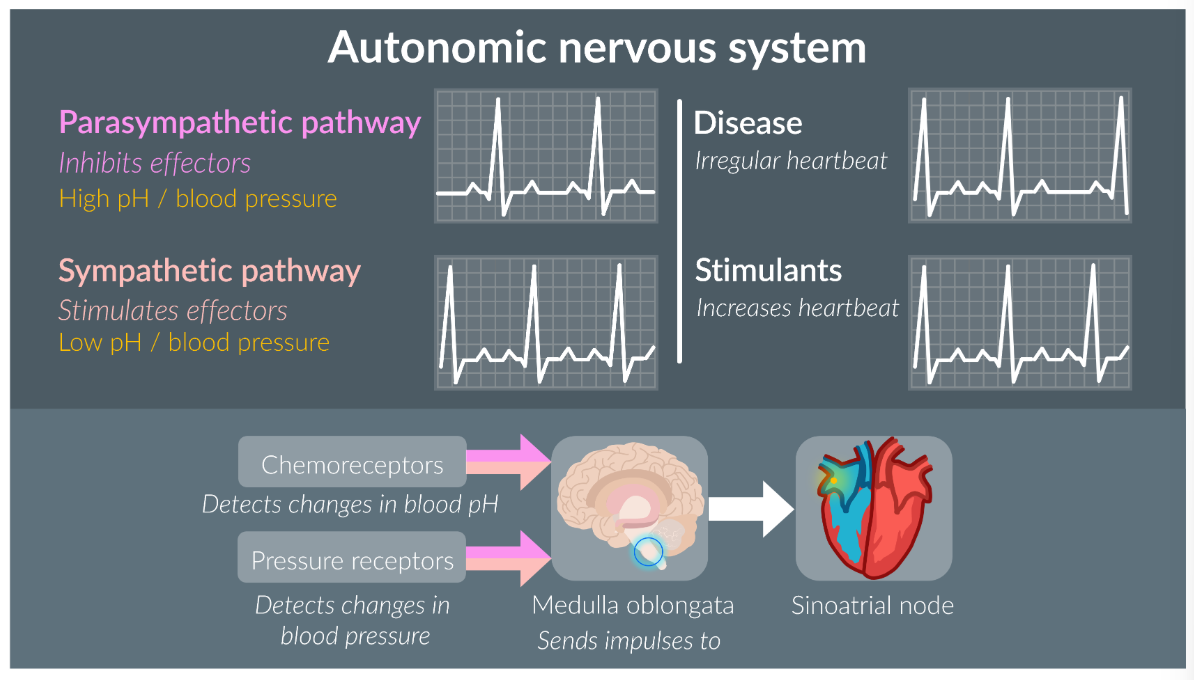
Changing heart rate
Control of heart rate is part of the autonomic nervous system (subconscious activites). Where the parasympathetic pathway causes a calming effect, and sympathetic pathways readies the body for fight or flight response.
So, when a receptor is detected, the electrical impulse passes through to the Medulla oblongata (cardiac control centre) and sends these impulses to the SAN, which changes the rate of impulses fired by the SAN.
There are two types of impulses that send impulses to the medulla:
Chemorecpetors: located in the walls of blood vessels, and detect changes in blood pH
When a low blood pH is detected (low oxygen, high carbon dioxide), the medulla obongata sends singles to the SAN, and this increases the heart rate.
Pressure receptors: located in the neck and heart, they detect changes in the blood pressure
When pressure receptors detect low blood pressure, the medualla obongata sends singles to the SAN to increase heart rate