Exam 4: Extensor Expansion (Module 8 Pt. 2)
1/23
There's no tags or description
Looks like no tags are added yet.
Name | Mastery | Learn | Test | Matching | Spaced | Call with Kai |
|---|
No analytics yet
Send a link to your students to track their progress
24 Terms
Finger Extension: Which joint seems to move the most in the initial stage of finger extension from a full fist?
PIP initiates, DIP joint follows
Stages of Digit Extension
PIP joint initiates digit extension
DIP joint follows into extension as the PIP joint continues to extend
Intrinsic muscles help to bring PIP & DIP joints into full extension
Finally, MCP joints fully extend w/ the digit
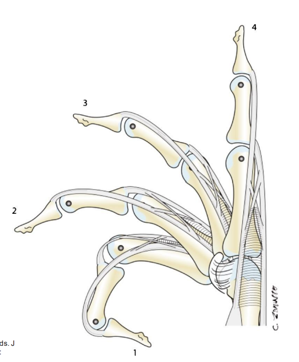
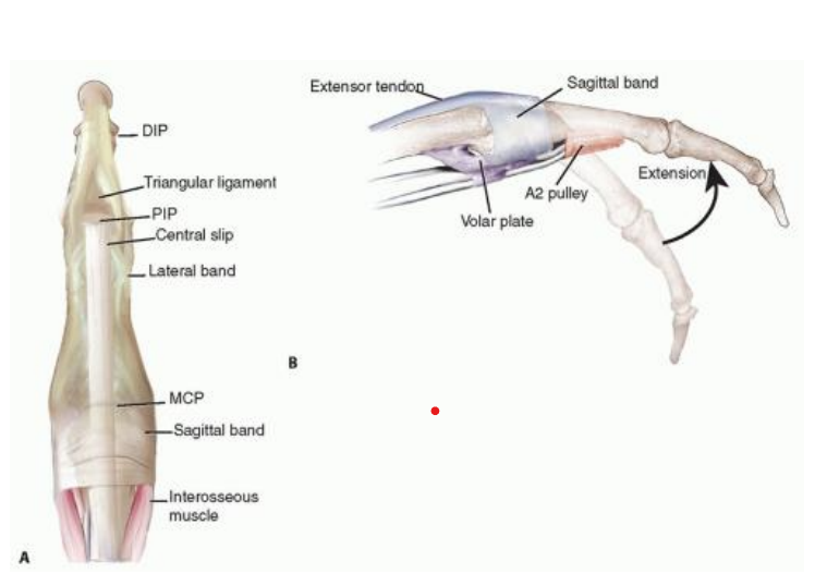
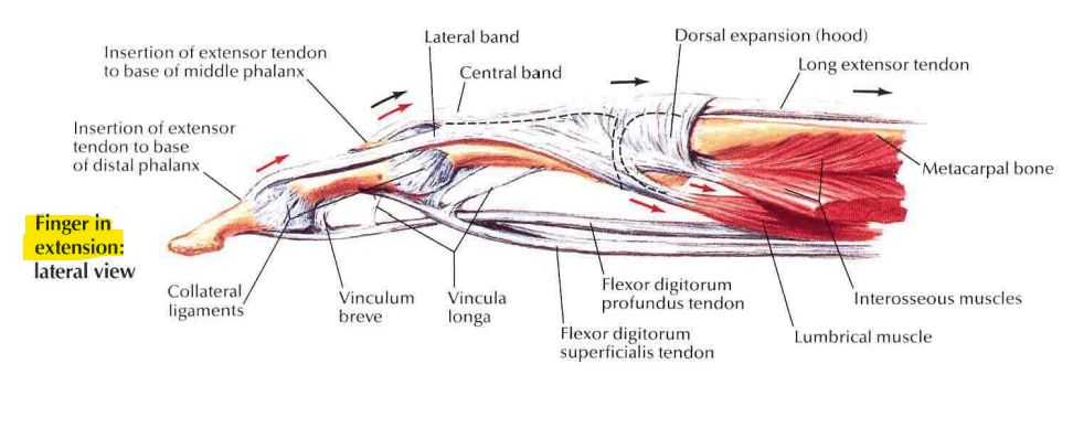
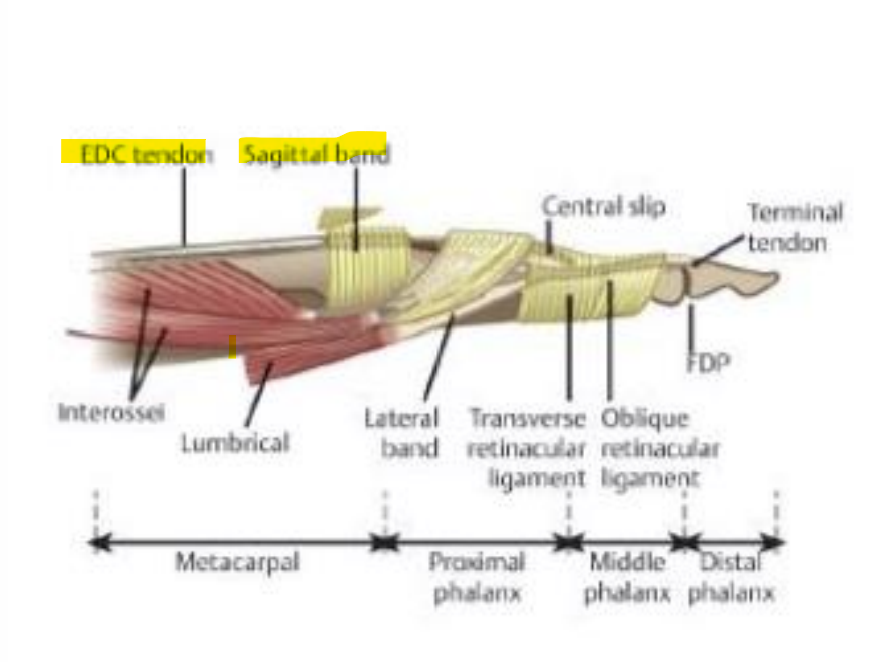
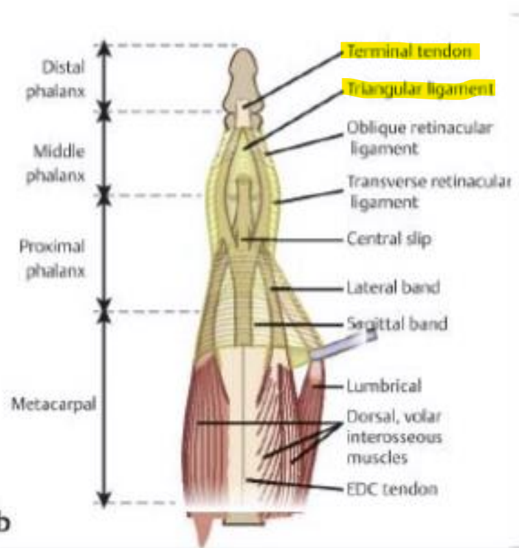
Components of the Extensor Expansion: finger extension at (3) joints…
MCP extension: EDC tendon & sagittal band
PIP extension: Central slip & lateral bands
DIP extension: Terminal tendon (bands become 1) & triangular ligament
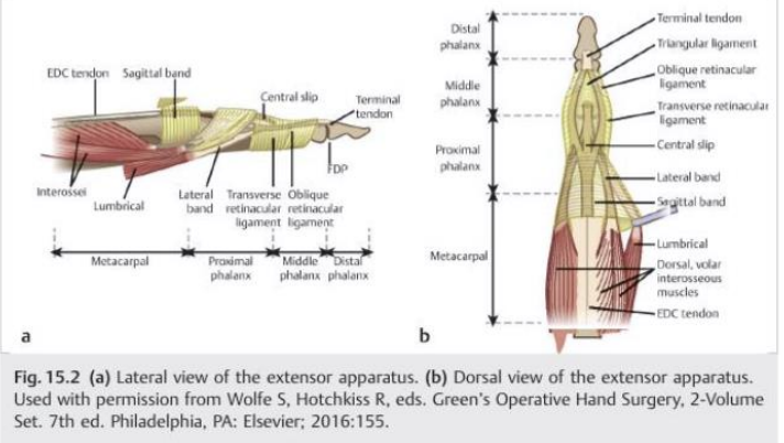
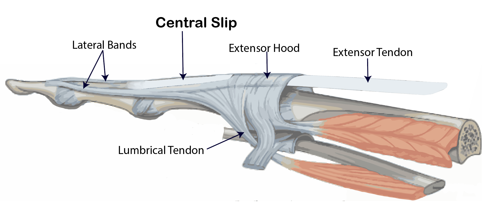
Extensor/Dorsal Hood
Triangular shaped web of CT structure that the EDC tendon & intrinsics (interossei & lumbricals) insert into
Facilitates & completes finger extension
Located between dorsum of MCP joint & dorsum of the proximal phalanx
MCP Joint: EDC Tendon & Sagittal Band
Sagittal bands: Originate from volar plate; wrap around MCP joint
Function: centralize EDC tendon
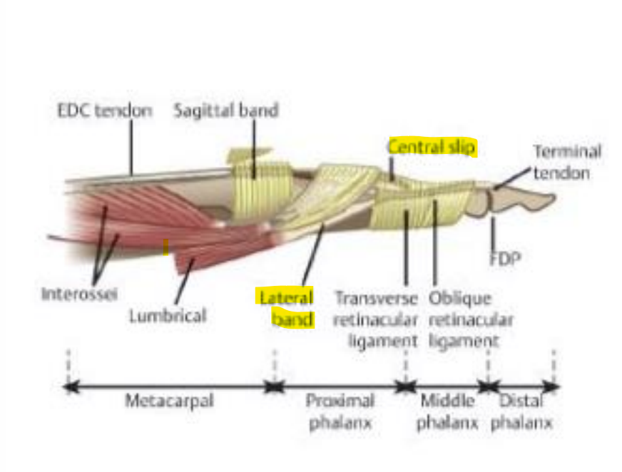
PIP Joint: Central Slip & Lateral Bands transfer force from the EDC tendon & intrinsic muscles to…?
Extend the PIP & DIP joints
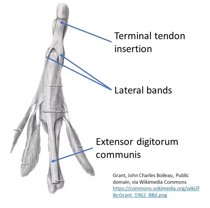
After leaving MCP joint, the EDC tendon splits into the central slip & 2 lateral bands
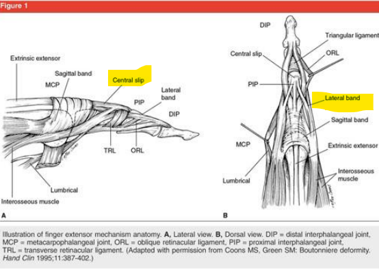
PIP Joint: Central Slip Function
PIP extension

PIP Joint: Lateral Bands Function
DIP extension
Transition between volar position (in flexion) to dorsal position (extension)
DIP Joint: Terminal Tendon
Lateral bands converge to form the terminal tendon @ dorsum of DIP joint
Function: DIP extension
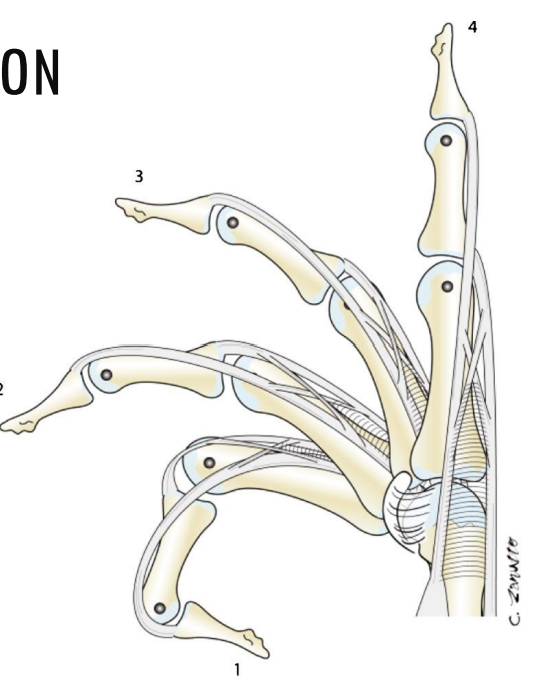
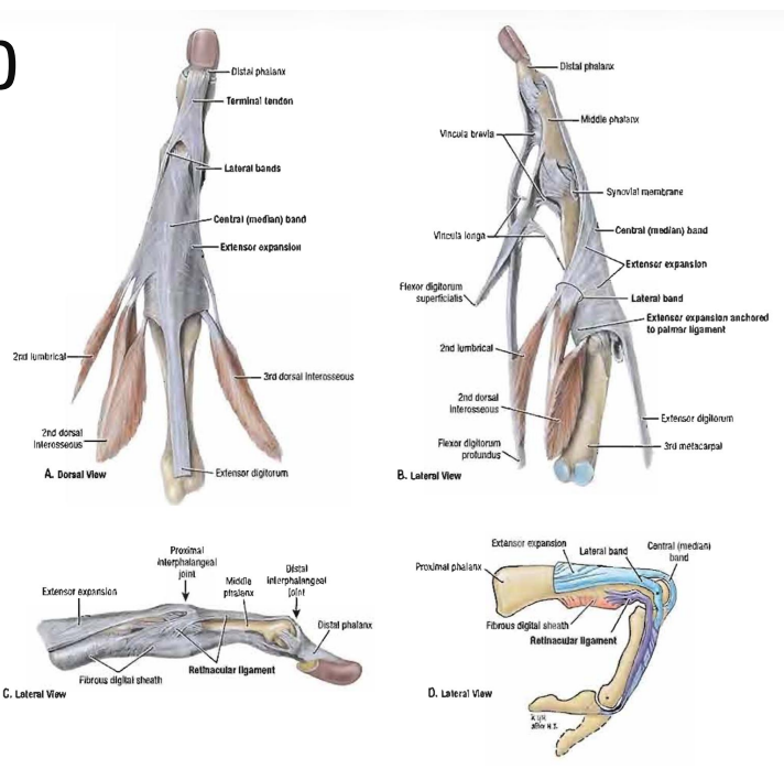
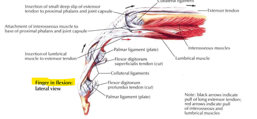
Digit Extension Mechanics
Central slip @ PIP joint initiates digit extension
Lateral bands dorsally migrate & extend DIP joint
Intrinsic muscles continue to extend both DIP & PIP joints through their insertions on the lateral bands
Sagittal bands extend the MCP joints as the EDC tendon migrates proximally
Lateral bands rest dorsally in extension (lateral view), and drops…
volar in flexion
Zones of extensor injuries are used to describe the area of…?
extensor injury (treatment & protocols differ)

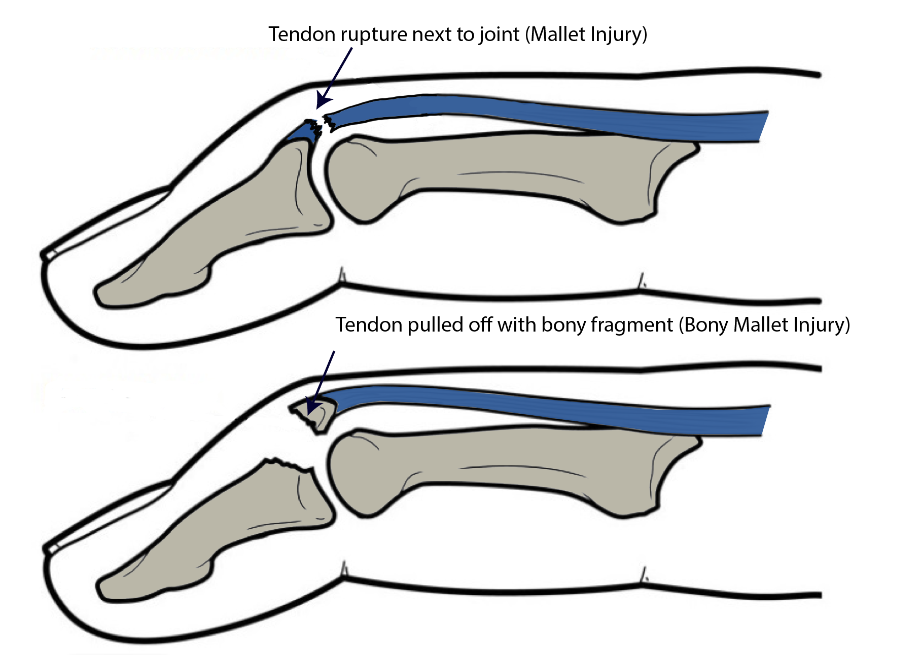
Zone I/1 Extensor Injury “Mallet Finger”
Distal to or @ DIP joint
MOI: direct trauma to fingertip, causing abrupt, forceful DIP joint flexion
Soft mallet: terminal tendon tear
Bony mallet: terminal tendon tear associated w/ avulsion fracture
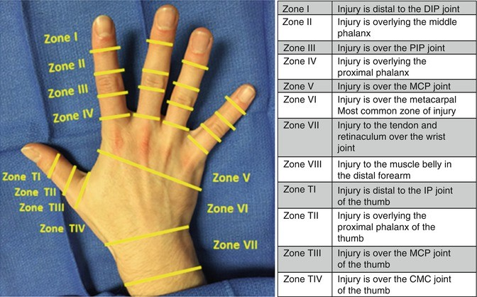
Zone II/2 Extensor Injury
Middle phalanx
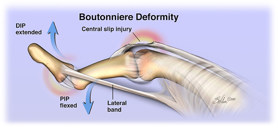
Zone III/3 Extensor Injury “Boutonniere”
PIP joint area
MOI: Blunt trauma to PIP joint, causing abrupt, forceful PIP flexion
Central slip tear
Lateral bands stay volar: pull PIP into flexion & DIP joint hyperextension
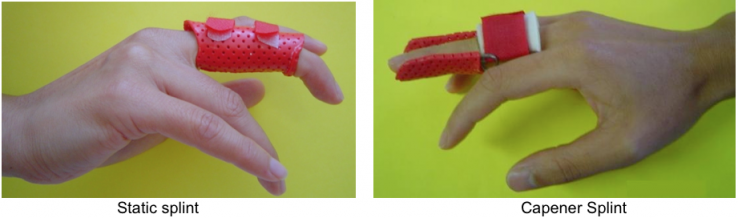
Wear static splint
Zone IV/4 Extensor Injury
Proximal phalanx
Zone V/5 Extensor Injury: Sagittal Band Injury
MCP joint area
If injured/attenuated, sagittal band no longer effective @ keeping EDC tendon centralized on dorsum of MCP joint in MCP flexion
Mallet Finger Conservative Management (Zone 1)
24/7 DIP joint extension orthosis for 6-8 wks, then gradual splint weaning & gentle AROM
If patient removes orthosis & starts moving fingertip, 6 wk wear time restarts!
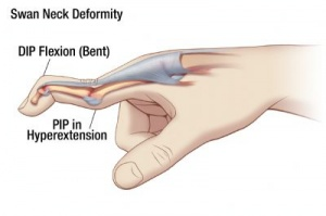
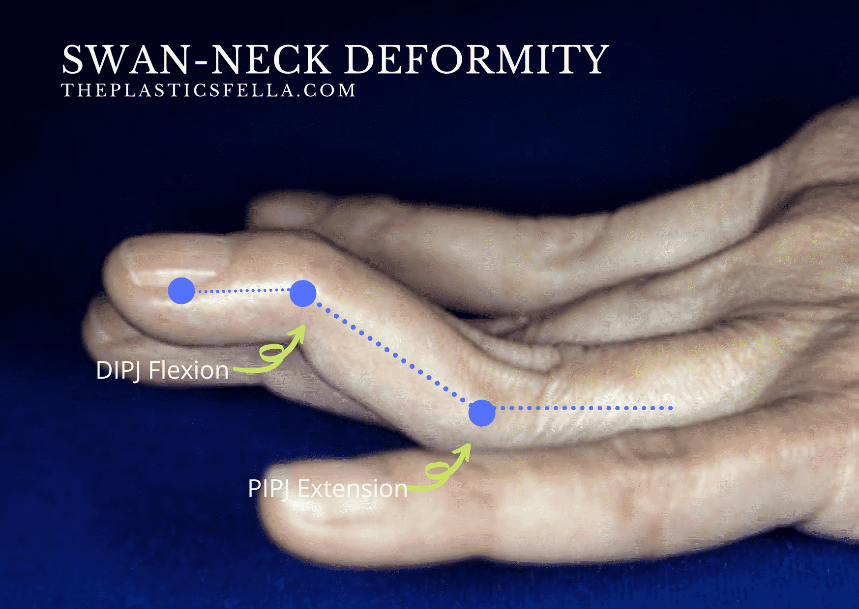
Zone III/3 Extensor Injury “Swan Neck Deformity”
PIP joint area
In people w/ joint hypermobility
Can occur after PIP dorsal dislocation
Volar plate insufficient @ limiting PIP joint hyperextension
Lateral bands sublux dorsally & become ineffective @ extending DIP joint
DIPJ flexion, PIPJ extension
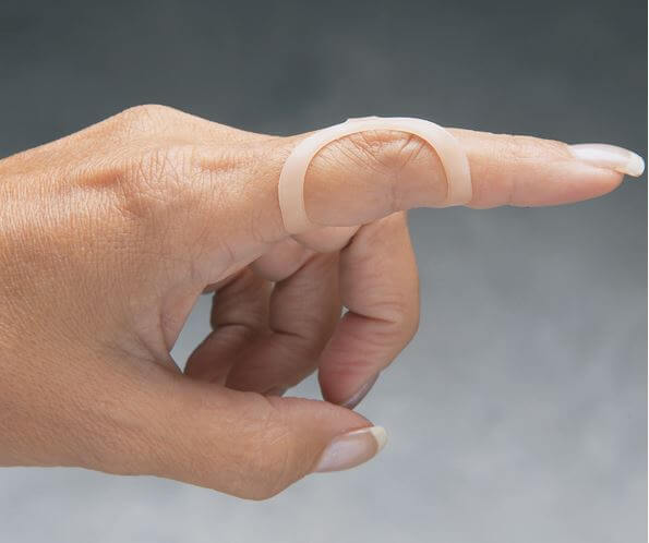
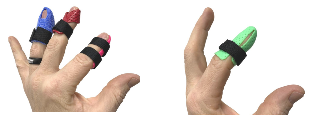
Zone III/3 Extensor Injury “Swan Neck” - Oval-8 ring splints manage this deformity by…
providing 3-point pressure system: prevents PIP (middle) joint from hyperextending
By positioning the oval band on top of the finger, they restrict backward bending + allows flexion & maintaining DIP joint extension.
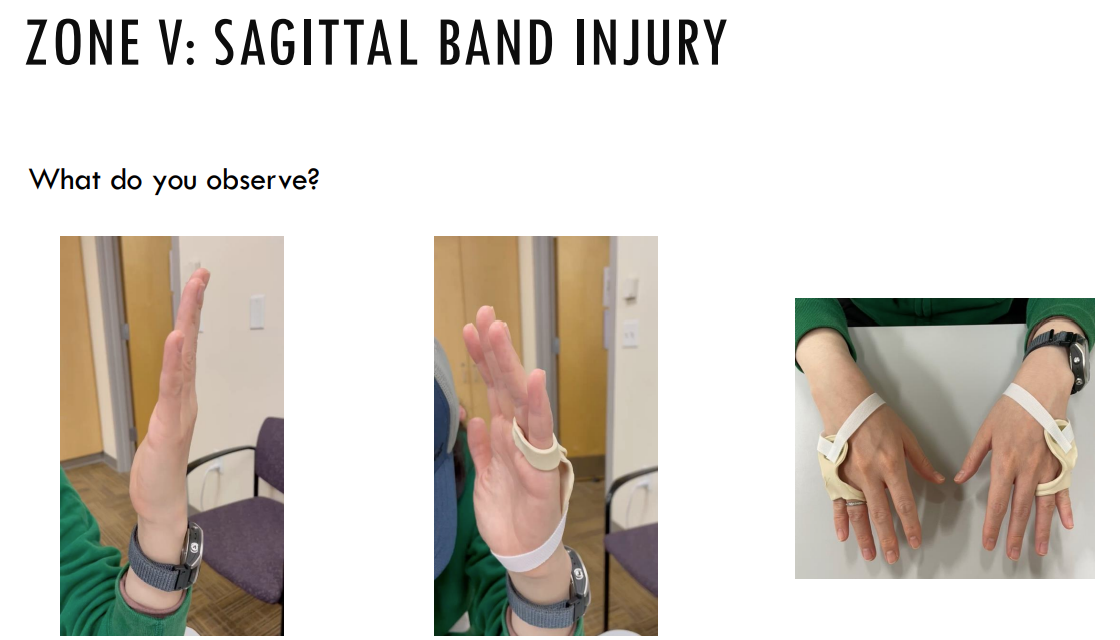
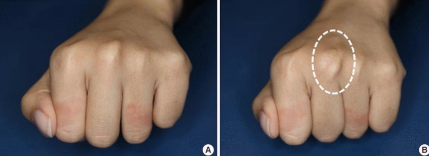
Zone V/5 Extensor Injury: Sagittal Band Injury “Boxer’s Knuckle” - What do you observe?
Present w/ pain, swelling, ecchymosis over MCP joint following trauma
Visible or palpable "snapping" or subluxation of the extensor tendon into the intermetacarpal space during flexion
Tendon typically deviates ulnarly during active extension
What structures are responsible for MCP extension?
EDC tendon & sagittal band
What structures are responsible for PIP extension?
Central slip & lateral bands
What structures are responsible for DIP extension?
Terminal tendon & triangular ligament