Biochemistry Chapter 4: 3D Structure of Proteins
1/41
There's no tags or description
Looks like no tags are added yet.
Name | Mastery | Learn | Test | Matching | Spaced | Call with Kai |
|---|
No analytics yet
Send a link to your students to track their progress
42 Terms
Levels of Protein Structure
1) Primary Structure
2) Secondary Structure
3) Tertiary Structure
4) Quaternary Structure
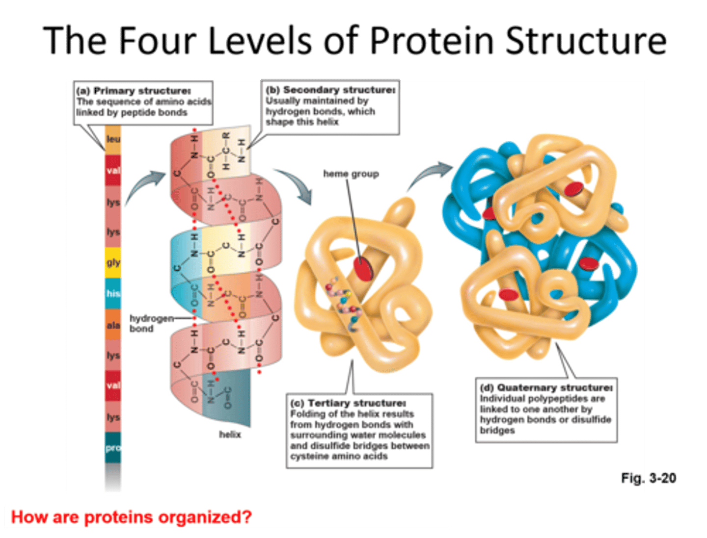
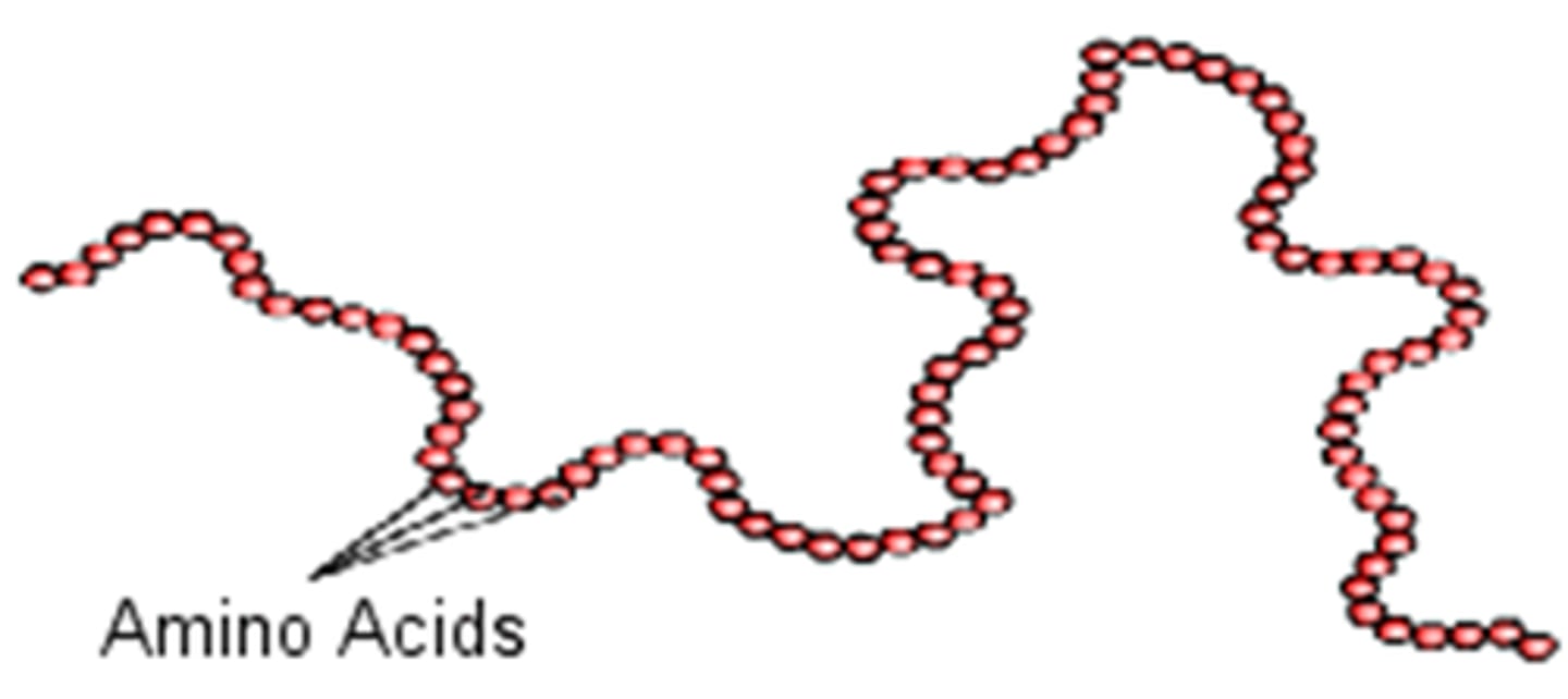
Primary Structure
Order (sequence) in which amino acids are covalently linked together.
The amino acid sequence helps determine the 3D conformation of a protein, which determines its properties
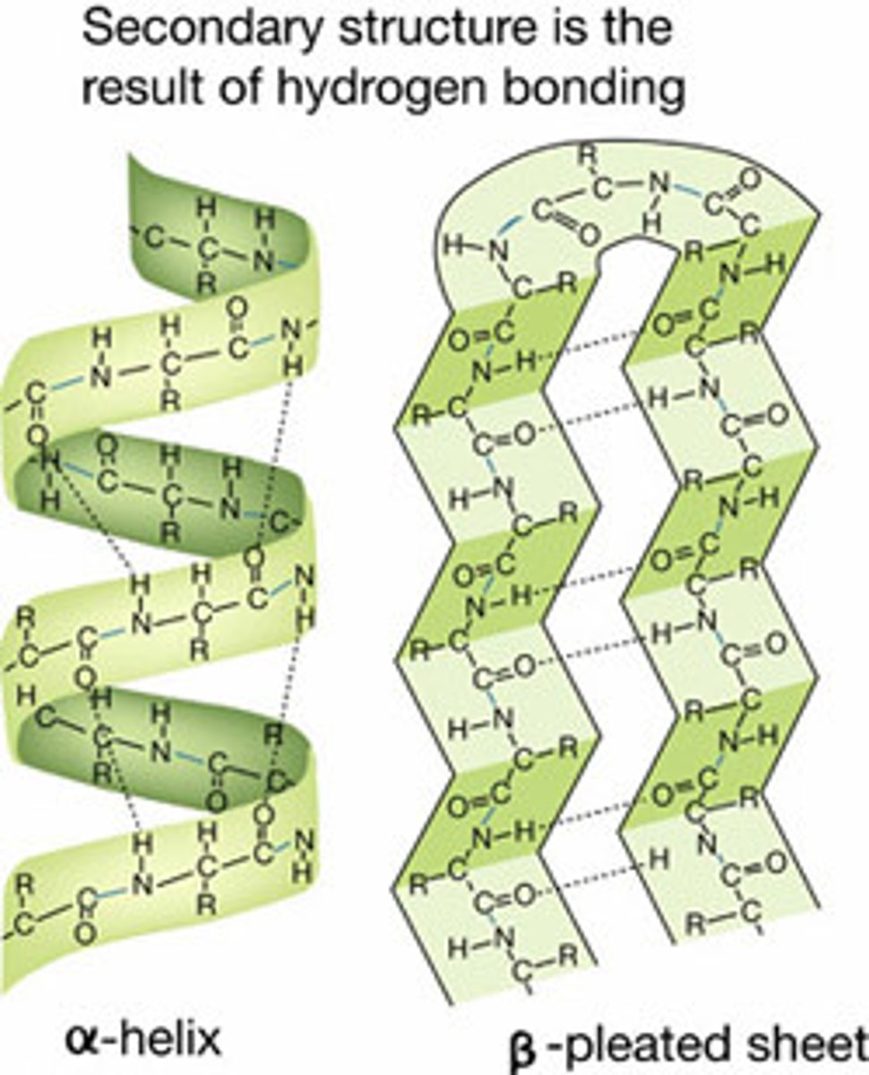
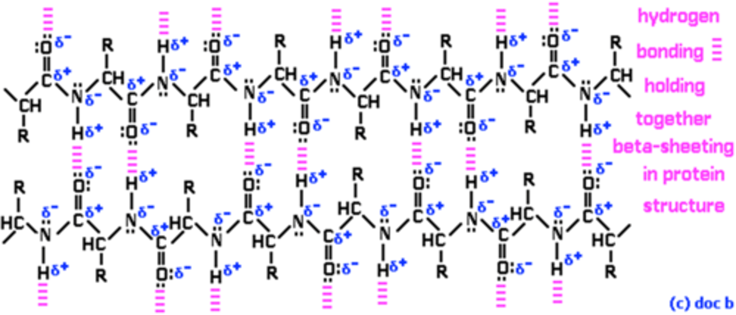
Secondary Structure
Ordered 3D arrangement in space of the backbone atoms in a polypeptide chain (there is hydrogen bonding between them). There are two types (arrangements):
1) Alpha-helixes (coils)
2) Beta-pleated sheets (antiparallel and straight)
- Perpendicular to the direction of the protein chain
Hydrogen-bonded arrangement of the polypeptide chain
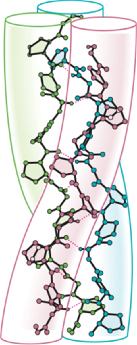
Supersecondary Structures
Result from the combination of alpha- and beta-strands.
It is basically a bunch of coils together.
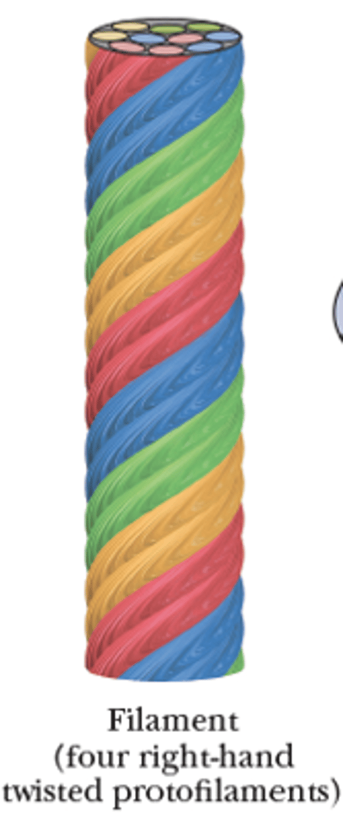
The collagen triple helix is an example of this structure, with all 3 individual collagen chains as helices that differ from the alpha helix
Fibrous Proteins
Contain polypeptide chains organized approximately parallel along a single axis.
Consists of long fibers or large sheets and are mechanically strong.
Ex: Collagen in connective tissue and blood vessels
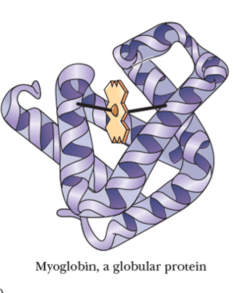
Globular Proteins
Proteins in which the backbone folds on itself to produce a more or less spherical shape.
Has compact structures
More active in transport
Tertiary Structure
3D arrangement of all atoms in a protein molecule, including those in side chains and in prosthetic groups (fully folded 3D shape of protein)
Includes the conformations of side chains and the positions of any prosthetic group
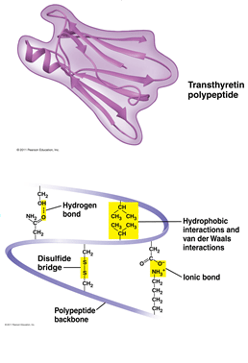
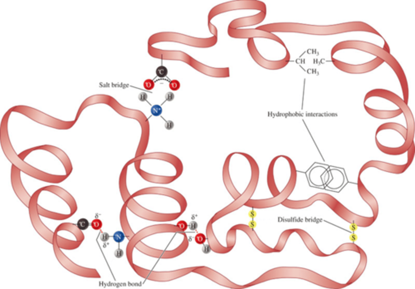
Types of Interactions Maintaining Tertiary Structure (hold it together)
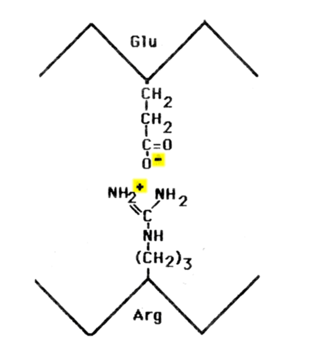
1) Salt Bridges between ionic side chains -COO- and -NH3+
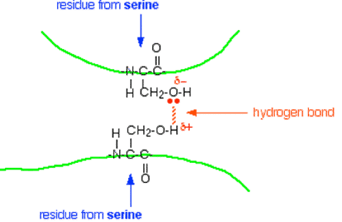
2) Hydrogen Bonds between polar residue side chains
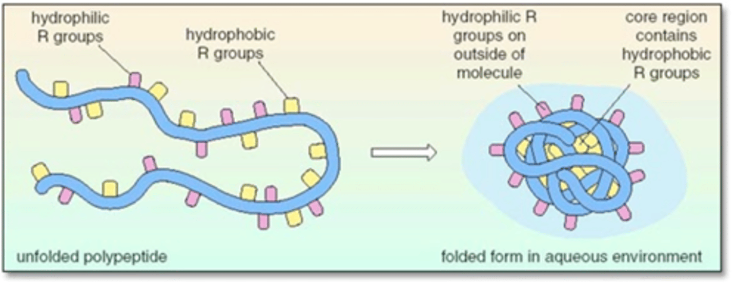
3) Hydrophobic Interactions -> 2 nonpolar (amino acid) groups are attracted by a mutual repulsion of water
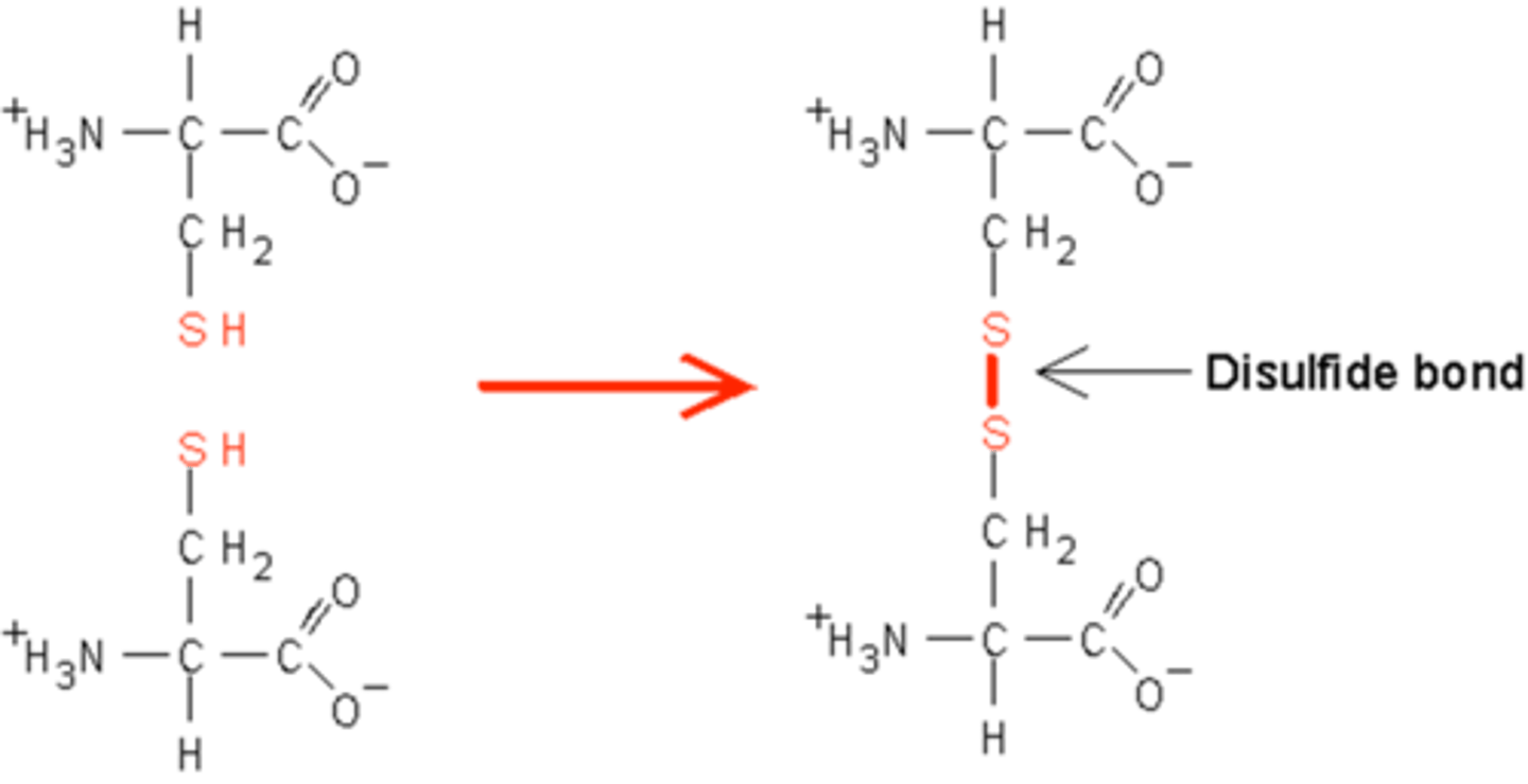
4) Disulfide Bridges between two cysteine residues
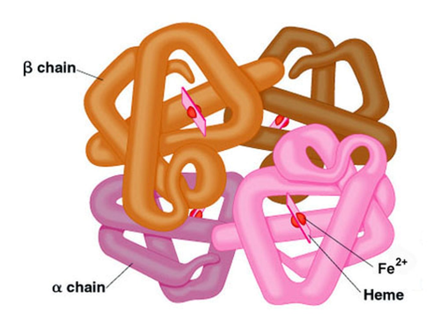
Quaternary Structure
Arrangement of subunits with respect to one another. Consists of more than one polypeptide chain
Subunits = individual parts of a large molecule
Ex: Hemoglobin
Important Interactions that Contribute to Protein Shapes:
1) Hydrogen-bonds along the backbone
Form between H from -NH- groups and O from -C=O groups
Important Interactions that Contribute to Protein Shapes:
2) Hydrogen-bonds of R groups with each other or with backbone
Some side chains can form hydrogen bonds, which helps "connect" different parts of the protein.
Important Interactions that Contribute to Protein Shapes:
3) Ionic Attractions between R groups (Salt Bridges)
Attraction between positive and negative charges on side chains
Important Interactions that Contribute to Protein Shapes:
4) Hydrophobic interactions between R groups
Hydrocarbon side chains are attracted to each other by weak dispersion forces
Creates a water-free "pocket" (like oil on water)
- Side chains go towards the middle to avoid water while hydrophilic parts go out.
Important Interactions that Contribute to Protein Shapes:
5) Covalent Sulfur-Sulfur Bonds (Disulfide Bonds)
Cysteine has a thiol group, -SH, that can react to form a sulfur-sulfur bond (disulfide)
This helps to connect two different protein chains or create a loop within the same chain
Ways to Determine Tertiary Structure
1) X-ray Crystallography -> Uses a perfect crystal in which all individual protein molecules have the same 3D structure and orientation.
- When a pure crystal is exposed to a beam of X rays, a diffraction pattern is produced
2) Nuclear Magnetic Resonance (NMR) Spectroscopy -> Large collection of data points are subjected to a computer analysis.
- Uses protein samples in aqueous solution
Oligomer
Molecule that is made up of a number of smaller subunits
Ex: Dimers (2), Trimers (3), Tetramers (4), etc.
Denaturation
Unraveling of the 3D structure of a macromolecule caused by the breakdown of noncovalent interactions that can result from:
1) Heat (a specific temperature)
2) Large changes in pH
3) Detergents, such as sodium dodecyl sulfate (SDS), which disrupt hydrophobic interactions
4) Urea and guanidine hydrochloride, which disrupt hydrogen bonding
5) Beta-mercaptoethanol (oxidizes/removes)
Overview of Protein Structure and Function (+ Interrelationships)
1) Primary Structure
- Amino acid sequence
- Results from covalent peptide bonds between amino acids
2) Secondary Structure
- Include alpha-helix and beta-sheet
- Hydrogen bonding between amide hydrogens and carbonyl oxygens of the peptide bonds
3) Tertiary Structure
- Overall folding of the entire polypeptide chain
- Interactions between different amino acid side chains
4) Quaternary Structure
- Concerned with topological, spatial arrangement of two or more polypeptide chains
- Involves both disulfide bridges and noncovalent interactions
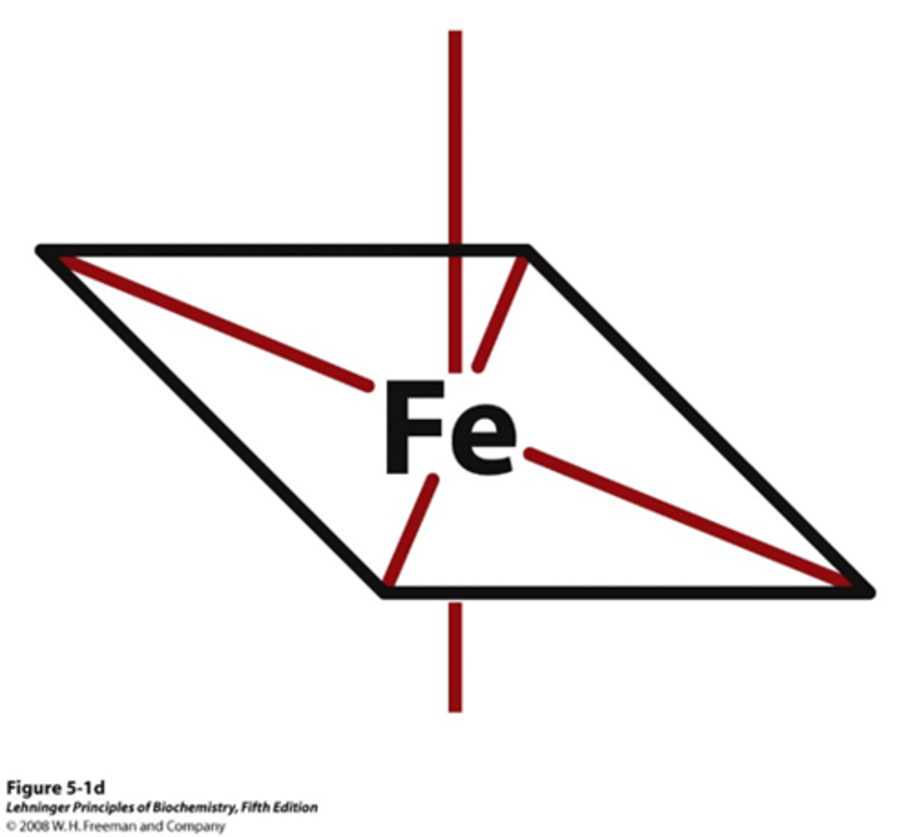
Heme
Consists of a complex organic ring structure, protoporphyrin IX, with a bound iron atom in its ferrous (Fe2+) state.
- The iron concentration within this is able to bond to oxygen by moving Histidine (His) in and out of the plane
Present in hemoglobin and myoglobin
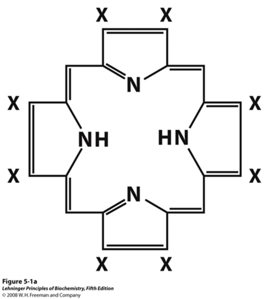
How many coordination bonds are there on the iron in the porphyrin ring system in heme?
6 sites:
- 4 are taken up by heme ring
- 2 are left free to bond to either: 1) a proximal histidine or 2) molecular oxygen
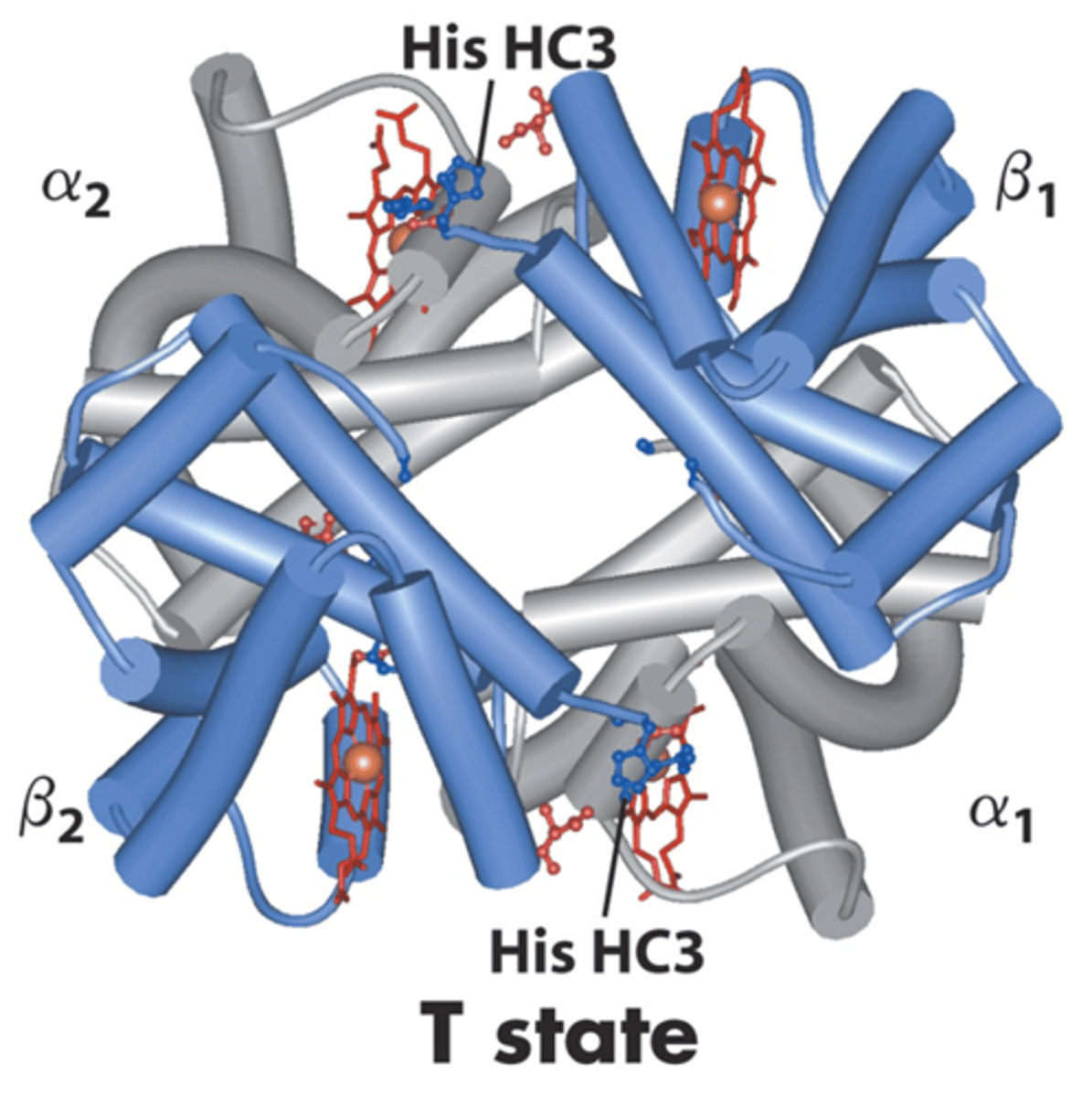
Hemoglobin
A tetramer (4 bonding sites (polypeptides) for oxygen for transport in the body)
Better for transport because of the multiple O2 binding sites
- Binds with high affinity in the lungs (high O2) and low affinity in tissues (low O2)
Myoglobin
A monomer (single bonding site (polypeptide) for oxygen to muscles)
It is insensitive to small changes in dissolved oxygen, so therefore better for storage
Distal Histidine (His)
NOT bonded to iron (only keeps the molecule in place)
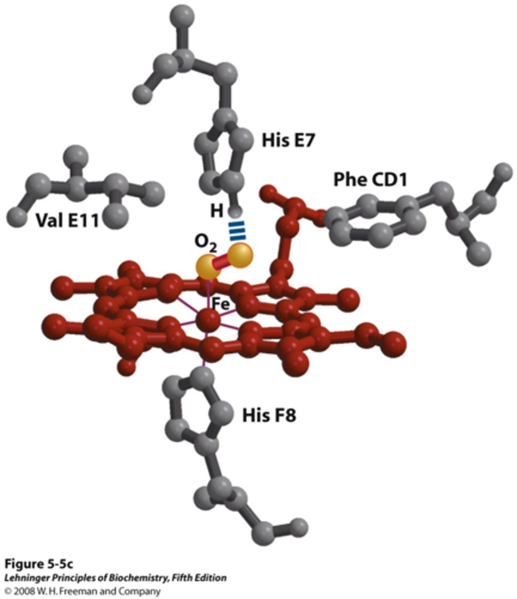
Proximal Histidine (His)
Attached directly to iron

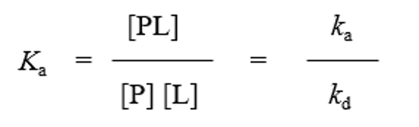
Association Constant (Ka)
A higher value corresponds to a higher affinity of the ligand for the protein

Dissociation Constant (Kd)
Reciprocal of Ka; the equilibrium constant for the release of ligand
A lower value corresponds to stronger binding affinity
Found closer on the Y-axis
What is Kd of the blue triangle protein?
~50

Which protein has greatest affinity for Lo?
Red square protein
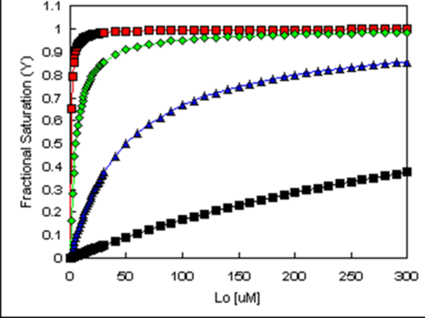
Which protein has least affinity for Lo?
Black square protein

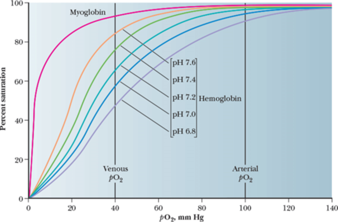
Oxygen to Myoglobin Curve
Hyperbolic
VERY high affinity to oxygen because of the single binding site
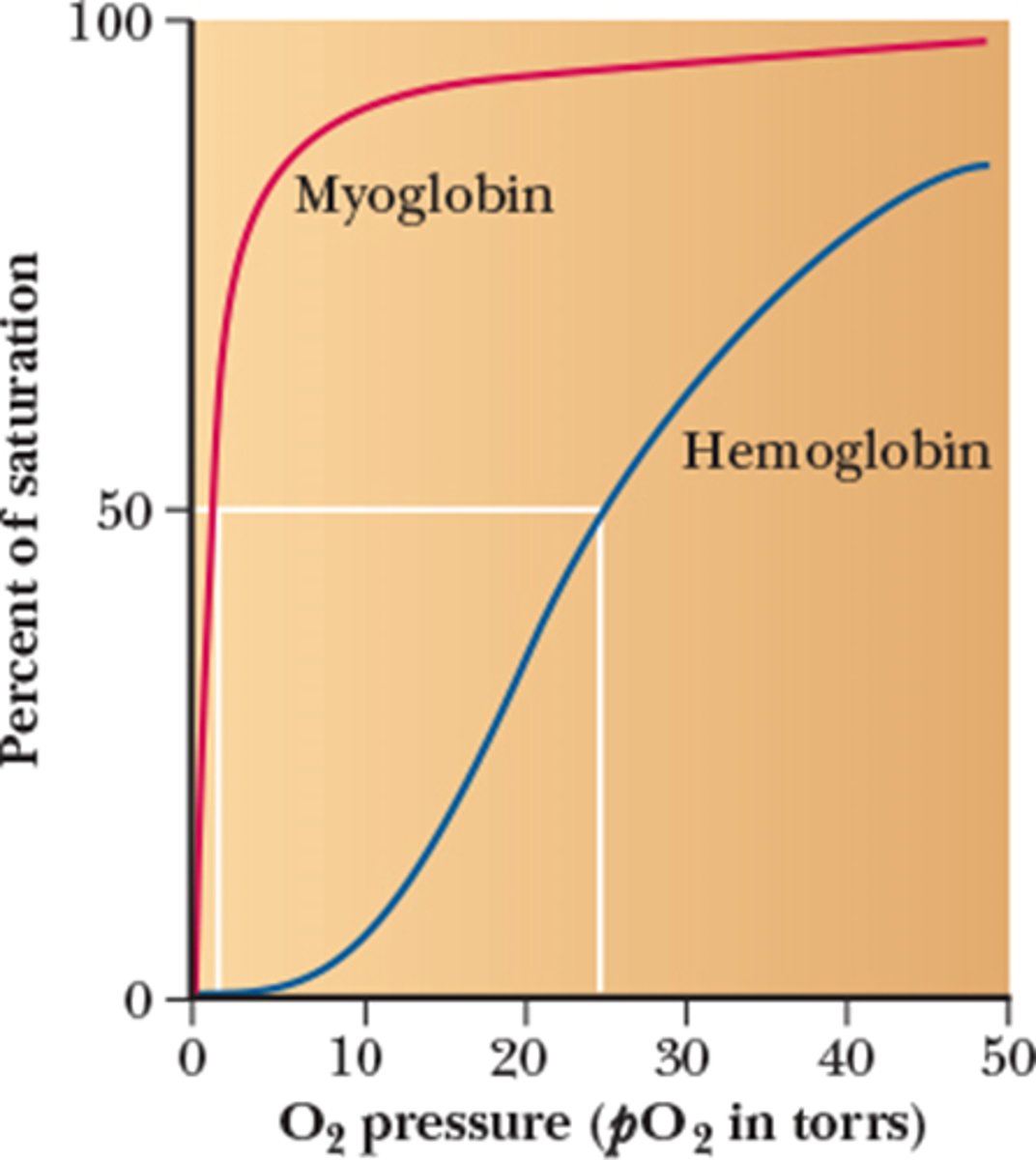
Oxygen to Hemoglobin Curve
Sigmoidal

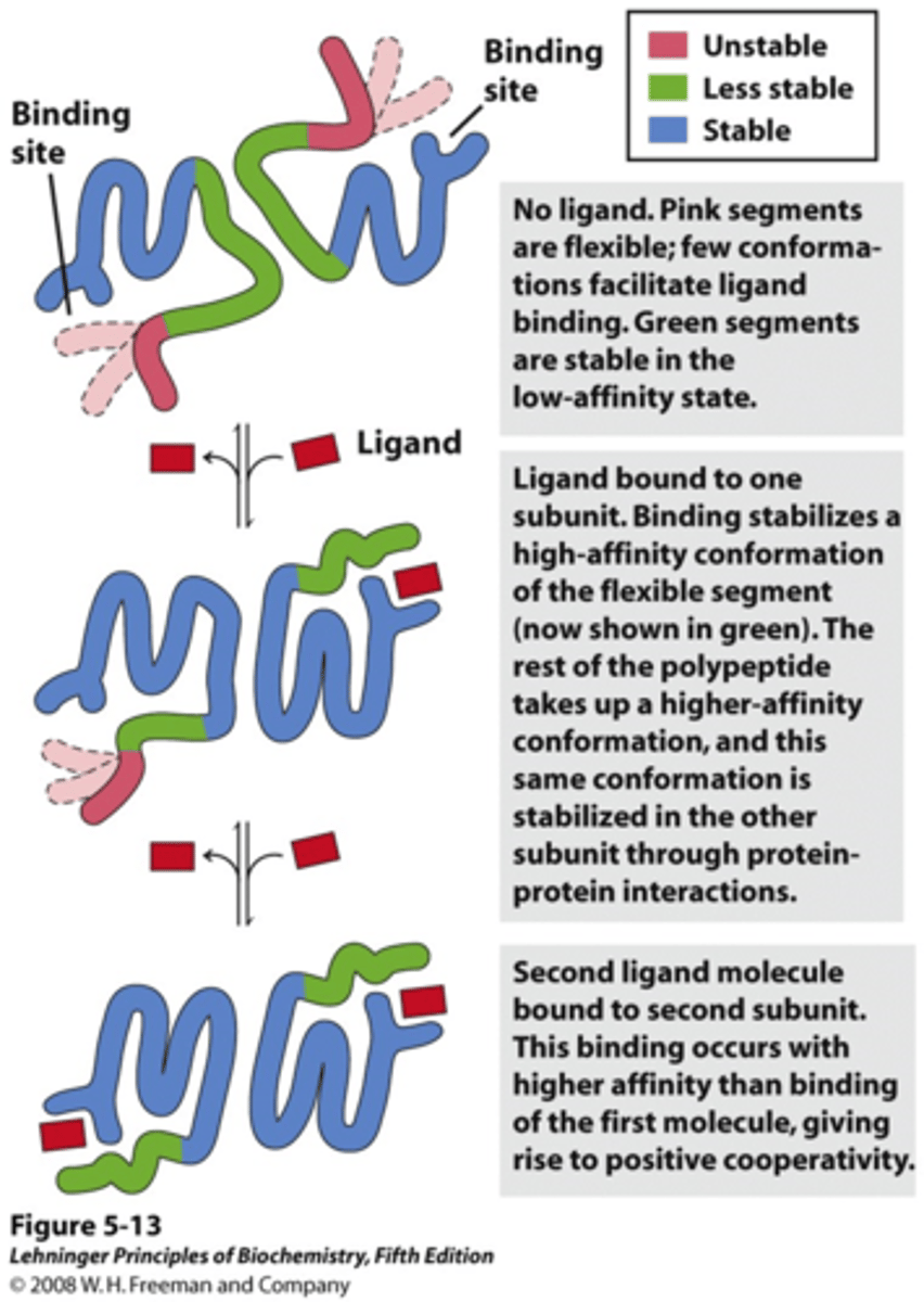
Positive Cooperativity
Binding of the first molecule makes binding of the second easier and so on, so the gradient of the curve steepens.

Myoglobin vs. Hemoglobin Summary
1) Myoglobin
- Function: Oxygen storage
- Requirement: Bind strongly to O2 at very low pressures
- Saturation: 50% at 1 torr partial pressure of O2
2) Hemoglobin
- Function: Oxygen transport
- Requirement: Bind strongly to O2 and release O2 easily
- Saturation: 100% when O2 pressure is 100 torr
Hemoglobin subunits are also structurally similar to myoglobin

Hemoglobin undergoes a transition from...
A low affinity state (T state) to a high affinity state (R state)
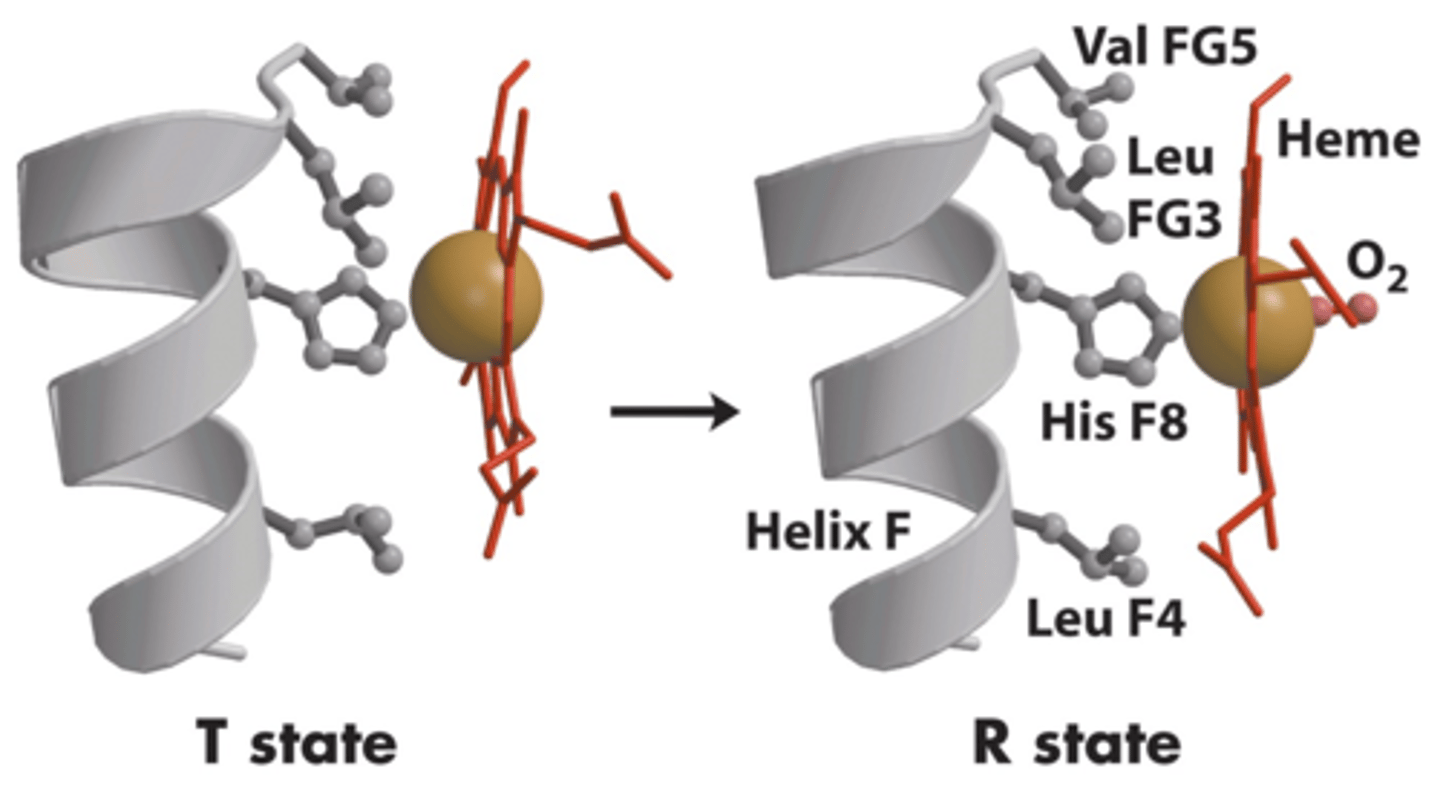
T-state
Tense state = O2 is NOT bound (does NOT accept O2)
Val also blocks O2 binding in this form
R-state
Relaxed state = O2 is bound (accepts O2)
Fe shifts into heme plane due to the movement of histidine, where it can bind to O2 better
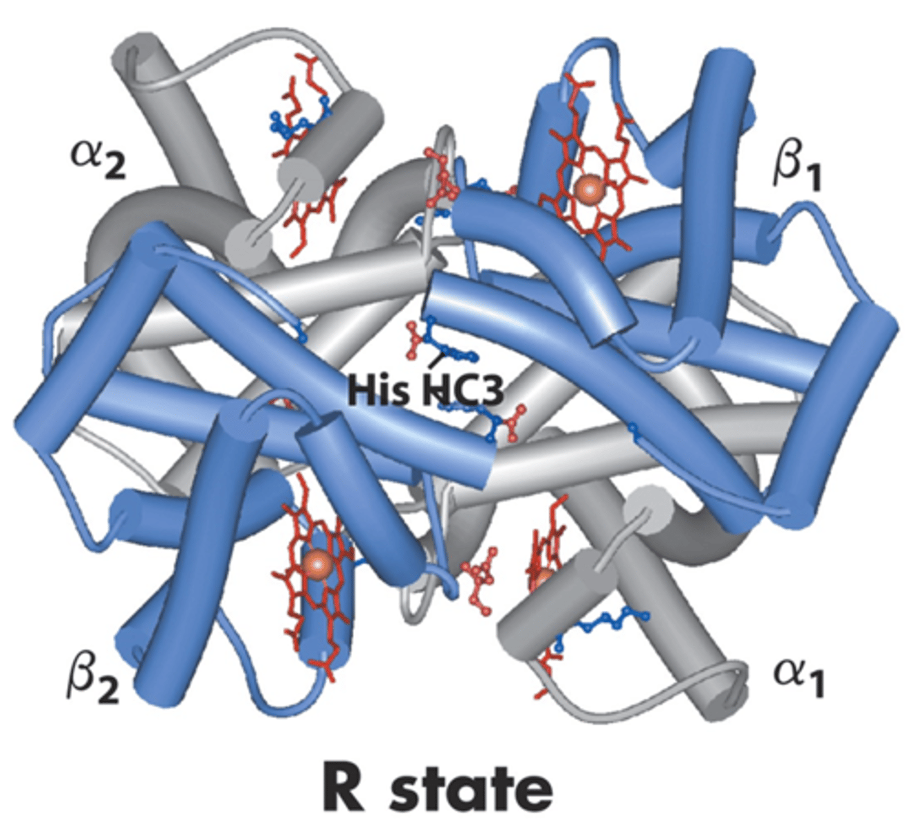
Structural stability is ______________ throughout a protein molecule
NOT uniform
The more bounded, the easier it is to bind (positive cooperativity)
How does pH affect binding affinity of hemoglobin?
Hemoglobin pH decreases over time (more acidic) because the more oxygenated Hb is, the stronger the acid since it has a lower pKa than its deoxygenated form.
- The Bohr Effect -> a decrease in the amount of oxygen associated with hemoglobin and other respiratory compounds in response to a lowered blood pH resulting from an increased concentration of carbon dioxide in the blood.
Myoglobin is NOT affected by pH change because it is only involved with storage, not transport.
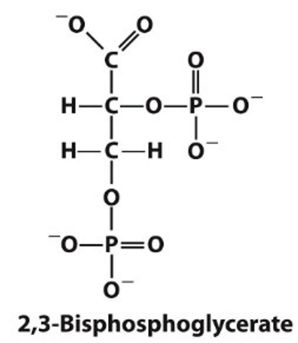
2,3-Biphosphoglycerate (BPG)
Helps regulate oxygen binding to hemoglobin by lowering the affinity
BPG greatly reduces the affinity of hemoglobin for oxygen, which is important in the physiological adaptation to the lower pO2 at high altitudes
- More red blood cells begin to be produced, which allows for more O2 to be collected
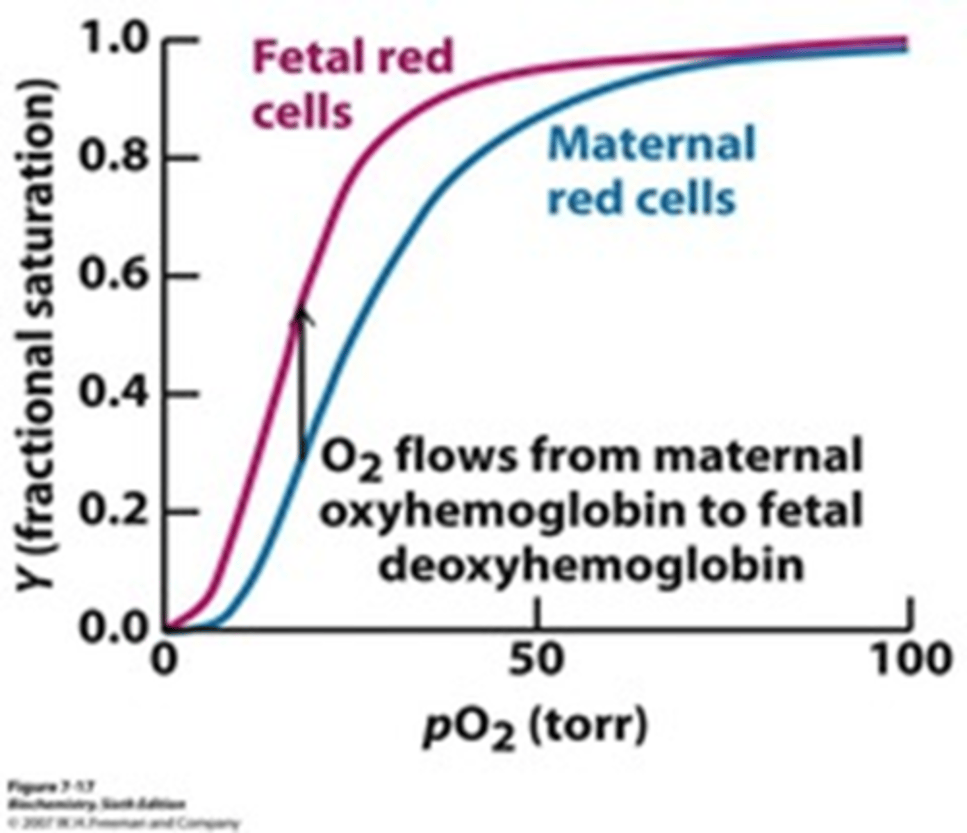
Fetal Hemoglobin
Receives O2 sourced from the mother's hemoglobin
Has HIGHER binding affinity than adult hemoglobin
Has 2 gamma and 2 alpha subunits (adult hemoglobin has 2 beta and 2 alpha subunits)
Exposure to CO of 0.1% for 1 hr leads to CO occupying 50% of heme sites in Hb—frequently fatal
How is it then, that a person whose blood is half-saturated with CO is practically helpless, but a person whose hemoglobin percentage diminished to 50% by anemia may be going about work his/her work as usual?
In anemia, the total hemoglobin is reduced, but the available hemoglobin still binds and releases oxygen normally, allowing partial oxygen delivery.
In CO poisoning, CO binds to hemoglobin with much higher affinity than oxygen, preventing oxygen from binding and impairing release to tissues. This leads to severe hypoxia even when hemoglobin levels appear normal.
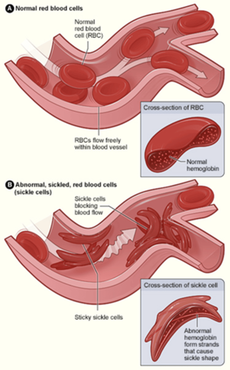
Sickle Cell Anemia
Genetic disease in which red blood cells do NOT bind O2 properly; take on sickle (rather than disc) shape and become trapped in capillaries
Caused by a single amino acid substitution mutation in the hemoglobin gene (Glu (hydrophilic/polar) -> Val (hydrophobic/nonpolar))
- This change causes hemoglobin to aggregate
Leads to a lack of O2
Dangerous/deadly when hemoglobin is deoxygenated