All Smarty PANCE EENT Blueprint lesson flashcard sets combined (Smarty PANCE)
1/498
There's no tags or description
Looks like no tags are added yet.
Name | Mastery | Learn | Test | Matching | Spaced | Call with Kai |
|---|
No analytics yet
Send a link to your students to track their progress
499 Terms
What is conjunctivitis?
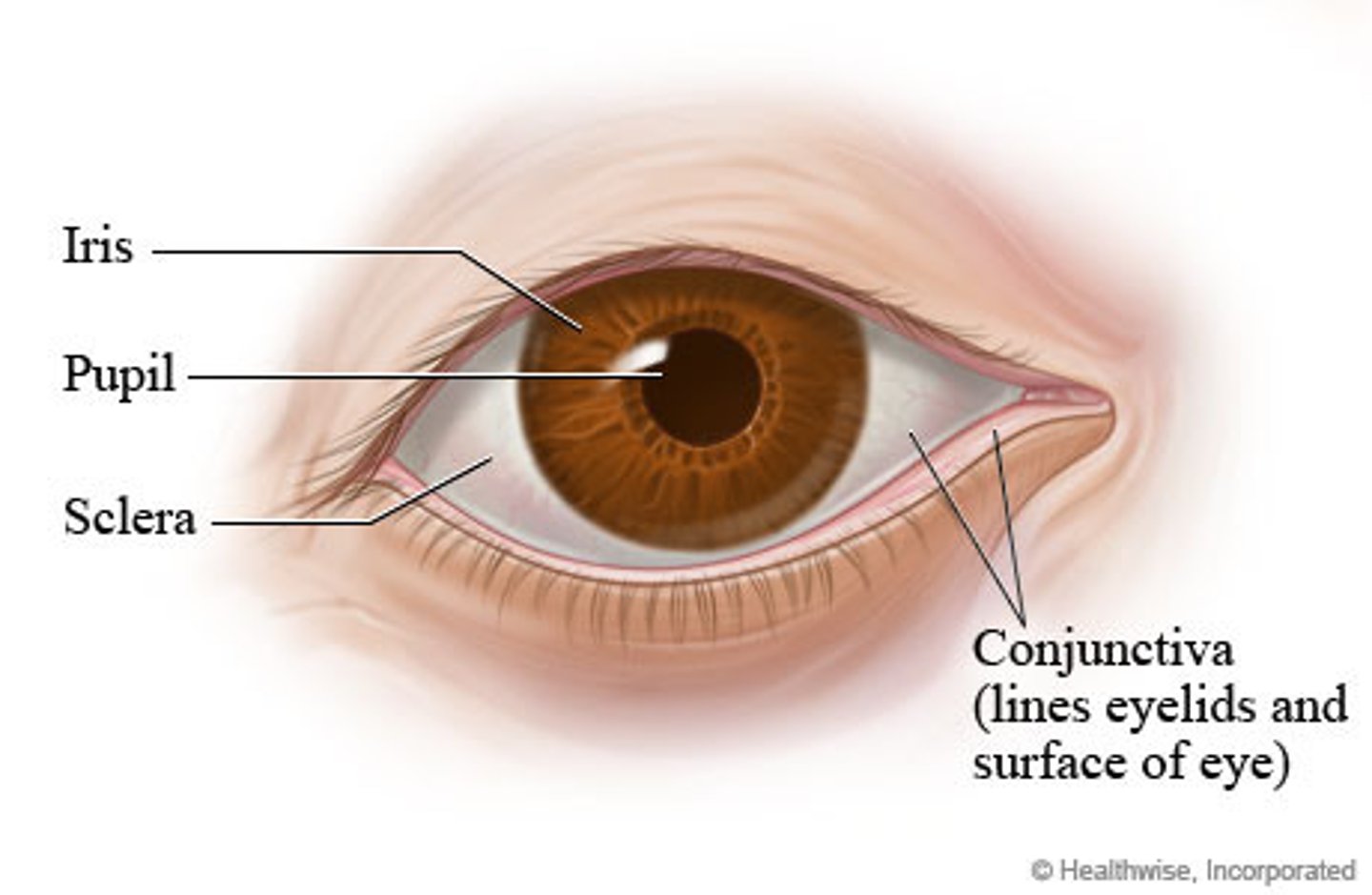
Conjunctivitis is inflammation of the conjunctiva, the outermost layer of the eye
The most common cause of conjunctivitis?
The most common cause is viral infection by adenovirus, but can also be caused by allergens or bacterial infection.
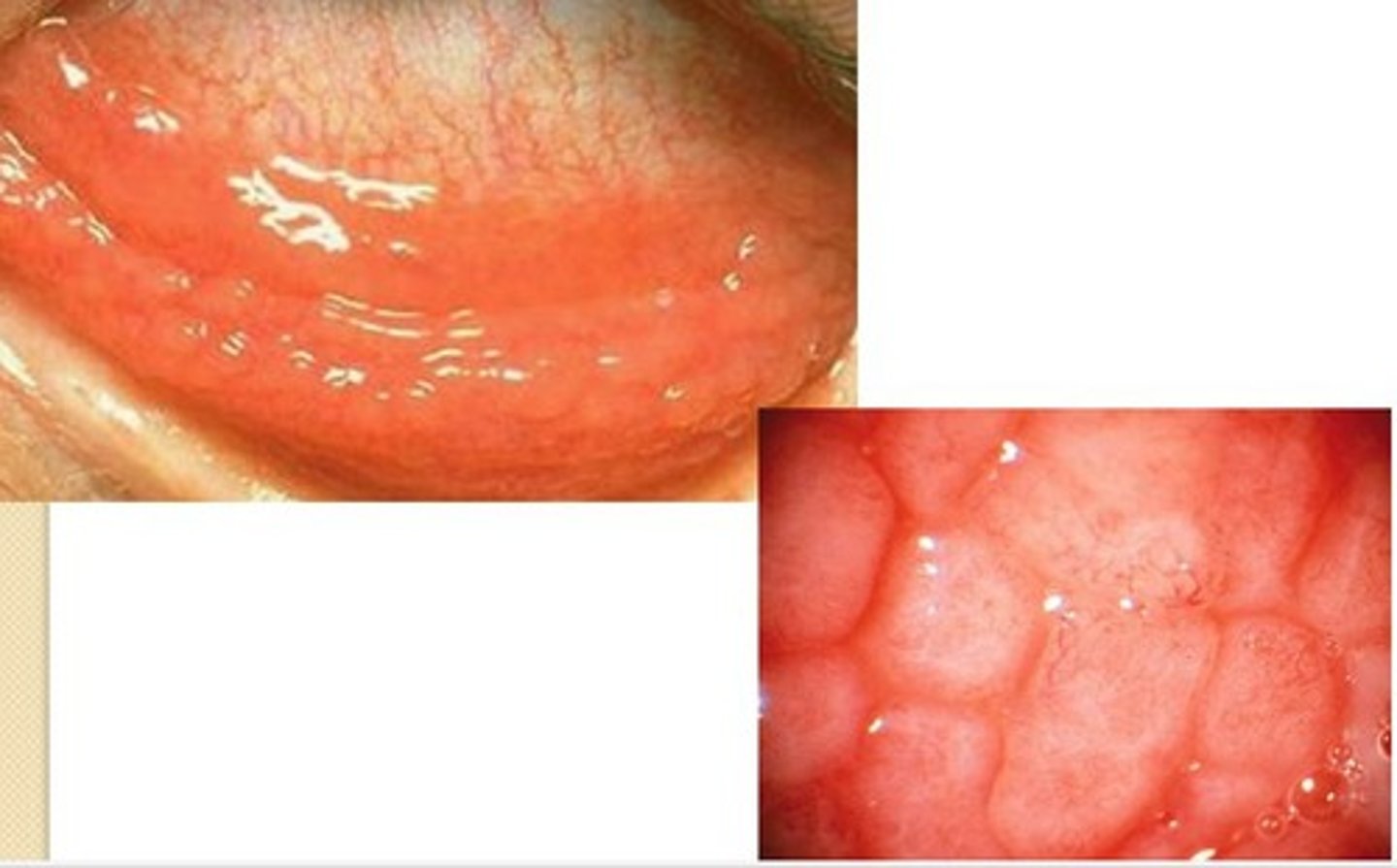
A patient presents with an itching, tearing, right eye. Upon examination large cobblestone papillae are found under the upper lid. What is the probable diagnosis?
Allergic conjunctivitis
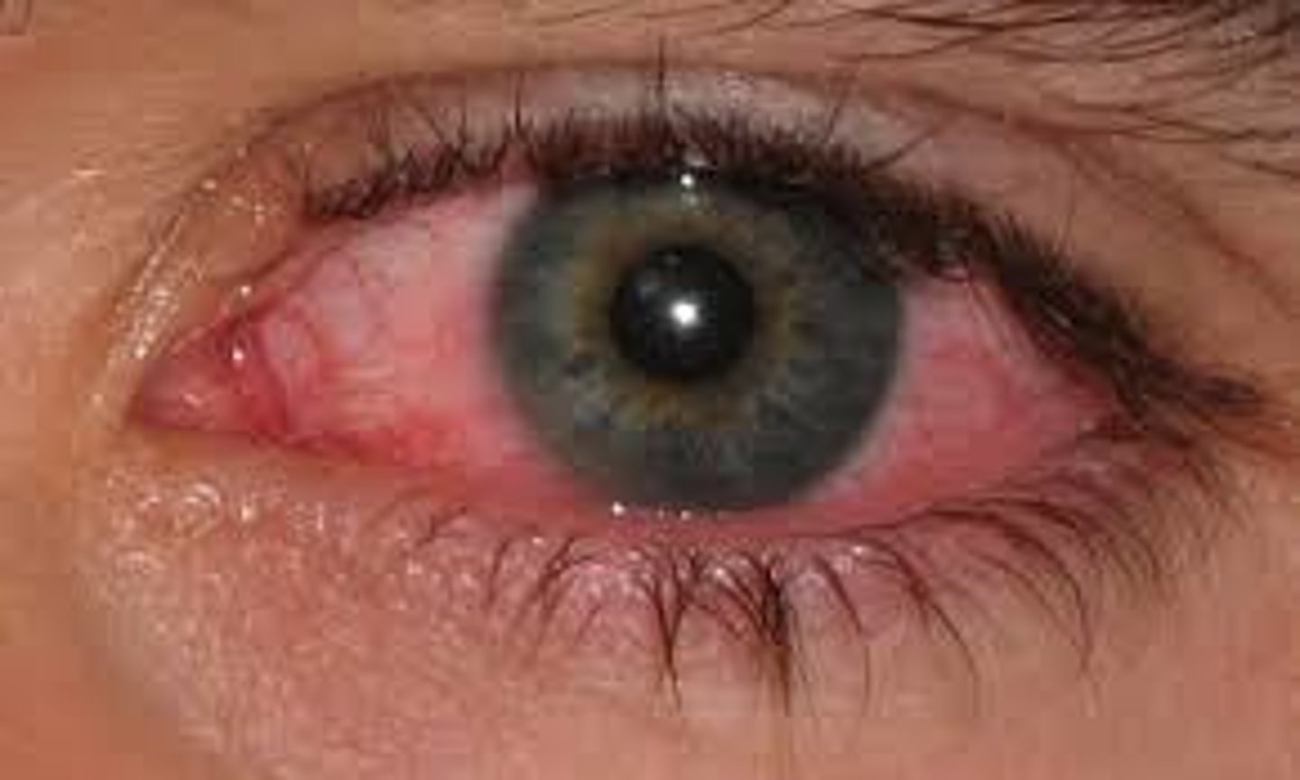
Pt will presents with bilateral preauricular lymphadenopathy, copious watery discharge, scant mucoid discharge. What is the probable diagnosis?
Viral conjunctivitis
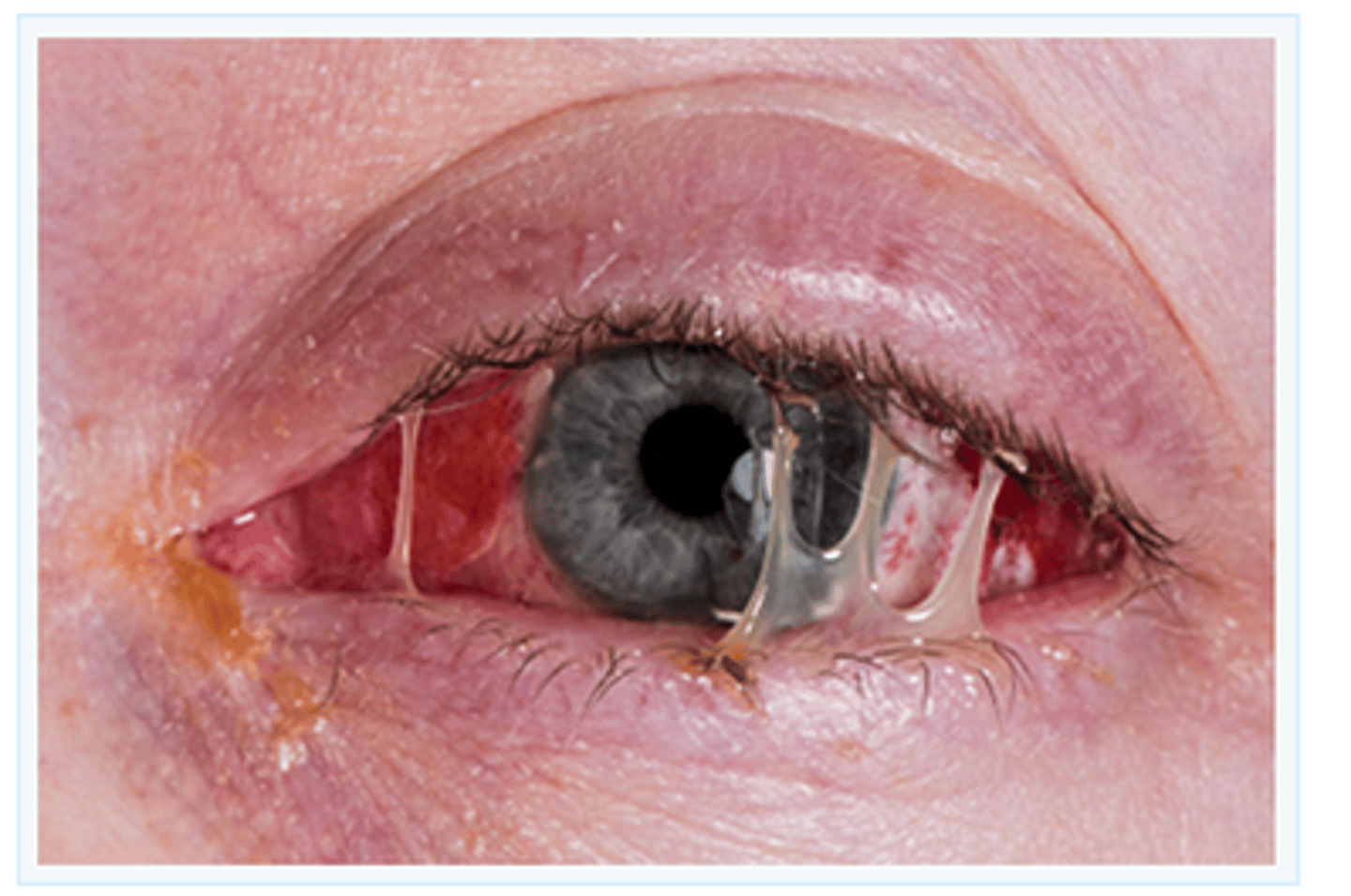
Pt will presents with acute onset of copious purulent discharge from both eyes. Eyes "glued" shut in the AM. There are NO visual changes and no ciliary injection. What is the probable diagnosis?
Bacterial conjunctivitis
What organisms are typically responsible for causing bacterial conjunctivitis?
Staphylococcus aureus, Streptococcus pneumoniae, Haemophilus influenzae.
Treatment of bacterial conjunctivitis?
Topical antibiotics: erythromycin, FQs, sulfonamides, aminoglycosides
Treatment for contact lens wearer?
Contact lenses use = pseudomonas. Treat with fluoroquinolone (ciprofloxacin/Ciloxan drops)
Treatment of viral conjunctivitis?
Viral conjunctivitis is self limiting. Treatment includes cool compresses, artificial tears, olopatadine (Patanol) for itchiness
Treatment of allergic conjunctivitis?
Systemic antihistamines and topical antihistamines or mast cell stabilizers. (Naphcon-A, Ocuhist, generics)
What condition is a seronegative arthritis that is often seen in combination with urethritis and conjunctivitis?
Reactive arthritis (post-infectious arthritis with urethritis and conjunctivitis was formerly known as Reiter syndrome). This condition is often precipitated by a sexually transmitted disease (often linked to Chlamydia trachomatis) or gastroenteritis and may present with lesions in multiple locations (mouth, penis, extremities), swollen toes and heel pain.
What is the most common type of gonorrhea infection that can be seen in newborns?
Conjunctivitis (purulent)
What is the treatment of neonatal conjunctivitis caused by Chlamydia trachomatis?
Oral erythromycin
What is a cataract?
A cataract is a clouding of the lens in the eye leading to a decrease in vision. It can affect one or both eyes.
Presentation of cataracts?
Cataracts are painless and often it develops slowly. Symptoms may include faded colors, blurry vision, halos around light, trouble with bright lights, and trouble seeing at night. This may result in trouble driving, reading, or recognizing faces
What are the risk factors for developing cataracts?
Risk factors for cataracts include age (usually > 60), smoking, ETOH, sunlight exposure, diabetes, metabolic syndrome, prolonged drug use (esp. glucocorticoids), radiation
What are the five most common causes of visual impairment in the elderly?
Presbyopia (farsightedness caused by loss of elasticity of the lens of the eye, occurring typically in middle and old age), cataracts, age-related macular degeneration (AMD), diabetic retinopathy, and glaucoma.
What are the leading causes of blindness in elderly Caucasians? How about elderly African Americans? And the overall leading cause of blindness worldwide?
● Age-related macular degeneration is the leading cause of blindness in elderly Caucasians.
● Glaucoma is the leading cause of blindness in elderly African Americans
● The overall leading cause of blindness worldwide is cataracts.
Diagnosis of cataracts?
● Visual acuity is tested
● Ophthalmoscope
● Slit-lamp microscope
*Cataracts typically have one of three components: nuclear sclerosis, cortical spoking, and posterior subcapsular haze
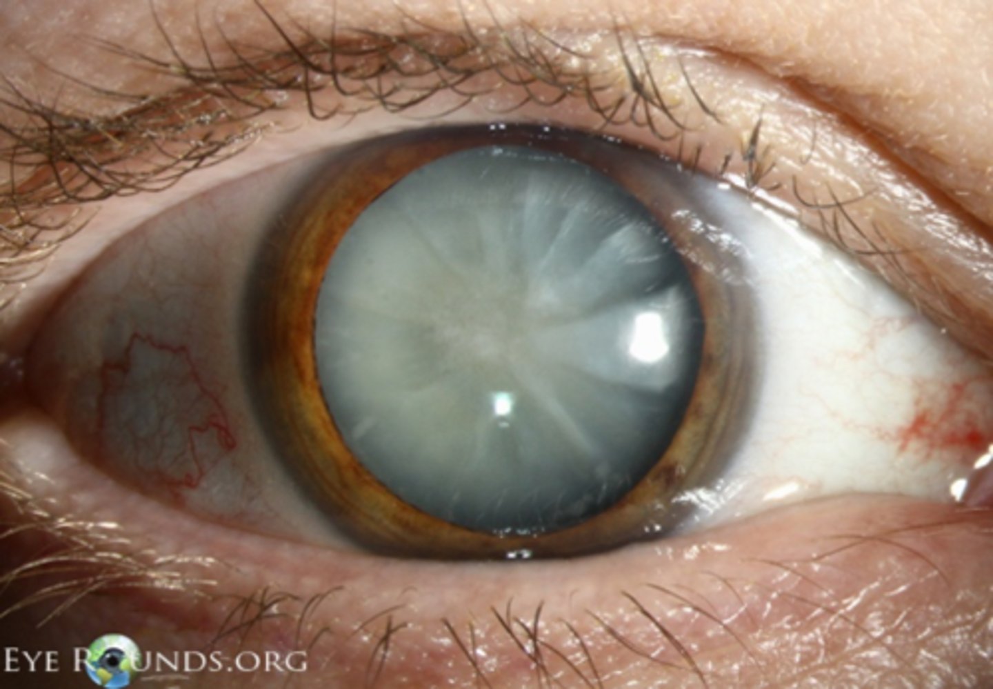
Nuclear Sclerosis (yellowing)?
Opaque nucleus, manifested as a yellow-brown hazy structure at the center of the lens on slit lamp examination
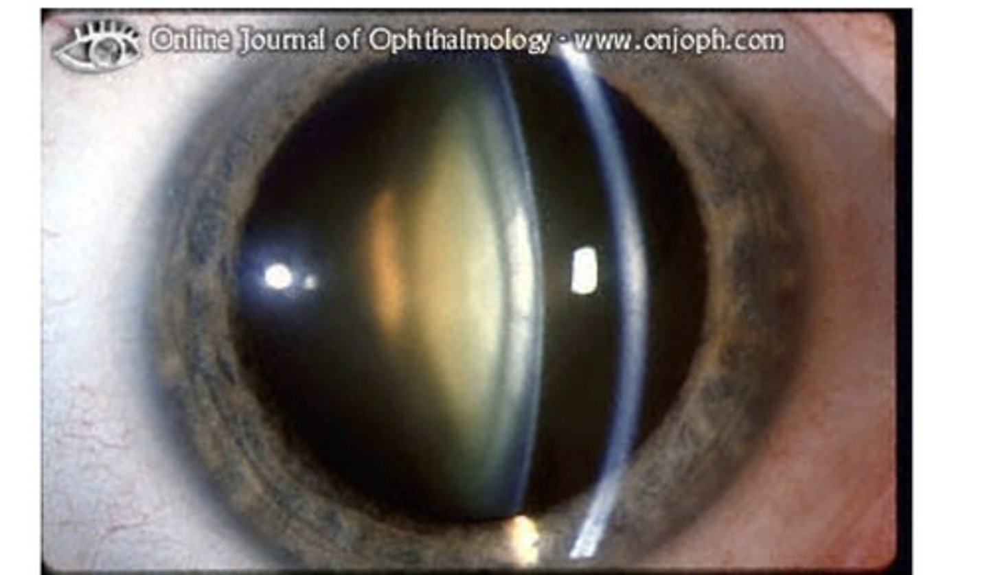
What is cortical spoking (CS)?
Cortical spoking cataract (CS) – Swelling of the cortex causing spoke/wedge-like peripheral cloudiness. Seen on slit lamp examination
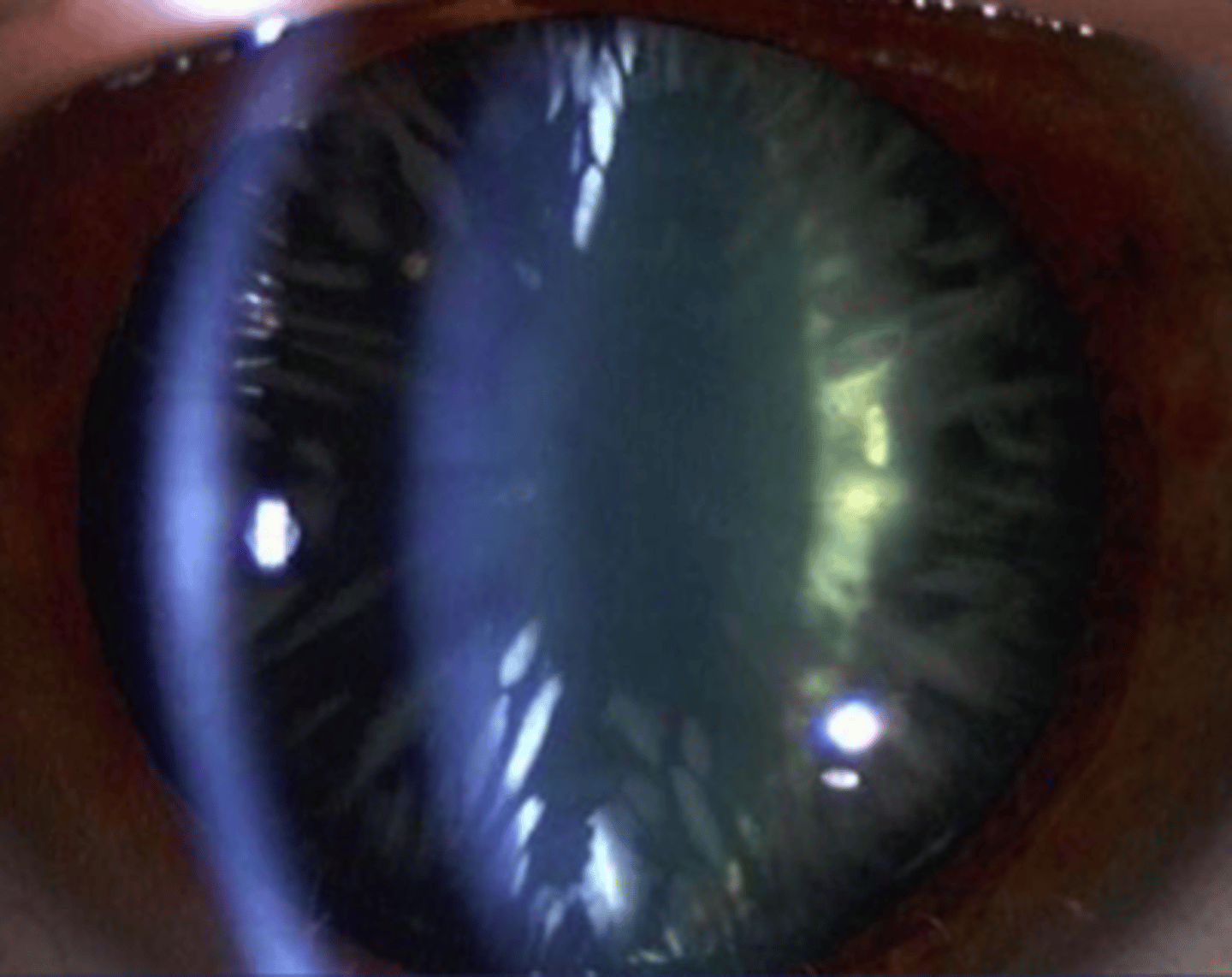
Posterior subcapsular haze?
A posterior subcapsular cataract reveals a "frost-like" haze just anterior to the posterior lens capsule which is the back surface of the lens on slit lamp examination
Name and describe the treatment of choice for functional vision impairment caused by cataracts.
Phacoemulsification where the cloudy lens is emulsified with an ultrasonic handpiece and aspirated from the eye. An artificial intraocular lens is then implanted into the space that used to contain the natural lens.
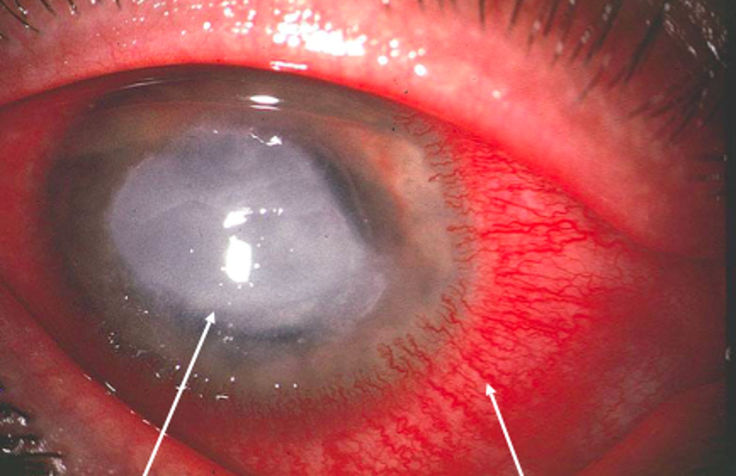
What is a corneal ulcer?
A corneal ulcer is an eye infection that causes an open sore on the cornea (the clear layer in front of the iris and pupil).
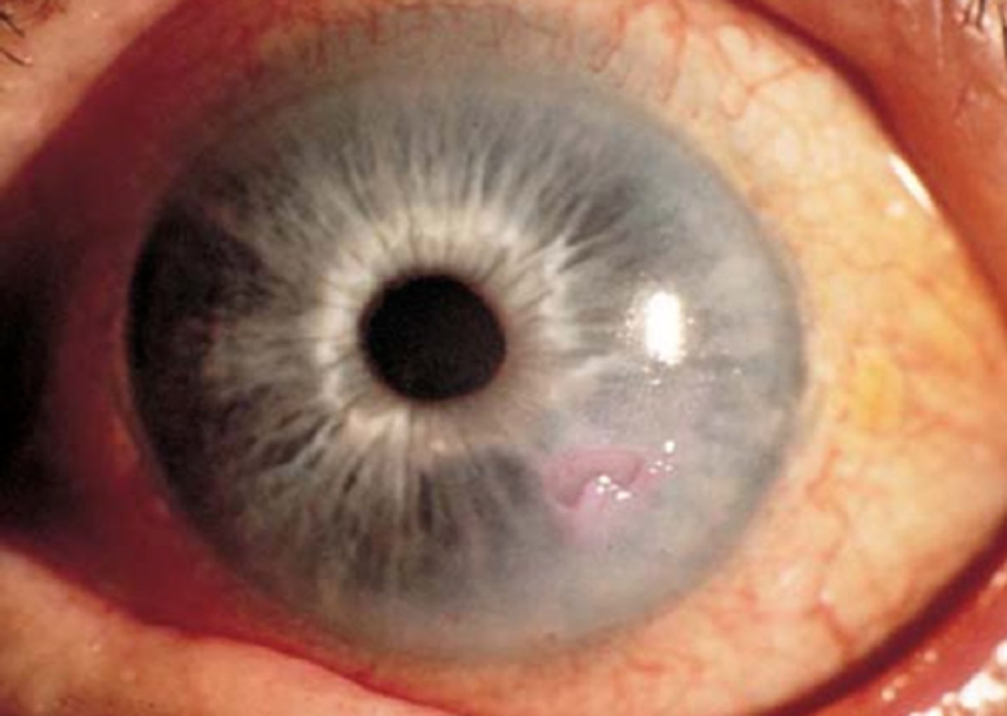
What causes a corneal ulcer?
Corneal ulcers can occur from direct injury to the eye, and usually represent an infection deeper in the cornea from a bacterial, fungal, or viral infection.
What are the symptoms of corneal ulcer?
Symptoms of a corneal ulcer are usually obvious, especially if the ulcer is deep. Because the cornea is very sensitive, corneal ulcers tend to produce severe pain. Vision is sometimes impaired, and the eye may be tearing and red. It may also hurt to look at bright lights.
How is a corneal ulcer diagnosed?
Fluorescein stain is diagnostic (ulcers will often appear round "ulcerated"- like an "ulcer" vs. dendritic like herpes). Corneal cultures should be obtained before starting antibiotics.
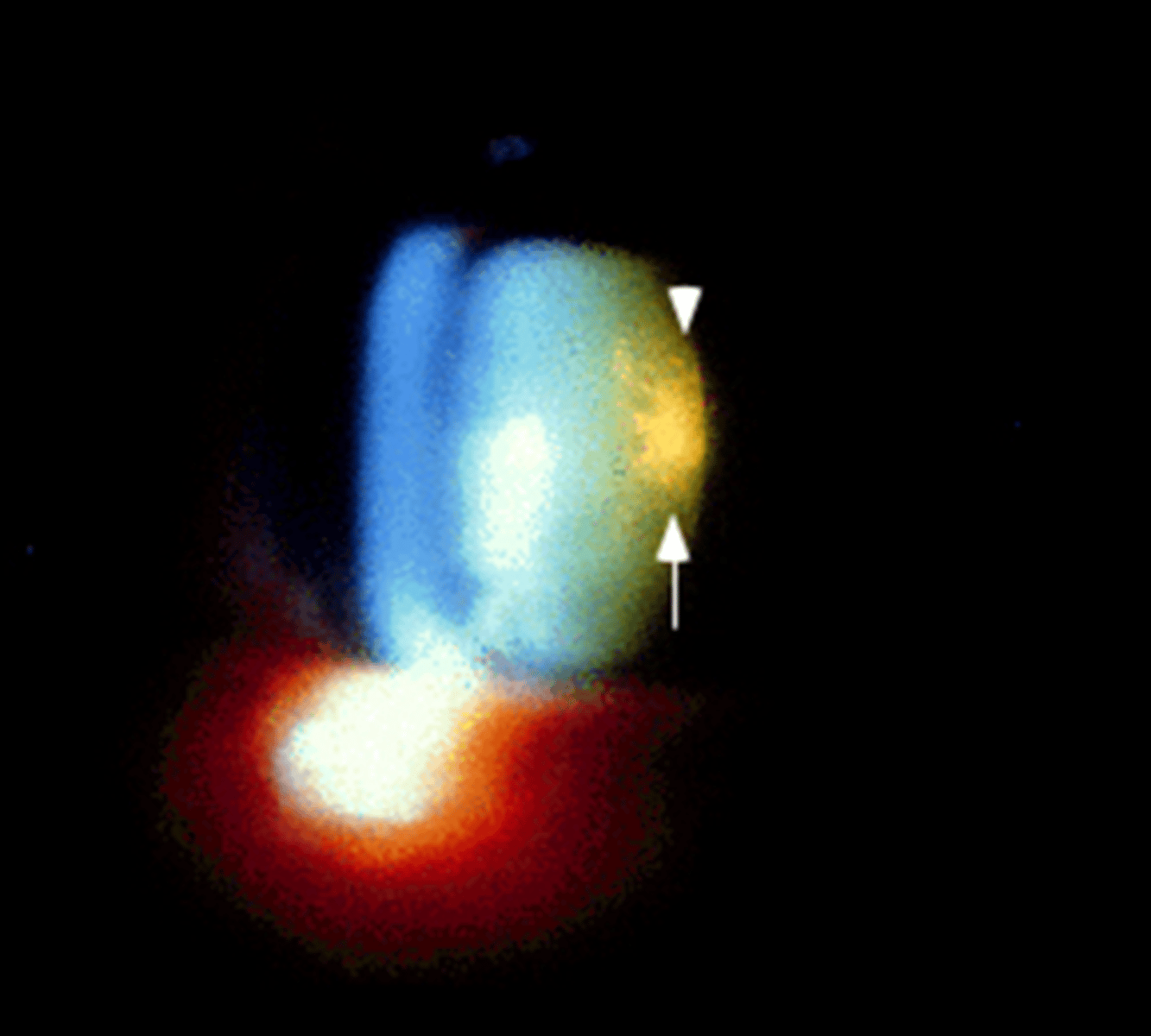
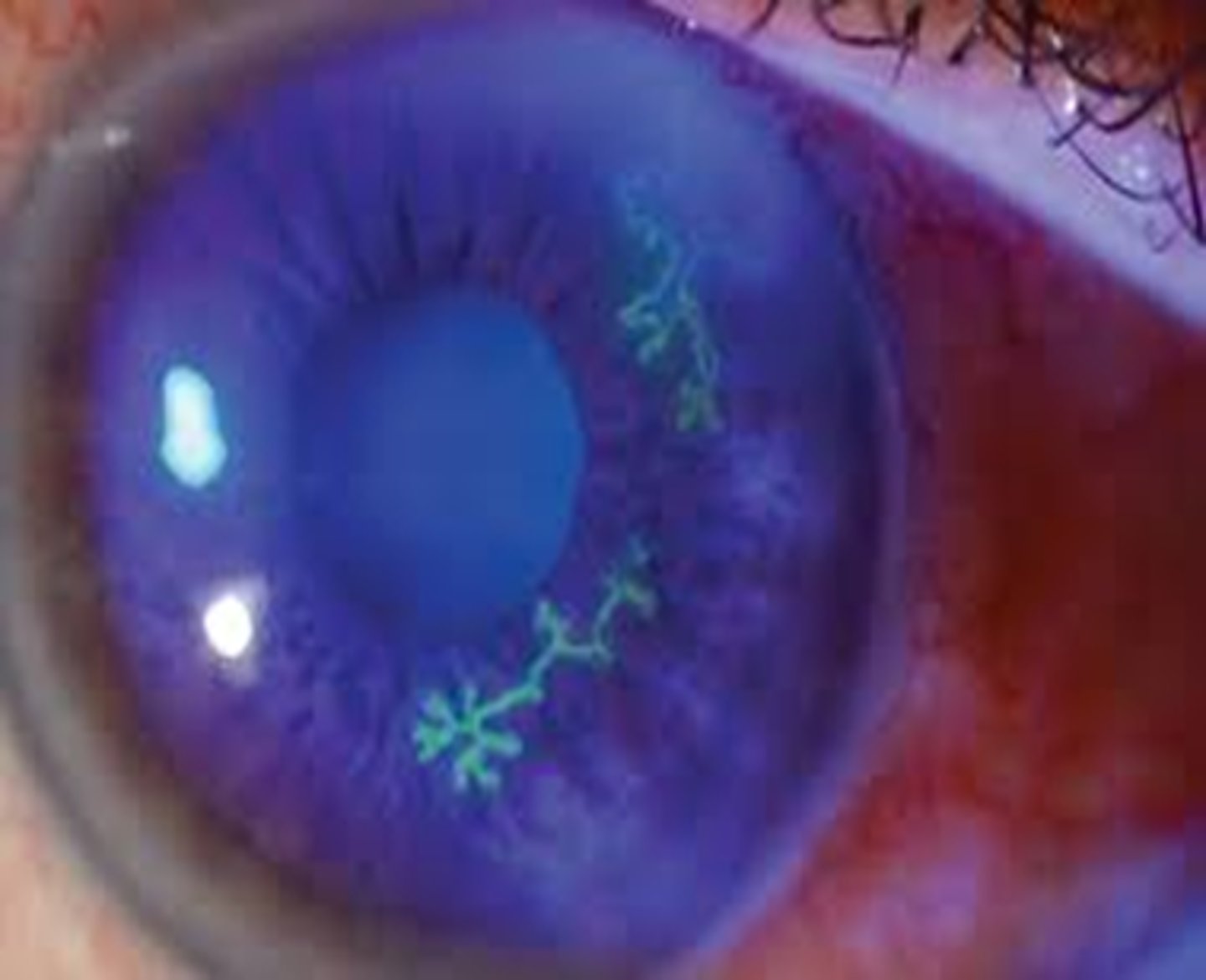
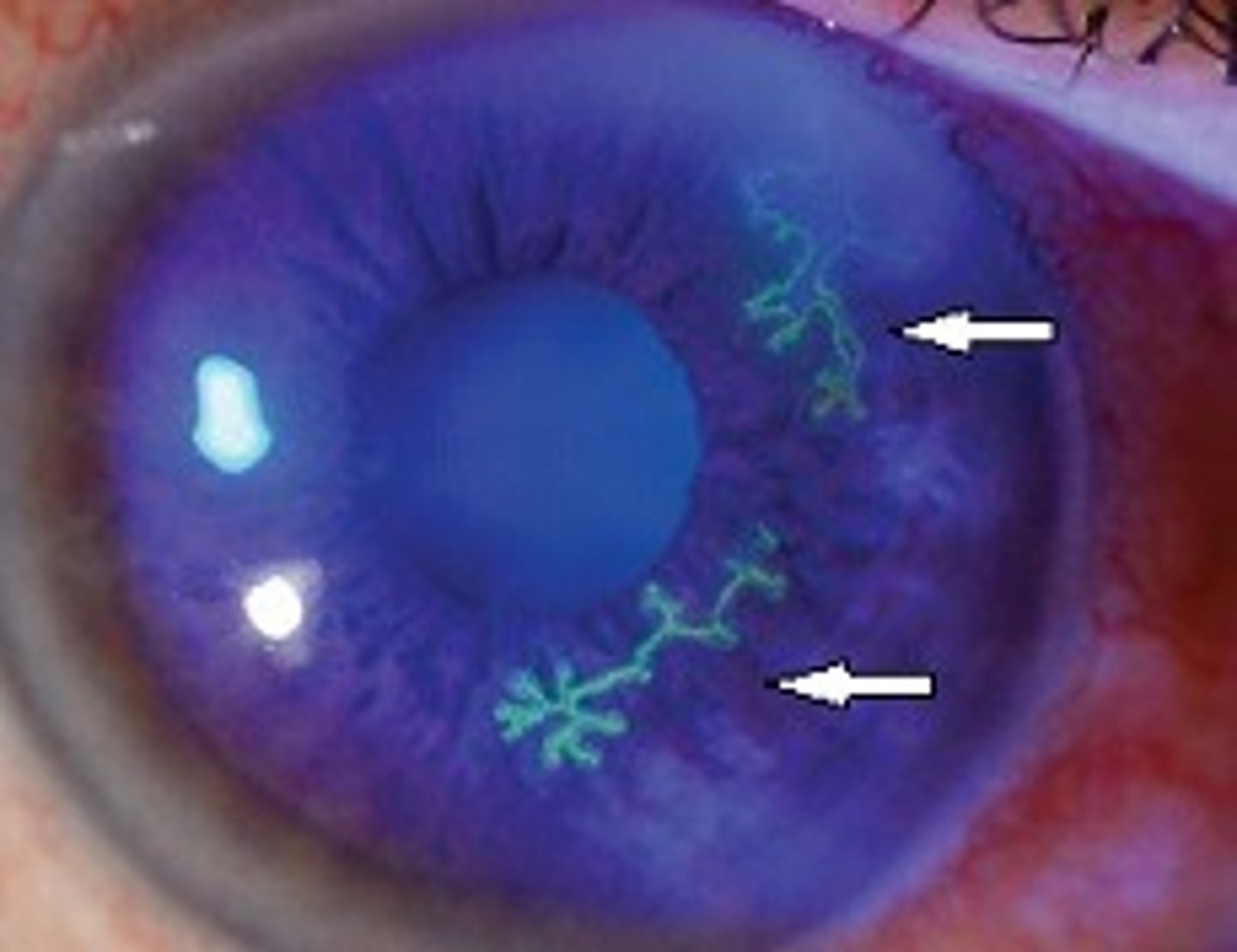
Dendritic ulcer with fluorescein stain?
Herpes Simplex Keratitis
Is a corneal ulcer an emergency?
A corneal ulcer is a medical emergency! All patients with corneal ulceration should be referred immediately to an ophthalmologist
Who is at greatest risk for development of a corneal ulcer?
Risk factor for contact lens wearers
Treatment of corneal ulcer?
Immediate referral - if immediate referral is not possible, it is reasonable to start antibiotics without delay. Appropriate ophthalmic antibiotics include ciprofloxacin 0.3%, ofloxacin 0.3%, gentamicin 0.3%, erythromycin 0.5%, polymyxin B/trimethoprim (Polytrim), and tobramycin 0.3%.
How long does it take for a corneal ulcer to heal?
Most appropriately treated corneal ulcers should improve within two to three weeks. Treatment may continue for longer to reduce the amount of potential scarring. Corneal ulceration is a serious condition, and with inadequate or no treatment, loss of vision and blindness may occur.
What is keratitis?
Inflammation of the clear tissue on the front of the eye (cornea)
What causes keratitis?
● Noninfectious keratitis can be caused by a minor injury, by wearing contact lenses too long, or by a foreign body in the eye.
● Infectious keratitis can be caused by bacteria, viruses, fungi, and parasites
Symptoms of keratitis?
Eye redness, pain, and blurred or decreased vision are common symptoms
Risk factors for keratitis?
Risks for keratitis include:
● Wearing contact lenses — especially sleeping in the lenses —increases the risk of both infectious and noninfectious keratitis
● Reduced Immunity
● Use of corticosteroid eye drops to treat an eye disorder
● Eye injury - If one of your corneas has been damaged from an injury in the past, you may be more vulnerable to developing keratitis.
What diagnostic finding is associated with bacterial keratitis?
The diagnostic finding in bacterial keratitis is a corneal opacity or infiltrate (typically a round white spot) in association with red-eye, photophobia, and foreign body sensation.
What diagnostic finding is associated with epithelial herpes keratitis?
Epithelial herpes keratitis is characterized by the presence of dendritic lesions
Treatment of keratitis?
Prompt ophthalmology consultation is needed to avoid loss of vision. Treatment includes medications such as antibiotics. In rare cases, antifungal drugs may be used.
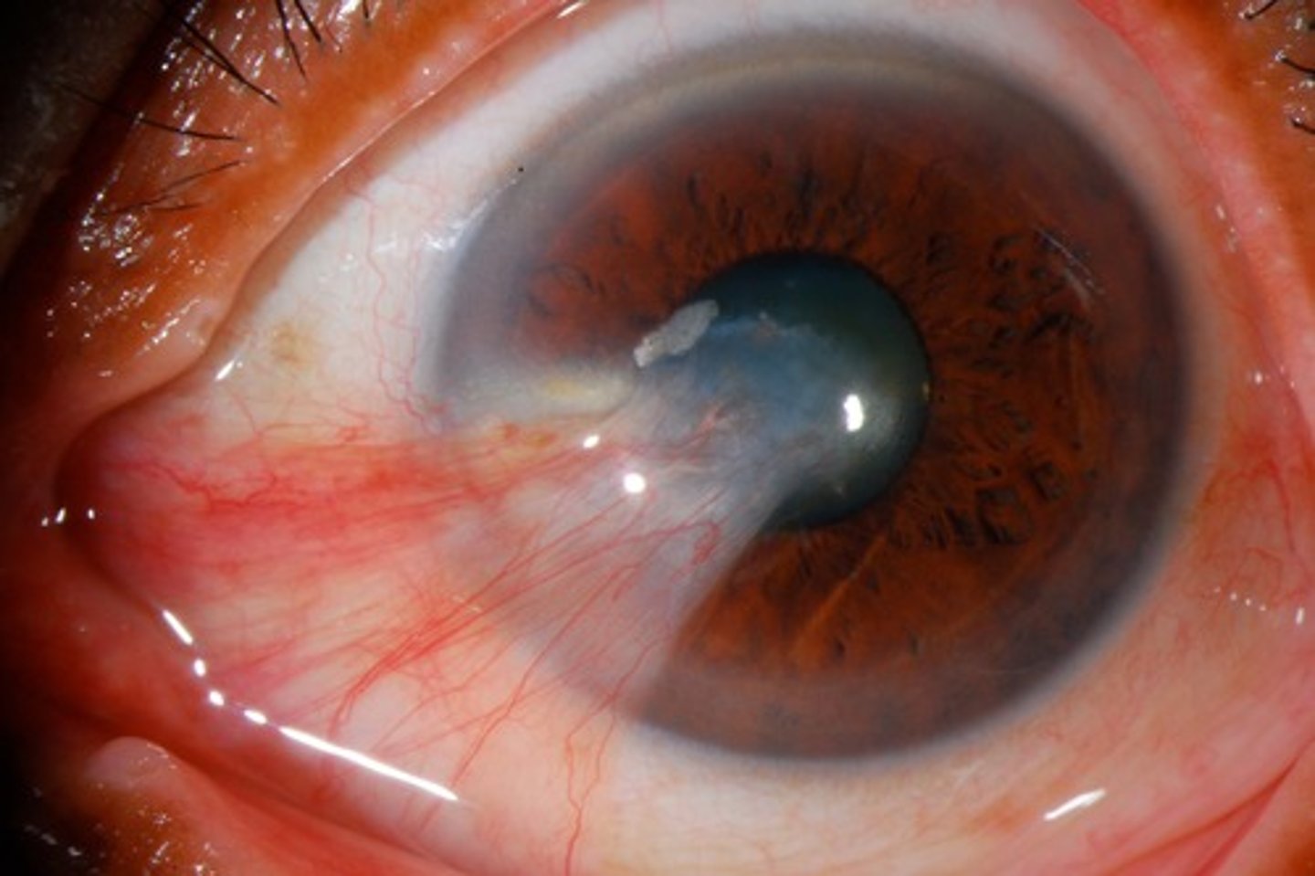
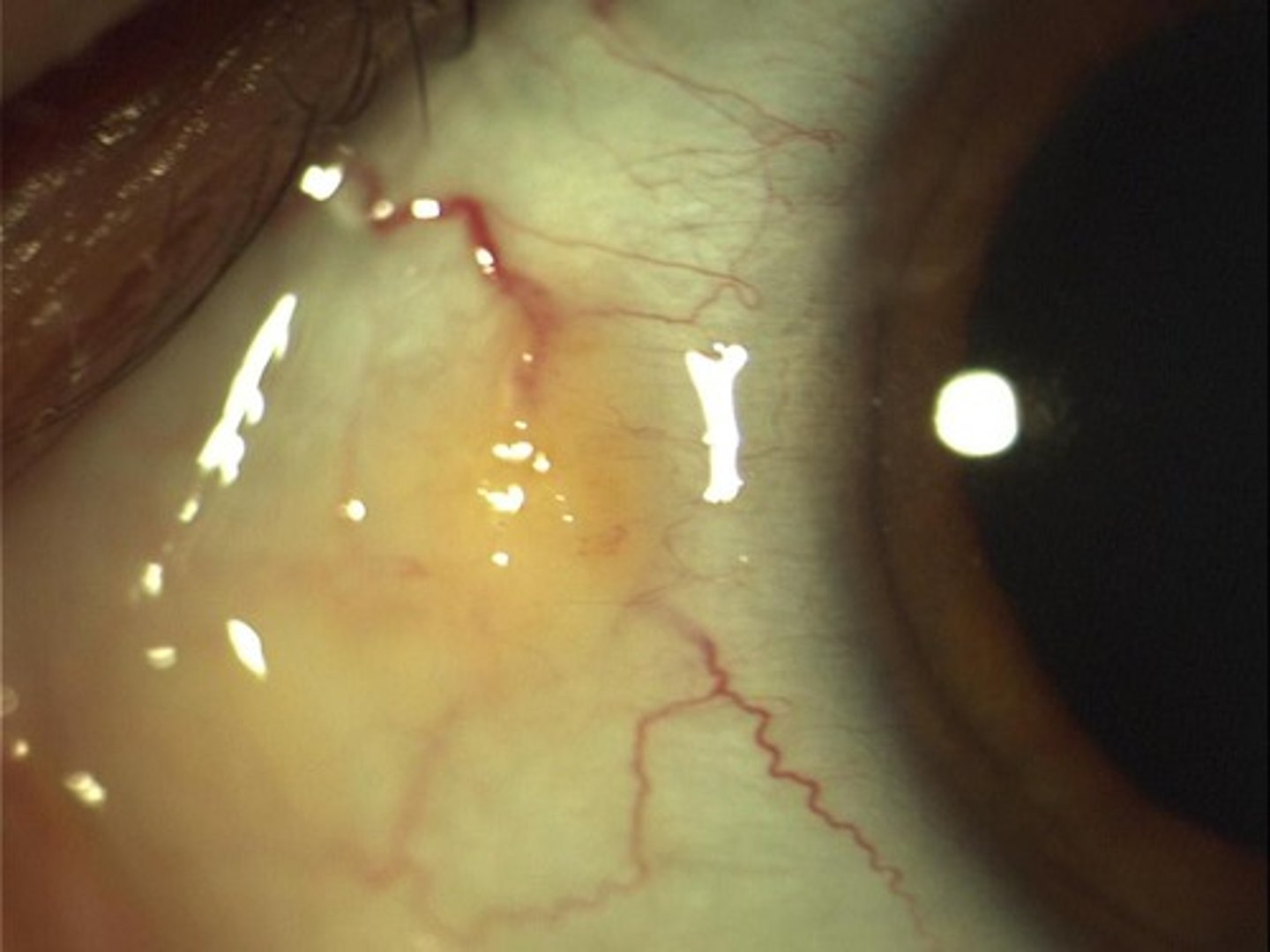
What is a pterygium?
A pterygium is a chronic growth over the medial or lateral aspect of the cornea approaching the pupil. "Pterygium" refers to the shape of the tissue, which looks like an insect wing.
What is a pinguecula?
A pinguecula is a yellowish nodule, particularly on the nasal aspect of the eye, but it may be lateral. It is often caused by wind and dust.
Pterygium vs pinguecula?
Pinguecula is a yellow, elevated nodule → DOES NOT GROW
Risk factors for pterygium?
Pterygium is associated with increased sun (UV) exposure and climates where there is wind, sand, and dust
What is the natural history of growth and progression of a pterygium?
Pterygium oscillate between active and inactive. Pterygium, when active, can grow over a period of several months to years. Activity is marked clinically by redness and localized thickening, which probably represent active inflammation. When inactive (white and flat), pterygium may remain static for decades with no measurable increase in size or clinical significance. It is unclear how pterygium converts from active to inactive, or if it can be reactivated.
What are the symptoms of a pterygium?
The most common symptoms caused by pterygium are redness and irritation. Visual impairment is less common
● In the absence of symptoms, patients may also report a change in the appearance of their eye, or pterygium may be noted incidentally on physical examination.
How is pterygium treated?
Patients with a small pterygium can be treated symptomatically for redness and irritation with artificial tears or other ocular lubricants
When is surgery indicated in the treatment of pterygium?
Surgery for pterygium is indicated in the following situations:
● Induced astigmatism that causes visual impairment
● Opacity in the visual axis
● Documented growth that is threatening to affect the visual axis via astigmatism or opacity
● Restriction of eye movement
● Significant cosmetic impact or intractable irritation
Prevention and prevention of recurrence of pterygium?
Exposure to ultraviolet (UV) light is an important risk for recurrence - Lubrication and protection with a hat and/or UV-blocking spectacles that fit closely, wrap around, or have side shields can prevent recurrence
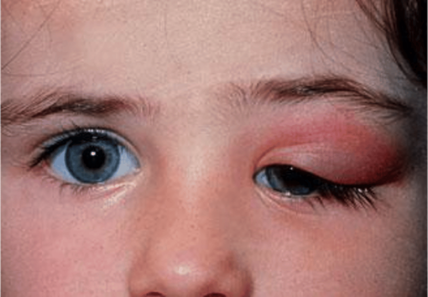
What is dacryoadenitis?
Dacryoadenitis is inflammation of lacrimal (tear-producing) glands usually caused by bacteria or a virus that initiates the inflammation (supratemporal)
What is dacryocystitis?
Dacryocystitis is infectious obstruction of the nasolacrimal duct (inferomedial region)
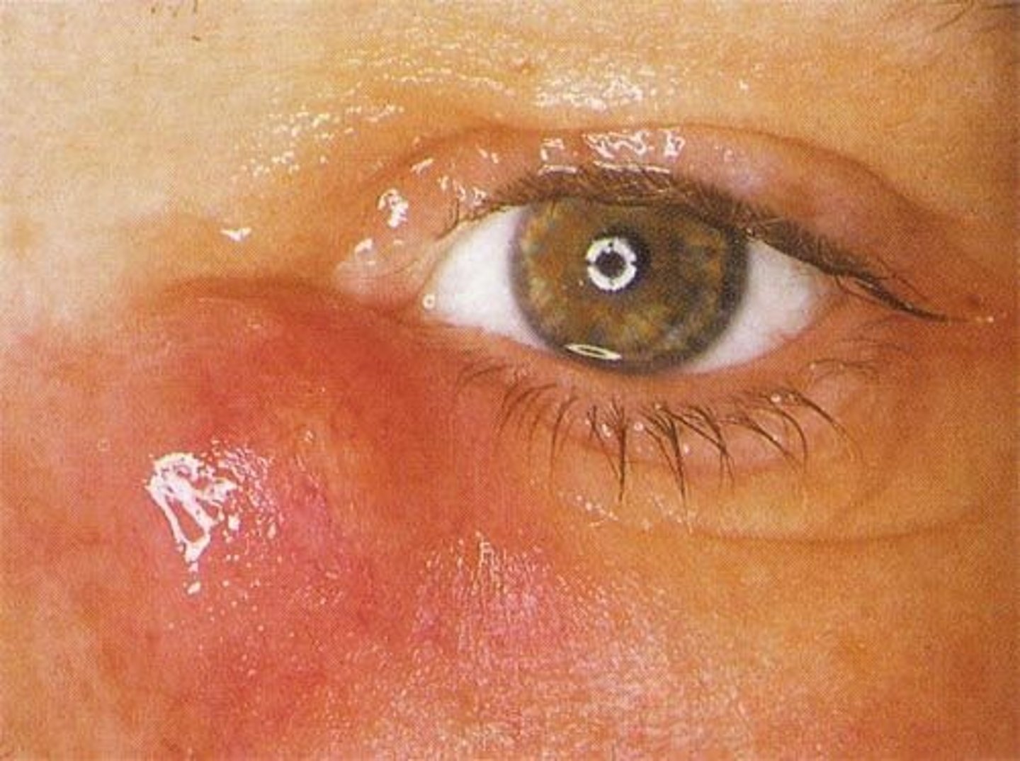
Is dacryocystitis and dacryoadenitis usually unilateral or bilateral?
Unilateral
Does dacryocystitis or dacryoadenitis appear medially?
On the boards you may be able to differentiate between the two based on the location: Dacryocystitis will be medial. Remember medial = C enter = dacroCystitis. Vs. dacryoAdenitis which up, up, and A way
Causes of acute dacryoadenitis?
Acute dacryoadenitis is most commonly due to viral or bacterial infection. Common causes include mumps, Epstein-Barr virus, staphylococcus, and gonococcus.
Causes of chronic dacryoadenitis?
Chronic dacryoadenitis is most often due to noninfectious inflammatory disorders. Examples include sarcoidosis, thyroid eye disease, and orbital pseudotumor.
Causes of dacryoadenitis?
Often caused by a stone, debris, or dacryostenosis (would be seen in small children) = congenital malformation or failure of duct to open
Diagnosis of dacryocystitis and dacryoadenitis?
The diagnosis is based on clinical observation. CT orbits if chronic
Treatment of dacryoadenitis?
If the cause of dacryoadenitis is a viral condition such as mumps, simple rest and warm compresses may be all that is needed. For other causes, the treatment is specific to the causative disease
Treatment of dacryocystitis?
● Acute dacryocystitis (< 3 months) is treated with systemic antibiotics
● Chronic dacryocystitis (> 3 months) typically presents with less inflammatory signs and requires surgical therapy for the underlying cause.
What is a chalazion?
A chalazion is noninfectious obstruction of a Meibomian gland causing extravasation of irritating lipid material in the eyelid soft tissues with focal secondary granulomatous inflammation
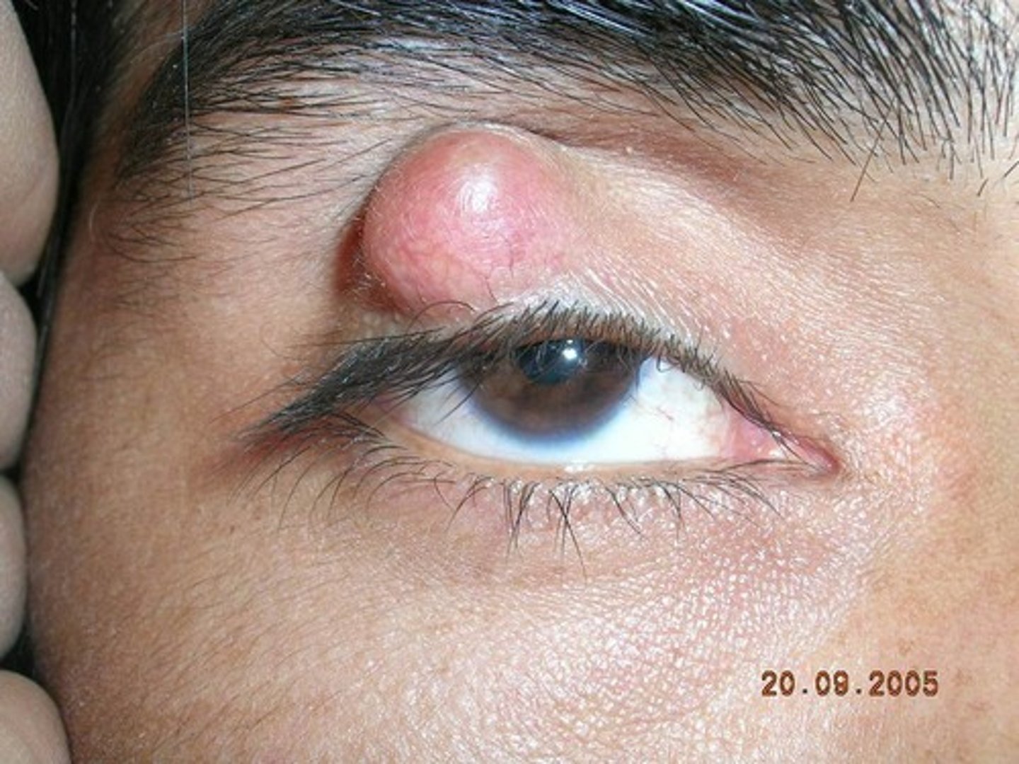
Do chalazion occur more commonly in the upper or lower eyelid?
Upper! Chalazion occur more commonly in the upper eyelid because of the presence of more sebaceous glands.
How do you differentiate a chalazion from a hordeolum (stye)?
Unlike a hordeolum (stye), a chalazion tends to have a more gradual onset, is less painful, and affects the middle part of the eyelid. Remember: "C" = Chalazion = Chronic and "Cold" (versus hordeolum which is "hot", acute and not chronic)
What is the treatment of chalazion?
Treat with warm compresses, and eyelid hygiene
Injection of corticosteroid or incision + curettage may be necessary in large ones affecting vision
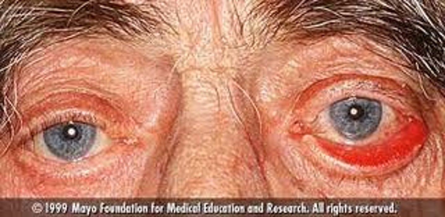
What is blepharitis?
Chronic inflammation of the eyelids without mass and without significant pain
What is the main cause of blepharitis?
Blepharitis is caused by dysfunctional Meibomian gland or staph infection
Is blepharitis usually unilateral or bilateral?
Bilateral. Blepharitis usually affects both eyes along the edges of the eyelids
What are two conditions associated with blepharitis?
Sborrhea and rosacea are associated with blepharitis
Diagnosis of blepharitis?
Slit lamp exam
Treatment of blepharitis?
Lid scrubs with diluted baby shampoo on cotton-tipped swab
● Massage to express gland
● Topical antibiotics if infection spreads
● Oral antibiotics for recalcitrant cases
What is ectropion?
Ectropion occurs with eversion (turning inside out) of the lower eyelid exposing the palpebral conjunctiva
What is entropion?
Entropion occurs when the eyelid turns in (inversion)
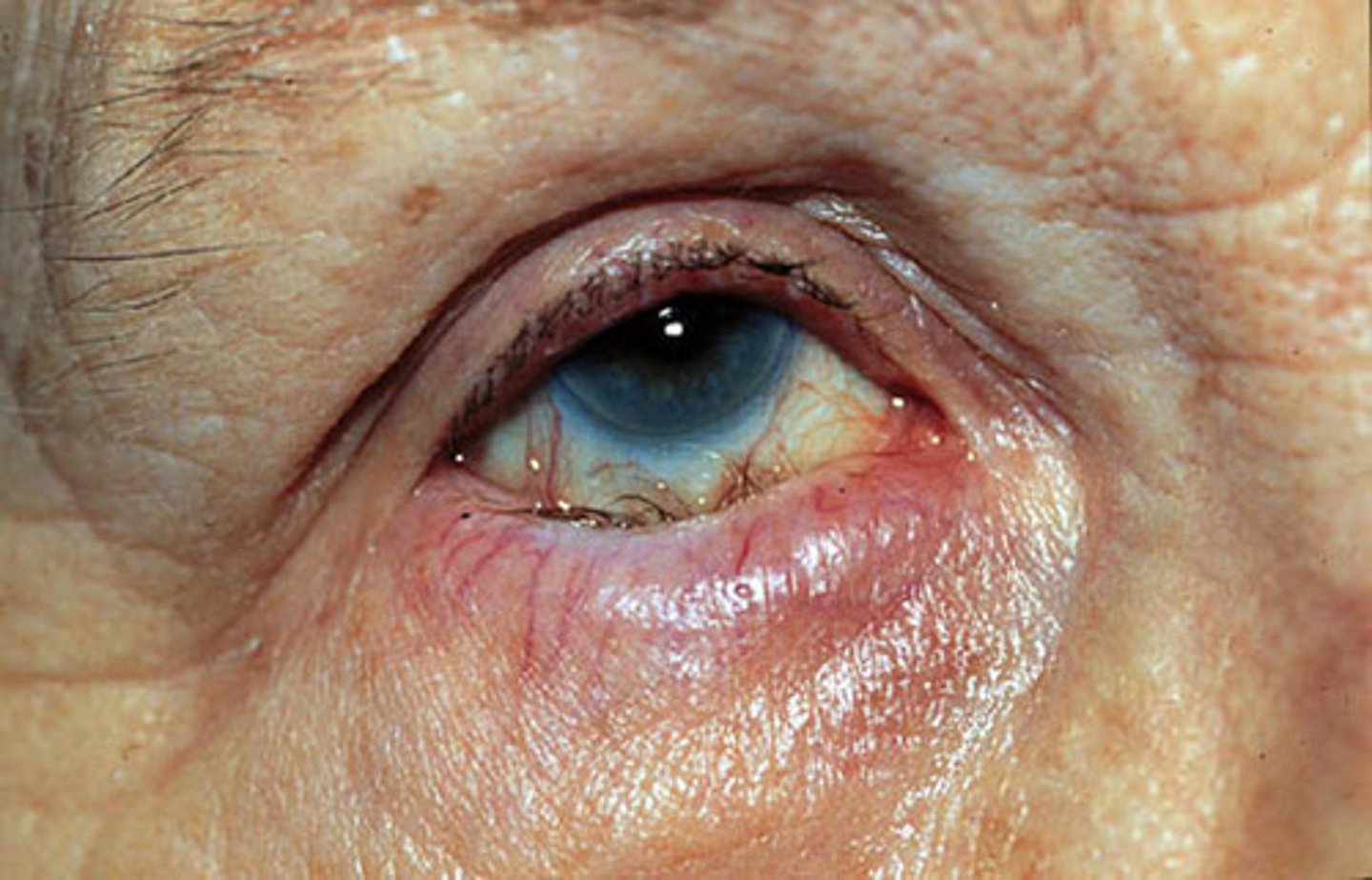
Symptoms of ectropion?
Symptoms of ectropion include tearing (due to poor drainage of tears through the nasolacrimal system, which may no longer contact the eyeball) and symptoms of dry eyes
Symptoms of entropion?
With an entropion the eyelashes rub against the eyeball and may lead to corneal ulceration and scarring. Symptoms can include foreign body sensation, tearing, and red eye
Diagnosis of entropion and ectropion?
The diagnosis is clinical
Treatment of ectropion?
Symptomatic treatment can include tear supplements and, at night, ocular lubricants; definitive treatment is surgery
Treatment of entropion?
Definitive treatment is surgery
What is a stye/hordeolum?
A painful red infection in a gland at the margin of the eyelid
Styes are usually caused by what infectious organism?
Staphylococcus aureus
What is a complication of a stye?
A complication of styes is the progression into a chalazion which is a cyst in the eyelid that can persist for a long time.
Is the onset of symptoms of a stye shorter or longer than that of a chalazion?
The onset of manifestations of a stye is usually shorter than that of a chalazion
What is the treatment of a stye?
Warm compress and topical antibiotics. A hordeolum that does not respond to hot compresses can be incised with a sharp, fine-tipped blade. Systemic antibiotics (eg, dicloxacillin or erythromycin 250 mg PO QID) are indicated when cellulitis accompanies a hordeolum
What is nystagmus?
Nystagmus is an involuntary, rapid and repetitive movement of the eyes. Usually, the movement is side-to-side (horizontal nystagmus), but it can also be up and down (vertical nystagmus) or circular (rotary nystagmus). The movement can vary between slow and fast, and it usually involves both eyes. It can have various causes.
Three types of nystagmus?
Horizontal, vertical, rotary
Two additional classification of nystagmus?
Nystagmus can be congenital and acquired
● Congenital nystagmus develops in infancy, usually between six weeks and three months of age. Congenital or inherited nystagmus is not typically associated with serious medical conditions.
● Acquired nystagmus occurs later in life and has a variety of causes, including an association with serious medical conditions
Gaze-evoked nystagmus?
Failure of gaze holding. Clinically, this usually manifests as a drift of the eyes toward the primary position. Most common and often benign.
Down/upbeat nystagmus?
CNS dysfunction
Vestibular horizontal nystagmus
Labyrinth or vestibular nerve dysfunction
A 34-year-old woman develops blurred vision associated with unilateral retro-orbital pain that is worse with eye movement. On physical examination, her visual acuity is decreased but extraocular eye movements are intact. Funduscopic examination reveals mild papillitis but no exudates or hemorrhages. What is the most likely diagnosis?
Optic neuritis
What is optic neuritis?
Acute inflammatory demyelination of the optic nerve
Presentation of optic neuritis?
Patients with optic neuritis will present with acute monocular vision loss and pain in the affected eye.
What is the most common cause of optic neuritis?
Multiple sclerosis is the most common cause and optic neuritis is often the initial presenting symptom. Infections, tumors, drugs, and toxins are other possible causes.
Optic neuritis is associated with what medication used in the treatment of tuberculosis?
Optic neuritis is associated with the use of ethambutol
Think ethambutol for "eyes"
How is optic neuritis diagnosed?
Optic neuritis is suspected in patients with characteristic pain and vision loss, particularly if they are young. Neuroimaging, preferably with gadolinium-enhanced MRI of the brain and orbits, is usually done and may show an enlarged, enhancing optic nerve. Fundoscopy will also demonstrate inflammation of the optic disc.
What is the treatment of optic neuritis?
Corticosteroids and other treatments can be given, particularly if multiple sclerosis is suspected.
What is papilledema?
Papilledema is swelling of the optic disk due to increased intracranial pressure
Papilledema is a sign of what?
Increased intracranial pressure (ICP)
Causes of papilledema?
Causes of papilledema include:
● Brain tumor or abscess
● Cerebral trauma or hemorrhage
● Meningitis
● Cavernous or dural sinus thrombosis
● Encephalitis
● Severe HTN
Diagnosis of papilledema?
Diagnosis is by ophthalmoscopy with further tests - Immediate neuroimaging and, if no mass lesion is seen, obtain CSF for analysis and measure CSF pressure with a lumbar puncture.
On lumbar puncture what confirms increased intracranial pressure?
Increased opening pressure with lumbar puncture confirms increased intracranial pressure
Management of papilledema?
Urgent treatment of the underlying disorder is indicated to decrease intracranial pressure. If intracranial pressure is not reduced, secondary optic nerve atrophy and vision loss eventually occur, along with other serious neurologic sequelae.
A child with a sinus infection presents with proptosis; a red, swollen eyelid; and an inferolaterally displaced globe. What is the diagnosis?
Orbital cellulitis and abscess associated with ethmoid sinusitis